Introduction
Non-small cell lung cancer (NSCLC) is the most
common type of lung cancer, and a major public health burden
(1). Therefore, the molecular
analysis of NSCLC is necessary to identify novel molecular targets
for determining the disease prognosis and informing the design of
targeted therapies.
There are 4 dickkopf (DKK) proteins: DKK1, 2, 3 and
4. DKK1 antagonizes the canonical Wnt signaling associated with the
pathogenesis of malignant tumors by forming a ternary complex with
LDL receptor-related proteins (2,3). High
levels of DKK1 expression have been observed in multiple lung
cancer cell lines and lung cancer tissue samples (4). DKK1 is expressed at very low levels in
normal adult human tissues, and higher levels in embryonic tissues
(5). The use of DKK1 and other
biomarkers may increase the sensitivity of lung cancer detection.
Our previous study demonstrated that the activity of the
DKK1 promoter was low in lung cancer cells (6). Thus, it is likely that enhancer elements
outside the basal promoter region contribute to its wide range of
expression patterns (7).
Promoters, located at the 5′ends of genes, surround
the transcriptional start site and act to initiate transcription
(8,9).
However, it is not fully understood how one transcriptional
regulatory element (TRE) can precisely define a human genetic
locus. Furthermore, not all functional elements have yet been
determined for each transcript. In general, both a promoter and its
associated transcript are embedded in a collection of positively
and negatively regulating elements, which can be positioned
anywhere in relation to the transcriptional start site, including
at 5′, 3′ and intronic locations, and may act across large segments
of DNA up to megabases in length (10). Therefore, the computational prediction
of functionally significant TREs is challenging. The Encyclopedia
of DNA Elements (ENCODE) project provides systematically mapped
regions of transcription, transcription factor association,
chromatin structure and histone modification (11,12). It
has integrated multiple technologies and approaches to identify and
define functional elements, including enhancer elements and
enhancer-blocking insulators. This collective effort may be used to
screen for potential evolutionarily conserved TREs of the
DKK1 promoter.
Regulatory elements have been characterized
downstream of a range of human genes, so analyzing multiple
intronic and extragenic DNase I hypersensitive sites (DHSs) at +27
kb around a gene in different epithelial cell types can identify
regions that are often associated with gene regulatory elements
(13,14). This strategy has previously been
successfully used to identify enhancers of the c-Myc and cystic
fibrosis transmembrane conductance regulator genes (15,16). Not
all DHSs contain TREs; they may be associated with structural
elements that function in chromatin organization instead, which may
have a major effect on the combination of DNA with transcriptional
regulation factors (17). Therefore,
the extent to which gene-specific combinatorial patterns of histone
modifications exist remains to be determined. Numerous covalent
histone modifications, including methylation and acetylation, which
occur mainly at N-terminal tails, affect gene transcription
(18). However, the extent to which
combinatorial patterns of histone modifications exist in the DHS
remains unknown. Histone modification is one of the most important
epigenetic factors in the regulation of gene expression (19,20). Thus,
active promoters are often marked by the acetylation of various
histone residues, including H3K4ac, H3K27ac and H3K9ac, or
methylation, including H3K4me1/2, H3K27me3 and H3K9me3 (21). This modification is largely conserved
across species.
With the development of methodology that can
evaluate regulatory elements in the whole genome (22), key regulatory elements of the
expression and function of one large proximal promoter fragment of
the DKK1 locus were investigated. The present study aimed to
comprehensively predict potential regulatory elements of the lung
cancer-specific DKK1 locus to identify widespread
enhancers.
Materials and methods
Cell lines and cell culture
A549, H446 and H460 human lung cancer cell lines,
and HEK293(T) cells were obtained from the American Type Culture
Collection (Manassas, VA, USA). The squamous cell carcinoma cell
line, Eca-109, and human umbilical vein endothelial cells (HUVECs)
were supplied by the Cell Bank of Type Culture Collection of the
Chinese Academy of Sciences (Shanghai, China). The 293 cells were
cultured in Dulbecco's modified Eagle's medium (DMEM; Gibco; Thermo
Fisher Scientific, Inc., Waltham, MA, USA) supplemented with 10%
fetal bovine serum (FBS; Gibco; Thermo Fisher Scientific, Inc.).
All other cells were cultured in RPMI 1640 medium (HyClone; GE
Healthcare Life Sciences, Logan, UT, USA) containing 10% FBS, and
all cells were maintained at 37°C in 5% CO2.
Computational prediction of TREs at
the DKK1 locus
To screen for evolutionarily conserved TREs in the
genetically identified DKK1 promoter, computational tools
were used to download and process information from the UCSC Genome
Browser (https://genome-cancer.ucsc.edu/), released by the
ENCODE project (23). A human genome
sequence of ~27 kb surrounding the lung cancer-specific DKK1
locus was downloaded. Enhancer predictions were performed by
separately analyzing histone modification patterns and the DHS of
DKK1 in A549 cells, HUVECs and normal human lung fibroblasts
(NHLFs). CCCTC binding factor (CTCF) is a multi-zinc finger protein
that binds to insulators (24). To
investigate whether histone methylation-defined chromatin domains
are demarcated by CTCF-bound insulators (25), the genome-wide distribution of CTCF
was determined in these cells. The enhancer modules were extended
to include the most adjacent blocks with high conservation scores
by using PhastCons conservation track and regulatory potential
track at the UCSC genome website. The top 12 enhancers located
within 3,500 bp upstream and 4,700 bp downstream of the DKK1
transcription start site in each cell were selected. Sequences and
detailed alignments are presented in Table I.
 | Table I.DKK1 distal promoter fragment
primers. |
Table I.
DKK1 distal promoter fragment
primers.
| Promoter
fragment | Length (bp) | Primer
sequences | Restriction
enzymes |
|---|
| DKK1-9
(−938-40) | 978 | F:
TCTCCACATTAGCCCACCAC |
HindIII/XhoI |
|
|
| R:
CTGCGGTCCCAGAGTCCT |
|
| DKK1-5
(533–40) | 573 | F:
ACTGCGACTCTAAAGGGTTAATG |
HindIII/XhoI |
|
|
| R:
CTGCGGTCCCAGAGTCCT |
|
| DKK1-3
(−350-40) | 390 | F:
CCCCTCGGCTCTGTAAAGTAT |
|
|
|
| R:
TGCGGTCCCAGAGTCCT |
|
| DKK1-2
(−282-40) | 320 | F:
CAAGTTCCCAGAGTTCCTGCT |
HindIII/XhoI |
|
|
| R:
CTGCGGTCCCAGAGTCCT |
|
| SV40 | 420 | F:
CGCAGCACCATGGCCTGA |
BgIII/Xho I |
|
|
| R:
TTGCAAAAGCCTAGGCCTCCA |
|
Strongest candidate TREs distally
regulate the DKK1 promoter
To interpret these datasets, it was confirmed that
the 12 candidate DNA sequences function as enhancers of
DKK1. Broad classes of chromatin states were distinguished,
referred to as promoters, enhancers, and insulators. To ensure that
elements capable of overcoming the effects of strong enhancers were
identified, the assay utilized an enhancer from the human β-globin
locus control region. Known as DHS II (HS2), this enhancer
functions in multiple cell lines, with multiple promoters (8,26).
Furthermore, the presence of HS2 provides a large window of
expression to reliably measure loss-of-function effects (27). The HS2 enhancer was incorporated into
silencer and TRE plasmids to confirm the ability to overcome strong
activating events, and as a measure of the interference between
promoter and enhancer interactions (28). However, it is not known whether HS2
has an enhancing effect on the DKK1 promoter. The expression
clones prepared for transfection are listed in Table II.
| Table II.Primer sequences of the 12 candidate
TREs. |
Table II.
Primer sequences of the 12 candidate
TREs.
| TRE | Locus | Length (bp) | Primer
sequences |
|---|
| TRE1 |
chr10:53,992,381–53,992,930 |
550 | F:
TGGTTCATATTTTGTTTTTCTTGTG |
|
|
|
| R:
ACCTAAGTTATTAAGTTTTGTCTCA |
| TRE2 |
chr10:54,016,954–54,017,517 |
565 | F:
TTTATGCTAAGACAAGGAGGTGT |
|
|
|
| R:
TTTTGAAGAATAAACATAACATGAGAA |
| TRE3 |
chr10:54,073,726–54,074,219 |
498 | F:
GCAAGGGCACCCAAGTTC |
|
|
|
| R:
CCAGAGCCATCATCTCAGAA |
| TRE4 |
chr10:54,078,638–54,079,344 |
707 | F:
AATGTCTGTTGTTGTTGCTGTG |
|
|
|
| R:
CCAGGCTCATTCTTATCAGTAG |
| TRE5 |
chr10:54,086,158–54,086,541 |
384 | F:
TTTTCATCCCTTTCCCTCACT |
|
|
|
| R:
GGGCAAGGAGAATCAGCTC |
| TRE6 |
chr10:54,128,561–54,128,810 |
250 | F:
CATGCCAGGCTCTCAGTAAG |
|
|
|
| R:
CTGTTGAGTCAGGGGTTTGG |
| TRE7 |
chr10:54,203,100–54,204,074 |
975 | F:
TCGCCTAGTGTATCTTTTAG |
|
|
|
| R:
TTTGTTGATATTTCATAATCATTGGA |
| TRE8 |
chr10:54,211,579–54,212,928 | 1,350 | F:
TATTCATTTGCATAAAAGATAAAGCC |
|
|
|
| R:
ATTTTCTTATTCATTCATTTCCTACCG |
| TRE9 |
chr10:54,218,904–54,219,195 |
293 | F:
GATGCTAAACATAGGTACTTTTGAA |
|
|
|
| R:
AATTTGACTATGGGCTTTTAGG |
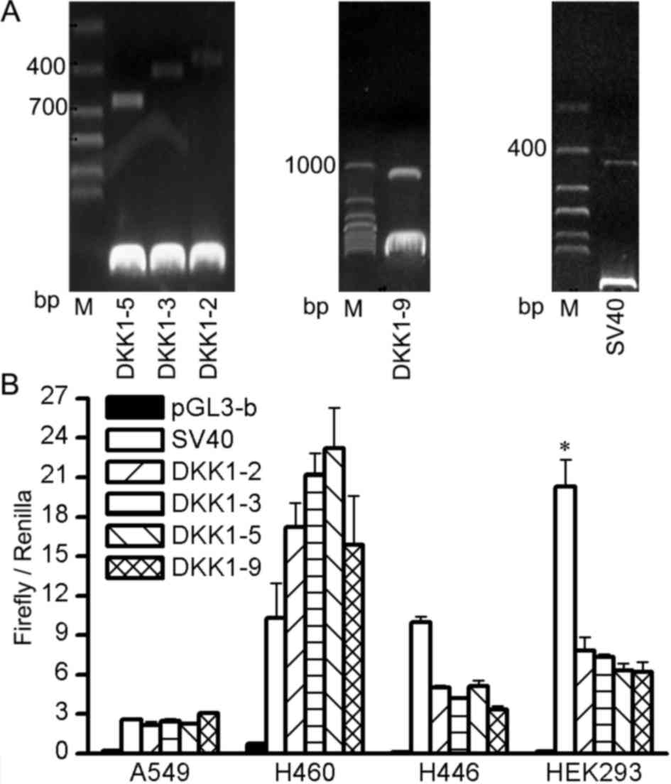
Construction of reporter plasmids
A total of 4 different fragments of the DKK1
promoter were amplified from A549 cell line genomic DNA, then
cloned into HindIII and XhoI sites upstream of the
firefly luciferase reporter gene in the pGL3-basic vector (Promega
Corporation, Madison, WI, USA) to produce pGL3-DKK1-2, pGL3-DKK1-3,
pGL3-DKK1-5 and pGL3-DKK1-9 constructs. The TK promoter-driven
Renilla luciferase reporter gene, derived from a commercial
vector (pRL-TK; Promega Corporation), was used to normalize for
firefly luciferase activity. As a control, the SV40
enhancer/promoter fragment was excised from pRL-TK and inserted in
forward and reverse orientations into the pGL3-basic plasmid at the
XhoI and BglII sites to produce pGL3-SV40. Genomic
DNA extraction, polymerase chain reaction (PCR) amplification and
concentration quantification were performed as previously described
(6). All primer sequences and cloning
sites are listed in Table I.
Subcloned fragments were confirmed by sequencing. Predicted TREs
were amplified by PCR according to the same protocol (6); Table II
presents the primer sequences. The products were ligated into the
multiple cloning site of the pGL3-DKK1-5 vector, upstream of a
candidate DKK1 promoter-driven luciferase reporter gene.
Plasmid transfection
Plasmids were purified using a Plasmid Midi kit
(Qiagen GmbH, Hilden, Germany) according to the manufacturer's
protocol. Cells were plated into 96-well plates at 8×10³
cells per well, 24 h prior to transfection, and were transfected at
80–90% confluence. The cells were co-transfected with the
recombined vectors carrying the firefly (50 ng) and Renilla
(1 ng) luciferase reporter genes. X-tremeGENE HP (Roche
Diagnostics, Basel, Switzerland) was added at 0.25 µl/well,
according to the manufacturer's protocol. The pGL3-basic vector (10
ng) and recombined pGL3-SV40 (10 ng) were used as negative
controls.
Firefly/Renilla luciferase
quantification
Luciferase activity was measured 48 h
post-transfection using Dual-Luciferase® Reporter Assay
System (Promega Corporation), according to the manufacturer's
protocol. Firefly luciferase reporter gene activity was normalized
to Renilla luciferase activity by calculating the
Renilla/firefly luciferase activity ratio for each
construct. The promoter control, DKK1-5, exhibited a
baseline level of luciferase activity, which was normalized to 1.
The activity of each test construct was calculated relative to the
DKK1 promoter construct. Transfections were performed in
triplicate, in 3 independent experiments.
Statistical analysis
The data are presented as the mean ± standard
deviation and were analyzed using SPSS 17.0 software (SPSS, Inc.,
Chicago, IL, USA) by one-way ANOVA with a Student-Newman-Keuls post
hoc test. P<0.05 was considered to indicate a statistically
significant difference.
Results
Identification of DKK1 DHSs
To identify evolutionarily conserved regulatory
elements, the genomic DNA sequence of A549 cells was downloaded
from the UCSC Genome Browser. DHSs were identified within the 27-kb
region of the human genome sequence surrounding DKK1 in the
non-cancer human cell lines, HUVECs and NHLFs. A total of 12
potential TREs were identified, the majority of which were located
within highly conserved sites of the DKK1 locus (Fig. 1A). The locations of these modules,
termed TRE 1–12, are presented in Table
II. The strongest signals, representing higher DNase I
hypersensitive sites and larger areas of histone interaction,
occurred around chr10: 54,073,726–54,074,219 (TRE3). Many of the
cancer-associated TREs are located within, or in close proximity
to, predicted DHSs, including TRE8, 10 and 12. TRE1, 2, 5 and 6
were associated with weak signaling compared with TRE3 and
TRE4.
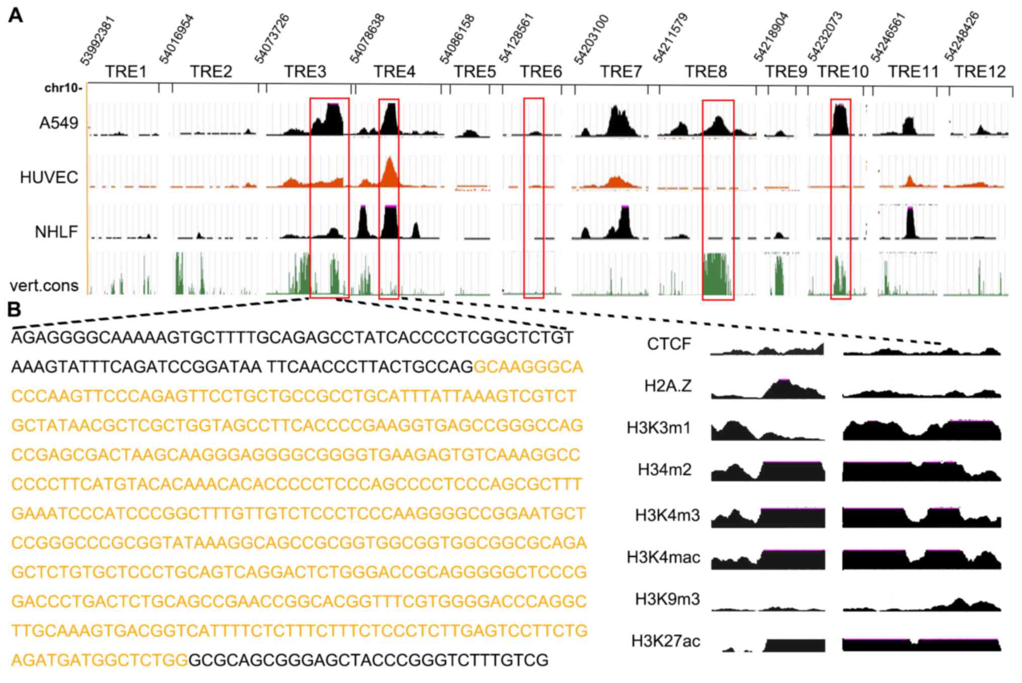 | Figure 1.Computationally predicted TREs in
DKK1, a lung cancer-associated gene. (A) A major DHS was
identified at the distal DKK1 promoter in 3 cell lines that
express the DKK1 gene, including a 100% vert.cons region.
(B) Histone modification patterns at the 2 DHSs of the DKK1
gene in A549; the highlighted sequence is the previously identified
TRE3. TRE, transcriptional regulatory element; DKK1,
dickkopf 1; DHS, DNase I hypersensitive sites; vert.cons, conserved
by vertebrates; chr, chromosome; CTCF, CCCTC binding factor;
HUVECs, human umbilical vein endothelial cells; NHLF, normal human
lung fibroblasts. |
Analysis of DKK1 histone modification
patterns
Fig. 1B illustrates
the histone modification patterns at the 2 hypersensitive sites of
the DKK1 gene in A549 cells. TRE maps of 6 histone
acetylation sites (H3K4me1, H3K4me2, H3K4me3, H3K4ac, H3K9ac and
H3K27ac), histone variant H2A.Z and a tissue-specific CTCF
recognition site in the data from 3 cell lines from ENCODE were
also analyzed, with no explicit knowledge of any previous
annotation (29). Data analysis
revealed H2A.Z, H3K4me1, H3K4me2, H3K4me3, H3K4ac and H3K27ac
signals in the intergenic region between TRE3 and 4, and TRE7, 8,
11 and 12 in both A549 and HUVEC cells, but not in NHLF cells.
Enrichment patterns of these modifications were mapped, and the
chromatin signatures for TRE1, 2, 5, 6, 9 and 10 were described.
H3K4me1, H3K4me2 and H3K27ac signals were highly localized to
intronic DHSs of TRE7 and 11 and 12 in HUVEC cells, while the other
signals were distributed over a broader region. Furthermore, the
significantly enriched CTCF signals at TRE6 and 10 suggest that
enhancer-blocking insulators flank the DKK1 locus.
DKK1-5 has high functionality as a
proximal promoter in different cells
Differences in relative luciferase activity were
observed among candidate promoters (Fig.
2). As was hypothesized, compared with an SV40 promoter, the
DKK1 promoter greatly increases luciferase activity in NSCLC
cell lines (A549 and H460) but not in SCLC cell lines (H446). In
contrast, DKK1 activity was lower than that of SV40 in
293(T) cells. There was no significant difference among the 4
segments (pGL3-DKK1-2, pGL3-DKK1-3, pGL3-DKK1-5 and pGL3-DKK1-9)
among the different cell types. Once length was taken into
consideration, the newly screened DKK1-5 fragment was determined to
have the most potential for application as a proximal promoter in
subsequent experiments.
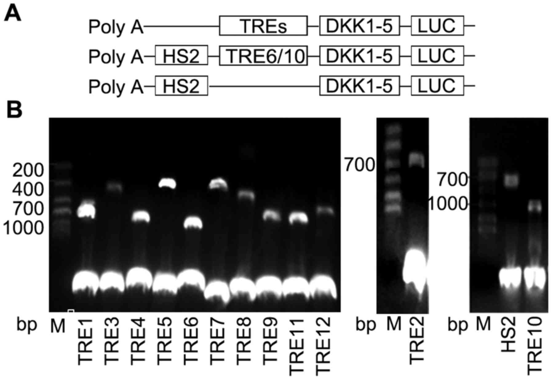
Construction of TRE-luciferase
plasmids
To determine whether the computationally identified
TREs possessed biological functionality, they were cloned upstream
of either a DKK1-5 promoter-driven luciferase reporter construct or
a native HS2 enhancer driven luciferase reporter construct
(Fig. 3A). The HS2 enhancer is well
characterized and has been used in similar reporter assays
(30–32). The candidate enhancers were cloned
into the enhancer site of the pGL3 DKK1-5 promoter vector. The
observed high CTCF signal predicted 2 insulating fragments, TRE6
and 10. As a silencing test, the insulator was subcloned upstream
of the DKK1-5 promoter and downstream of the HS2 enhancer to
determine whether the elements had a cooperative effect on
luciferase expression (Fig. 3A). The
reconstructed TRE reporter plasmids were validated by restriction
enzyme digestion (Fig. 3B).
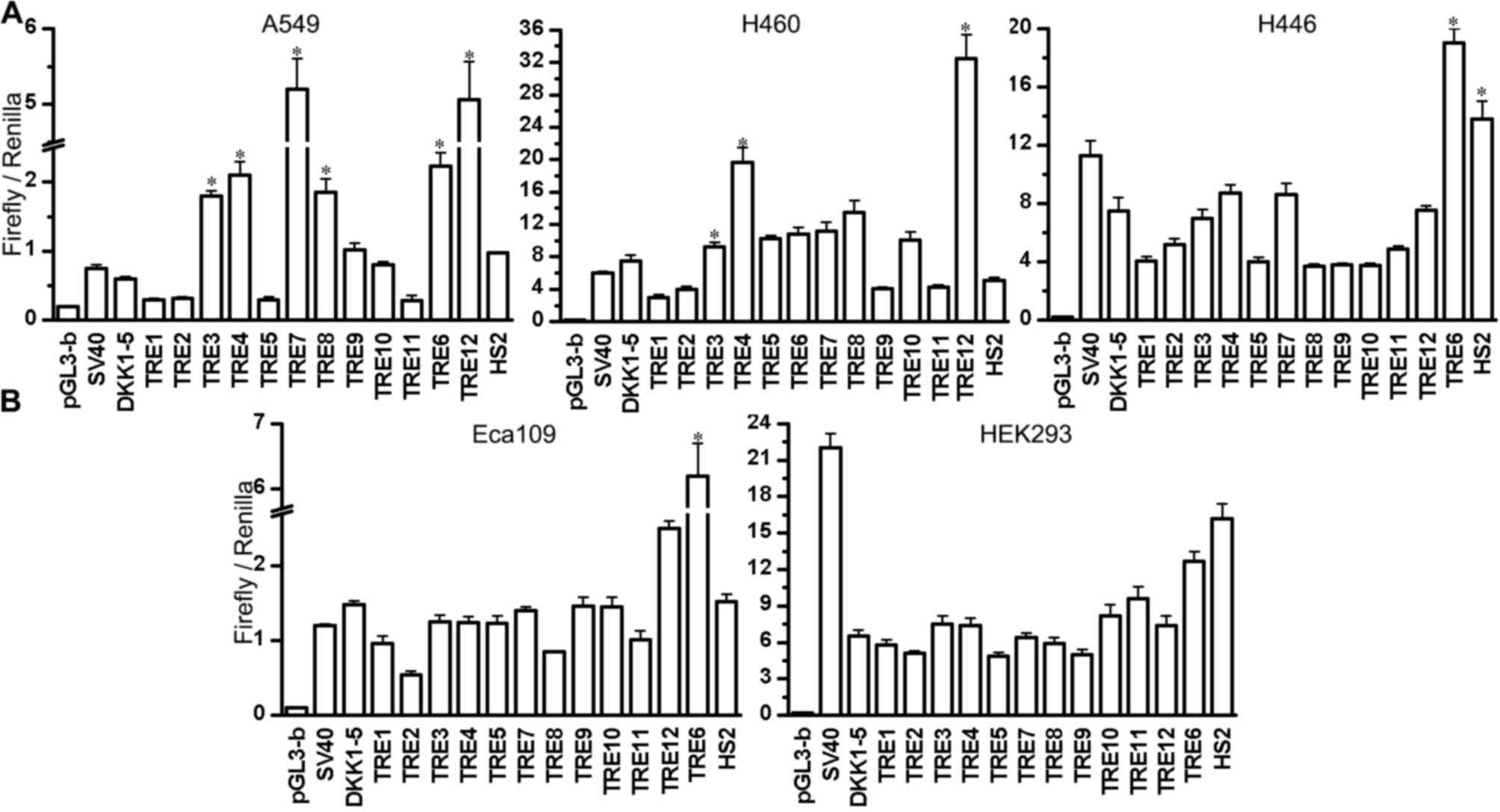
Functional validation of
transcriptional regulatory elements predicted at the DKK1
locus
The relative luciferase activity of
TRE-DKK1-luciferase constructs was investigated in A549 and H460
NSCLC cells, the small cell lung cancer cell line H446, the
esophageal cancer cell line Eca-109, and 293 cells (Fig. 4). The enhancer-control revealed the
upper threshold of expression contributed by the HS2 enhancer, and
was used as a normalization control. Luciferase reporter assays in
A549 cells revealed that TRE7 and 12 act as strong transcriptional
enhancers, leading to an 8-fold increase in DKK1 promoter
activity. TRE3, 4, 6 and 8 also increased the activity of the
DKK1 promoter in A549 cells, reflecting the DHS and histone
modification results (Fig. 1).
Importantly, HS2 together with TRE6 increased DKK1 promoter
activity in A549 and H446 cells, whereas HS2 together with TRE10
and HS2 alone had a minor or no effect. This was also observed in
H460 cells, albeit to a lesser extent. This suggests that TRE6 and
TRE10 do not act as a silencer in A549, H446 and H460 cells. In
H460 cells, it was demonstrated that TRE3, 4 and 12 significantly
increased the activity of the DKK1 promoter, while TRE6, 7
and 8 had little effect. Furthermore, TRE3, 4, 7 and 12 had no
effect on DKK1 activity in H446 cells. The majority of the
putative enhancers did not significantly stimulate the DKK1
promoter in Eca-109 or 293, except for TRE6 which resulted in high
DKK1 promoter activity in Eca-109 cell line.
Collectively, these results demonstrate that the 6
identified DNA sequences act as efficient TREs. TRE3, 4, 6, 7, 8
and 12 significantly increased the activity of the DKK1
promoter in A549 cells, and TRE7 increased the activity to the
highest degree. TRE3 and 4 increased promoter activity in H460
cells, while TRE12 showed a strong effect in A549, H460 and Eca-109
cells. Furthermore, TRE7 and 8 did not increase promoter activity
in H460 cells, suggesting that TRE3 and 4 may have
NSCLC-specificity.
Discussion
DKK1 has been reported to be a biomarker in lung
cancer, with high levels of specificity and expression (4). However, our previous study demonstrated
low DKK1 promoter activity in lung cancer cells (6). In the present study, 12 TREs were
predicted using DHS and histone modification analysis of the lung
cancer-specific DKK1 locus in A549 cells using the UCSC
genome browser. Several of these protocols were applied to
distinguish between active and poised enhancers. The functionality
of the TREs was investigated using a dual-luciferase reporter assay
in 5 different cell types. It was demonstrated that several of
these DHS regions possess cooperative enhancer activity in
vitro.
It has been identified that specific TREs may be
associated with low promoter activity (7,33).
However, to the best of our knowledge, no TREs were previously
defined for the lung cancer-specific expression of DKK1.
Methods to detect DNA sequence elements on a genome-wide scale and
to examine the role of different one-dimensional regulatory signals
remain a challenge to develop (34,35).
However, this work uses a computational approach to predict TREs
that contain cell line-specific long-range interactions between
enhancers and promoters. The present study also presents methods to
distinguish between different classes of functional TREs, which may
contribute to the elucidation the mechanisms for cell type-specific
gene expression.
The majority of predicted enhancers are distal to
core promoters, and can be detected by locating tissue-specific
DHSs within and around the gene (36). Previous studies have demonstrated that
a number of histone modification patterns of promoters, including
histone acetyltransferase p300, are enriched at the enhancer site
in fetal mouse tissues (37,38). Thus, it is likely that TREs are highly
evolutionary conserved and are marked by histone acetylation
(39) or the binding of coactivator
proteins such as p300 and MED1 (26).
The effect of insulators at human enhancers may contribute to the
understanding of how enhancers function in tissue-specific gene
regulation (40). H3K4me1/2, H3K27ac,
H3K9ac and H3K4ac modifications are associated with active
enhancers (39,41). The histone variant H2A.Z associates
with functional regulatory elements, and was previously
demonstrated to be enriched in active promoters and strong
enhancers, while the insulator binding protein, CTCF, marks the
boundaries of histone methylation domains (42). However, in the present study,
H3K9me1/3 and H3K27me3 signals were modestly elevated at silent
promoters, and little change was observed in the intergenic regions
of the cell lines. By integrating the predictions of DHSs and
selected histone modifications in A549, HUVEC and NHLF cell ENCODE
datasets, 12 candidate DNA sequences were proposed as potential
TREs. A set of cis-acting elements were characterized within the
DKK1 locus, and demonstrated that enhancer elements located
within cancer-associated regions can regulate DKK1 promoter
activity (Fig. 4). As the activity of
the DKK1 promoter was only increased in NSCLC cells, it is
likely that the most crucial sequences (TRE3, 4, 7 and 8) are
NSCLC-specific.
An important aspect of this study was its initial
predictive framework for the computational prediction of
enhancer-promoter interactions from datasets. The observed changes
in reporter activity indicate that these constructive TREs may have
potential regulatory ability. However, it should be noted that only
the specific TREs of DKK1 expression in cell lines were
examined in the present study, and that additional in vivo
studies are required to verify the results. Finally, the power of
these techniques as a strategy to predict and identify TREs on the
basis of their epigenetic characteristics, independent of motifs or
other sequence features, was demonstrated in the present study,
with particular respect to DHS mapping and histone acetylation
patterns. A detailed enhancer map may be used to inform future
therapeutic genome editing (43), as
demonstrated by a recent report applying the CRISPR-Cas9 system to
enhancers in situ, which confirmed that the BCL11A enhancer
may be a novel target for intractable diseases (44).
In conclusion, the present study confirms the value
of using computational tools to predict TREs, and provides the
first insight into the intronic cis-acting elements that control
the regulation of DKK1 expression. A systematic strategy to
predict cell line-specific enhancer-promoter interactions using a
minimal dataset is proposed. Furthermore, the present study
suggests that the active global enhancer network offers important
insights into cancer development and potentially, targeted
therapy.
Acknowledgements
The authors would like to thank Mr Li and Dr. Duan
(Department of Nuclear Medicine & Institute of Anesthesiology
and Pain, Taihe Hospital, Hubei University of Medicine) for the
reagent gifts, and for assistance with cell culture in our
laboratory. Their ideas also provided a valuable added dimension to
the study.
Funding
The present study was supported by the National
Natural Science Foundation of China (grant no., 81401447), the
Science and Technology Development Foundation of Shiyan City (grant
no., 16Y16) and the Key Discipline Project of Hubei University of
Medicine.
Availability of data and materials
The datasets used and/or analyzed during the current
study are available from the corresponding author on reasonable
request.
Authors' contributions
ZP and YG designed the study, interpreted the
experimental results and modified the manuscript. XD and JZ
constructed plasmids, interpreted results and drafted the
manuscript. YG and ZP prepared the figures. RW and YC performed
statistical analysis. WL and FL did computational prediction of
TREs at the DKK1 locus. HZ and YY cultured cells and measured
luciferase activity. All authors have read and approved the
manuscript.
Ethics approval and consent to
participate
Not applicable.
Consent for publication
Not applicable.
Competing interests
The authors declare that they have no competing
interests.
References
|
1
|
Siegel RL, Miller KD and Jemal A: Cancer
statistics, 2015. CA Cancer J Clin. 65:5–29. 2015. View Article : Google Scholar : PubMed/NCBI
|
|
2
|
Hadjihannas MV, Brückner M, Jerchow B,
Birchmeier W, Dietmaier W and Behrens J: Aberrant Wnt/beta-catenin
signaling can induce chromosomal instability in colon cancer. Proc
Natl Acad Sci USA. 103:10747–10752. 2006. View Article : Google Scholar : PubMed/NCBI
|
|
3
|
Niehrs C: Function and biological roles of
the Dickkopf family of Wnt modulators. Oncogene. 25:7469–7481.
2006. View Article : Google Scholar : PubMed/NCBI
|
|
4
|
Yamabuki T, Takano A, Hayama S, Ishikawa
N, Kato T, Miyamoto M, Ito T, Ito H, Miyagi Y, Nakayama H, et al:
Dikkopf-1 as a novel serologic and prognostic biomarker for lung
and esophageal carcinomas. Cancer Res. 67:2517–2525. 2007.
View Article : Google Scholar : PubMed/NCBI
|
|
5
|
Forget MA, Turcotte S, Beauseigle D,
Godin-Ethier J, Pelletier S, Martin J, Tanguay S and Lapointe R:
The Wnt pathway regulator DKK1 is preferentially expressed in
hormone-resistant breast tumours and in some common cancer types.
Br J Cancer. 96:646–653. 2007. View Article : Google Scholar : PubMed/NCBI
|
|
6
|
Xu R, Guo LJ, Xin J, Li WM, Gao Y, Zheng
YX, Guo YH, Lin YJ, Xie YH, Wu YQ and Xu RA: Luciferase assay to
screen tumour-specific promoters in lung cancer. Asian Pac J Cancer
Prev. 14:6557–6562. 2013. View Article : Google Scholar
|
|
7
|
Zhang C, Dai H and de Crombrugghe B:
Characterization of Dkk1 gene regulation by the osteoblast-specific
transcription factor Osx. Biochem Biophys Res Commun. 420:782–786.
2012. View Article : Google Scholar : PubMed/NCBI
|
|
8
|
Chung JH, Bell AC and Felsenfeld G:
Characterization of the chicken beta-globin insulator. Proc Natl
Acad Sci USA. 94:575–580. 1997. View Article : Google Scholar : PubMed/NCBI
|
|
9
|
Niu J, Wang J, Hu H, Chen Y, An J, Cai J,
Sun R, Sheng Z, Liu X and Lin S: Cross-talk between freezing
response and signaling for regulatory transcriptions of MIR475b and
its targets by miR475b promoter in Populus suaveolens. Sci Rep.
6:206482016. View Article : Google Scholar : PubMed/NCBI
|
|
10
|
Naumova N, Smith EM, Zhan Y and Dekker J:
Analysis of long-range chromatin interactions using Chromosome
Conformation Capture. Methods. 58:192–203. 2012. View Article : Google Scholar : PubMed/NCBI
|
|
11
|
Sanyal A, Lajoie BR, Jain G and Dekker J:
The long-range interaction landscape of gene promoters. Nature.
489:109–113. 2012. View Article : Google Scholar : PubMed/NCBI
|
|
12
|
Ecker JR, Bickmore WA, Barroso I,
Pritchard JK, Gilad Y and Segal E: Genomics: ENCODE explained.
Nature. 489:52–55. 2012. View
Article : Google Scholar : PubMed/NCBI
|
|
13
|
Deng T, Zhu ZI, Zhang S, Postnikov Y,
Huang D, Horsch M, Furusawa T, Beckers J, Rozman J, Klingenspor M,
et al: Functional compensation among HMGN variants modulates the
DNase I hypersensitive sites at enhancers. Genome Res.
25:1295–1308. 2015. View Article : Google Scholar : PubMed/NCBI
|
|
14
|
Sherwood RI, Hashimoto T, O'Donnell CW,
Lewis S, Barkal AA, van Hoff JP, Karun V, Jaakkola T and Gifford
DK: Discovery of directional and nondirectional pioneer
transcription factors by modeling DNase profile magnitude and
shape. Nat Biotechnol. 32:171–178. 2014. View Article : Google Scholar : PubMed/NCBI
|
|
15
|
Sotelo J, Esposito D, Duhagon MA, Banfield
K, Mehalko J, Liao H, Stephens RM, Harris TJ, Munroe DJ and Wu X:
Long-range enhancers on 8q24 regulate c-Myc. Proc Natl Acad Sci
USA. 107:3001–3005. 2010. View Article : Google Scholar : PubMed/NCBI
|
|
16
|
Uslu VV, Petretich M, Ruf S, Langenfeld K,
Fonseca NA, Marioni JC and Spitz F: Long-range enhancers regulating
Myc expression are required for normal facial morphogenesis. Nat
Genet. 46:753–758. 2014. View
Article : Google Scholar : PubMed/NCBI
|
|
17
|
Khurana E, Fu Y, Chakravarty D, Demichelis
F, Rubin MA and Gerstein M: Role of non-coding sequence variants in
cancer. Nat Rev Genet. 17:93–108. 2016. View Article : Google Scholar : PubMed/NCBI
|
|
18
|
Wang Z, Zang C, Rosenfeld JA, Schones DE,
Barski A, Cuddapah S, Cui K, Roh TY, Peng W, Zhang MQ and Zhao K:
Combinatorial patterns of histone acetylations and methylations in
the human genome. Nat Genet. 40:897–903. 2008. View Article : Google Scholar : PubMed/NCBI
|
|
19
|
Schones DE and Zhao K: Genome-wide
approaches to studying chromatin modifications. Nat Rev Genet.
9:179–191. 2008. View
Article : Google Scholar : PubMed/NCBI
|
|
20
|
Zhou VW, Goren A and Bernstein BE:
Charting histone modifications and the functional organization of
mammalian genomes. Nat Rev Genet. 12:7–18. 2011. View Article : Google Scholar : PubMed/NCBI
|
|
21
|
Ernst J and Kellis M: Large-scale
imputation of epigenomic datasets for systematic annotation of
diverse human tissues. Nat Biotechnol. 33:364–376. 2015. View Article : Google Scholar : PubMed/NCBI
|
|
22
|
Gallo SM, Gerrard DT, Miner D, Simich M,
Des Soye B, Bergman CM and Halfon MS: REDfly v3.0: Toward a
comprehensive database of transcriptional regulatory elements in
Drosophila. Nucleic Acids Res. 39:(Database Issue). D118–D123.
2011. View Article : Google Scholar : PubMed/NCBI
|
|
23
|
ENCODE Project Consortium: A user's guide
to the encyclopedia of DNA elements (ENCODE). PLoS Biol.
9:e10010462011. View Article : Google Scholar : PubMed/NCBI
|
|
24
|
Bell AC, West AG and Felsenfeld G: The
protein CTCF is required for the enhancer blocking activity of
vertebrate insulators. Cell. 98:387–396. 1999. View Article : Google Scholar : PubMed/NCBI
|
|
25
|
Cuddapah S, Jothi R, Schones DE, Roh TY,
Cui K and Zhao K: Global analysis of the insulator binding protein
CTCF in chromatin barrier regions reveals demarcation of active and
repressive domains. Genome Res. 19:24–32. 2009. View Article : Google Scholar : PubMed/NCBI
|
|
26
|
Petrykowska HM, Vockley CM and Elnitski L:
Detection and characterization of silencers and enhancer-blockers
in the greater CFTR locus. Genome Res. 18:1238–1246. 2008.
View Article : Google Scholar : PubMed/NCBI
|
|
27
|
Talbot D and Grosveld F: The 5′HS2 of the
globin locus control region enhances transcription through the
interaction of a multimeric complex binding at two functionally
distinct NF-E2 binding sites. EMBO J. 10:1391–1398. 1991.PubMed/NCBI
|
|
28
|
Elnitski L, Miller W and Hardison R:
Conserved E boxes function as part of the enhancer in
hypersensitive site 2 of the beta-globin locus control region role
of basic helix-loop-helix proteins. J Biol Chem. 272:369–378. 1997.
View Article : Google Scholar : PubMed/NCBI
|
|
29
|
Heintzman ND, Stuart RK, Hon G, Fu Y,
Ching CW, Hawkins RD, Barrera LO, Van Calcar S, Qu C, Ching KA, et
al: Distinct and predictive chromatin signatures of transcriptional
promoters and enhancers in the human genome. Nat Genet. 39:311–318.
2007. View
Article : Google Scholar : PubMed/NCBI
|
|
30
|
Wu C, Chen Z, Dardalhon V, Xiao S,
Thalhamer T, Liao M, Madi A, Franca RF, Han T, Oukka M and Kuchroo
V: The transcription factor musculin promotes the unidirectional
development of peripheral Treg cells by suppressing the TH2
transcriptional program. Nat Immunol. 18:344–353. 2017. View Article : Google Scholar : PubMed/NCBI
|
|
31
|
Fleetwood MR, Ho Y, Cooke NE and Liebhaber
SA: DNase I hypersensitive site II of the human growth hormone
locus control region mediates an essential and distinct long-range
enhancer function. J Biol Chem. 287:25454–25465. 2012. View Article : Google Scholar : PubMed/NCBI
|
|
32
|
Jane SM, Ney PA, Vanin EF, Gumucio DL and
Nienhuis AW: Identification of a stage selector element in the
human gamma-globin gene promoter that fosters preferential
interaction with the 5′HS2 enhancer when in competition with the
beta-promoter. EMBO J. 11:2961–2969. 1992.PubMed/NCBI
|
|
33
|
Mina M, Magi S, Jurman G, Itoh M, Kawaji
H, Lassmann T, Arner E, Forrest AR, Carninci P, Hayashizaki Y, et
al: Promoter-level expression clustering identifies time
development of transcriptional regulatory cascades initiated by
ErbB receptors in breast cancer cells. Sci Rep. 5:119992015.
View Article : Google Scholar : PubMed/NCBI
|
|
34
|
Schoenfelder S, Furlan-Magaril M, Mifsud
B, Tavares-Cadete F, Sugar R, Javierre BM, Nagano T, Katsman Y,
Sakthidevi M, Wingett SW, et al: The pluripotent regulatory
circuitry connecting promoters to their long-range interacting
elements. Genome Res. 25:582–597. 2015. View Article : Google Scholar : PubMed/NCBI
|
|
35
|
Wittkopp PJ and Kalay G: Cis-regulatory
elements: Molecular mechanisms and evolutionary processes
underlying divergence. Nat Rev Genet. 13:59–69. 2011. View Article : Google Scholar : PubMed/NCBI
|
|
36
|
Barski A, Cuddapah S, Cui K, Roh TY,
Schones DE, Wang Z, Wei G, Chepelev I and Zhao K: High-resolution
profiling of histone methylations in the human genome. Cell.
129:823–837. 2007. View Article : Google Scholar : PubMed/NCBI
|
|
37
|
Visel A, Prabhakar S, Akiyama JA, Shoukry
M, Lewis KD, Holt A, Plajzer-Frick I, Afzal V, Rubin EM and
Pennacchio LA: Ultraconservation identifies a small subset of
extremely constrained developmental enhancers. Nat Genet.
40:158–160. 2008. View Article : Google Scholar : PubMed/NCBI
|
|
38
|
Blow MJ, McCulley DJ, Li Z, Zhang T,
Akiyama JA, Holt A, Plajzer-Frick I, Shoukry M, Wright C, Chen F,
et al: ChIP-Seq identification of weakly conserved heart enhancers.
Nat Genet. 42:806–810. 2010. View
Article : Google Scholar : PubMed/NCBI
|
|
39
|
Heintzman ND, Hon GC, Hawkins RD,
Kheradpour P, Stark A, Harp LF, Ye Z, Lee LK, Stuart RK, Ching CW,
et al: Histone modifications at human enhancers reflect global
cell-type-specific gene expression. Nature. 459:108–112. 2009.
View Article : Google Scholar : PubMed/NCBI
|
|
40
|
Wendt KS, Yoshida K, Itoh T, Bando M, Koch
B, Schirghuber E, Tsutsumi S, Nagae G, Ishihara K, Mishiro T, et
al: Cohesin mediates transcriptional insulation by CCCTC-binding
factor. Nature. 451:796–801. 2008. View Article : Google Scholar : PubMed/NCBI
|
|
41
|
Ernst J, Kheradpour P, Mikkelsen TS,
Shoresh N, Ward LD, Epstein CB, Zhang X, Wang L, Issner R, Coyne M,
et al: Mapping and analysis of chromatin state dynamics in nine
human cell types. Nature. 473:43–49. 2011. View Article : Google Scholar : PubMed/NCBI
|
|
42
|
Rada-Iglesias A, Bajpai R, Swigut T,
Brugmann SA, Flynn RA and Wysocka J: A unique chromatin signature
uncovers early developmental enhancers in humans. Nature.
470:279–283. 2011. View Article : Google Scholar : PubMed/NCBI
|
|
43
|
Pougach K, Voet A, Kondrashov FA,
Voordeckers K, Christiaens JF, Baying B, Benes V, Sakai R, Aerts J,
Zhu B, et al: Duplication of a promiscuous transcription factor
drives the emergence of a new regulatory network. Nat Commun.
5:48682014. View Article : Google Scholar : PubMed/NCBI
|
|
44
|
Canver MC, Smith EC, Sher F, Pinello L,
Sanjana NE, Shalem O, Chen DD, Schupp PG, Vinjamur DS, Garcia SP,
et al: BCL11A enhancer dissection by Cas9-mediated in situ
saturating mutagenesis. Nature. 527:192–197. 2015. View Article : Google Scholar : PubMed/NCBI
|