Introduction
As the main histological subtype of liver cancer,
hepatocellular carcinoma (HCC) is the third-leading cause of
cancer-associated mortality worldwide (1,2). There are
>700,000 incidences of morality due to HCC annually (3,4). China in
particular has a high incidence rate of HCC, accounting for over
half of new cases and mortalities, with >422,100 HCC-associated
mortalities in 2015 (5,6). Extensive studies have shown that the
long-term prognosis of HCC remains dismal following radical
excision owing to the high frequencies of tumor recurrence and
distant metastases (6). Therefore, it
is necessary to identify novel prognostic factors to guide clinical
disease management following surgical resection.
A-kinase anchoring protein 1 (AKAP1, also known as
AKAP121 and AKAP149) is a scaffold protein that integrates protein
kinase A (PKA) and other signaling components to the outer
mitochondrial membrane (7). Recent
studies have revealed the roles of AKAP1 in regulating
mitochondrial function, oxidative metabolism and cell survival
(8,9).
AKAP1 recruits the PKA holoenzyme to specific subcellular sites and
substrates, which is critical for the physiological actions of the
kinase (10). AKAP1 also interacts
with protein-tyrosine phosphatase D1 (PTPD1), a non-receptor
tyrosine phosphatase that activates Src tyrosine kinase by binding
with it (11).
In the heart, AKAP1 deficiency promotes cardiac
mitochondrial aberrations and mitophagy, which enhances infarct
size, pathological cardiac remodeling and increases the mortality
rate from ischemic conditions (9). A
more recent study (12) also
demonstrated that AKAP1 is a transcriptional target of Myc and
supports the growth of cancer cells. Upregulation of AKAP1 in
high-grade human tumors is associated with enhanced mechanistic
target of rapamycin (mTOR) activation and reduced patient survival.
Knockdown of AKAP1 inhibited the mTOR pathway and impaired
glioblastoma growth. However, the expression pattern and
significance of AKAP1 in HCC remains to be investigated.
Despite the growing evidence of a link between AKAP1
and cancer, little is known about the expression level of AKAP1 and
its clinicopathological significance in human HCC. The present
study evaluated AKAP1 expression in paired tumorous and
non-tumorous HCC samples. Next, the prognostic value of AKAP1 was
determined in patients with HCC who previously underwent
hepatectomies.
Patients and methods
Patients and samples
Formalin-fixed, paraffin-embedded HCC tissues from
141 male and 17 female patients (age range, 18 to 78 years; median
age, 50 years) who underwent curative resection at Eastern
Hepatobiliary Surgery Hospital (Shanghai, China) between September
2006 and July 2011 were randomly retrieved. Another 30 pairs of
fresh HCC tissues and the corresponding adjacent non-tumorous
tissues (peritumoral tissues) were collected (26 male and 4 female
patients; age range, 32 to 72 years; median age, 51 years) for
reverse transcription-quantitative polymerase chain reaction
(RT-qPCR). All human sample collection procedures were approved by
the Biomedical Ethics Committee of Eastern Hepatobiliary Surgery
Hospital (Shanghai, China). Informed consent for tissue banking and
future medical research was obtained from each participant prior to
surgery. The diagnosis of HCC was confirmed by pathological tests
in all patients. The clinical staging was determined by the
Tumor-Node-Metastasis (TNM) classification system (13). All patients were followed up until
December 2014. The overall survival (OS) time was defined as the
interval between the dates of surgery and death, and the
disease-free survival (DFS) time was defined as the interval
between the dates of surgery and first incidence of recurrence. If
recurrence was not diagnosed, patients were censored at the date of
mortality or the last follow-up.
Public database
A publically available cohort of human HCC patients
from the National Center for Biotechnology Information Gene
Expression Omnibus (GEO) dataset GSE45436 (https://www.ncbi.nlm.nih.gov/geo/query/acc.cgi?acc=GSE45436)
was used. This dataset included data from 39 samples of normal
liver tissues and 95 samples of HCC tissues.
RT-qPCR
Total RNA was extracted from the fresh samples using
TRIzol reagent (Invitrogen; Thermo Fisher Scientific, Inc.,
Waltham, MA, USA), following manufacturer instructions. RNA was
then reversed transcribed to cDNA with Superscript III RT
(Invitrogen; Thermo Fisher Scientific, Inc.) and random primers.
Levels of AKAP1 and 18S were measured by SYBR Green qPCR Master Mix
(Takara Bio, Inc., Otsu, Japan) using an ABI PRISM7300HT Sequence
Detection system (Applied Biosystems; Thermo Fisher Scientific,
Inc.). The 18S rRNA expression level in the corresponding tissue
was used as an internal control. Primers were designed as follows:
AKAP1 forward, 5′-GCTTACGGCTTGTACCTGAAG-3′ and reverse,
5′-ATGGTGCTCTTGGAAATACGC-3′; and 18S forward,
5′-CGGCTACCACATCCAAGGAA-3′ and reverse, 5′-GCTGGAATTACCGCGGCT-3′.
The PCR reactions were performed under the following conditions:
Initial denaturation at 95°C for 15 min, followed by 40 cycles of
95°C for 30 sec, 60°C for 20 sec and 72°C for 30 sec. All samples
were run in triplicate, and the data were normalized to the 18S
internal controls. The relative AKAP1 expression levels were
calculated based on the 2−ΔΔCq method (14).
Histopathological and
immunohistochemical evaluation
The formalin-fixed paraffin-embedded HCC tissue
blocks were cut into 5 µm-thick sections. Then sections were
deparaffinized at room temperature by 100% xylene (2 times, 5 min
each) and rehydrated using a graded alcohol series (2 changes of
100% ethanol for 5 min each; 95 and 70% ethanol for 5 min each).
Following incubation with 3% hydrogen peroxide for 20 min at room
temperature, to quench endogenous peroxidase activity, the sections
were processed for heat-induced antigen retrieval in sodium citrate
buffer (pH=6.0). After several washes with 0.01 M phosphate buffer,
the sections were incubated with 10% goat serum (Beyotime Institute
of Biotechnology, Shanghai, China) for 30 min at room temperature
to block non-specific binding. Next, the slides were incubated with
primary anti-AKAP1 antibody (cat no. 5203; dilution, 1:200; Cell
Signaling Technology, Inc., Danvers, MA, USA) overnight at 4°C.
After the primary antibody was washed off, anti-rabbit
peroxidase-conjugated secondary antibody (cat no. sc-2357;
dilution, 1:500; Santa Cruz Biotechnology, Inc., Dallas, TX, USA)
incubation was performed for 30 min at room temperature, and then
DAB reagent (Dako; Agilent Technologies, Inc., Santa Clara, CA,
USA) was utilized for detection. The staining of slides was
observed and images captured using an Olympus microscope (IX-70
Olympus Corporation, Tokyo, Japan). All sections were evaluated by
two independent pathologists who were blind to the
clinicopathological data of the patients.
The percentages of positive tumor cells were
semi-quantitatively graded as follows: 0, <5%; 1, 5–25%; 2,
26–50%; and 3, >50%. The staining intensity of tumor cells was
scored as follows: 0, negative staining; 1, weak staining; 2,
moderate staining; and 3, strong staining. The two scores were
multiplied to obtain the final immunoreactive score. High
expression of AKAP1 in tumor cells was defined as an immunoreactive
score ≥4.
Statistical analysis
Each experiment was performed in triplicate and data
are presented as the mean ± standard deviation. SPSS version 16
(SPSS, Inc., Chicago, IL, USA) was used for statistical analysis.
χ2 tests were applied to determine statistical
significance. Kaplan-Meier and Cox regression analyses were used to
perform survival analysis. P<0.05 was considered to indicate a
statistically significant difference.
Results
Increased expression of AKAP1 in
patients with HCC
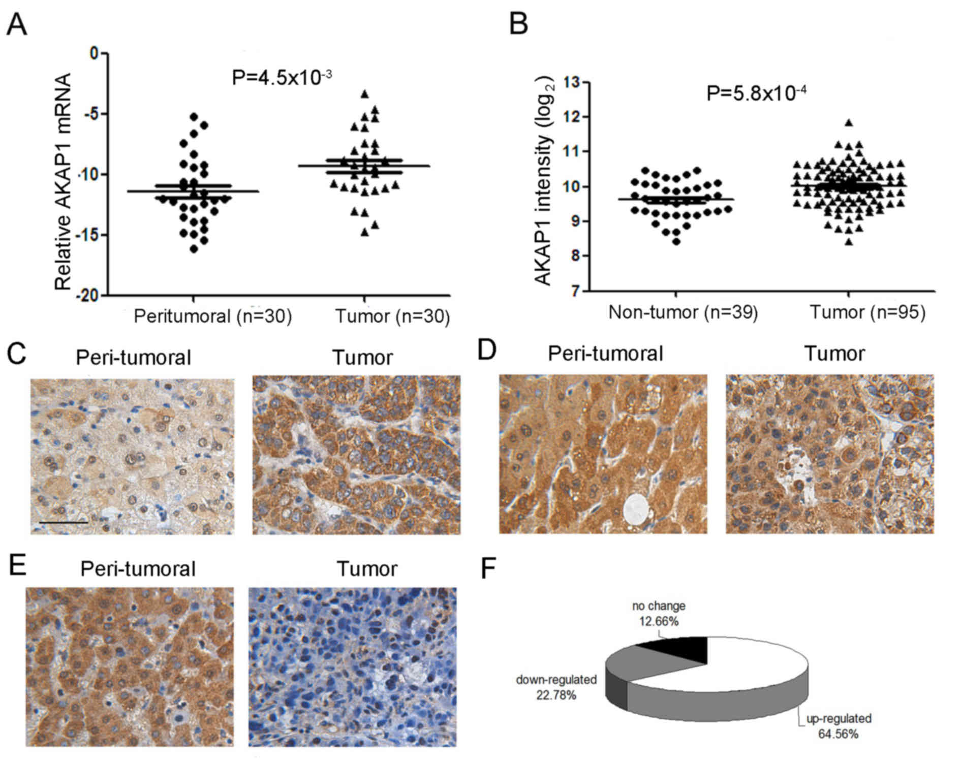
To determine the expression of AKAP1 in patients
with HCC, RT-qPCR assays were performed for 30 pairs of HCC tissues
and the corresponding adjacent non-tumorous tissues (peritumoral
tissues). As shown in Fig. 1A, AKAP1
mRNA levels were significantly higher in HCC tissues. Consistently,
analysis of public microarray data (GEO accession number GSE45436)
also revealed that AKAP1 is significantly upregulated in tumor
samples (Fig. 1B). To investigate
AKAP1 expression in HCC tissues further, its expression was
assessed in 158 cases of HCC and matched adjacent tissues by
immunohistochemical (IHC) staining. As shown in Fig. 1C-E, the expression pattern of AKAP1
varies significantly among the HCC samples. The expression of AKAP1
was significantly increased in HCC samples relative to paired
non-cancerous tissues in from 64.6% (102/158) of patients with HCC
(Fig. 1F).
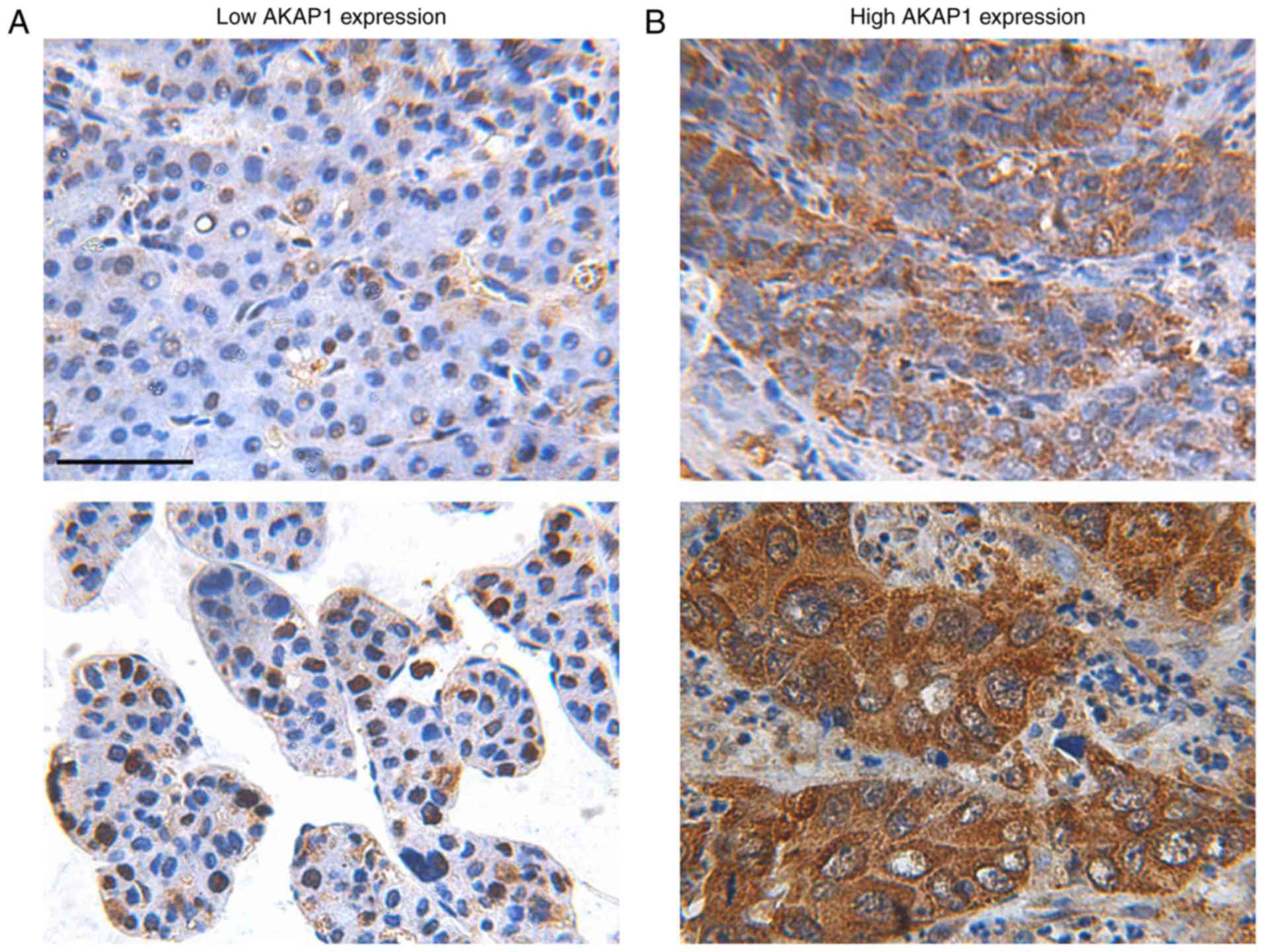
High AKAP1 expression is associated
with aggressive clinicopathological features
Based on IHC staining intensity and percentages of
positive tumor cells (Fig. 2), the
patients were subdivided into two groups: High (n=107) and low
(n=51) AKAP1 expression groups. As depicted in Table I, AKAP1 expression levels were
significantly higher in HCC patients with increased tumor size
(P=0.024), portal venous invasion (P=0.00498), and late TNM stage
(P=0.0296). These data indicate that high AKAP1 expression is
associated with aggressive clinicopathological features.
 | Table I.Association between AKAP1 expression
and clinicopathological characteristics in 158 hepatocellular
carcinoma patients. |
Table I.
Association between AKAP1 expression
and clinicopathological characteristics in 158 hepatocellular
carcinoma patients.
| Variables | All cases | Low AKAP1 | High AKAP1 | P-value |
|---|
| Total | 158 | 51 | 107 |
|
| Sex |
|
|
| 0.414 |
|
Female | 17 | 4 | 13 |
|
| Male | 141 | 47 | 94 |
|
| Age, years |
|
|
| 0.847 |
|
<50 | 73 | 23 | 50 |
|
| ≥50 | 85 | 28 | 57 |
|
| HBV infection |
|
|
| 0.944 |
| Yes | 139 | 45 | 94 |
|
| No | 19 | 6 | 13 |
|
| Liver cirrhosis |
|
|
| 0.420 |
| Yes | 112 | 34 | 78 |
|
| No | 46 | 17 | 29 |
|
| AFP, ng/ml |
|
|
| 0.828 |
| ≥200 | 91 | 30 | 61 |
|
|
<200 | 67 | 21 | 46 |
|
| Tumor
multiplicity |
|
|
| 0.325 |
|
Single | 55 | 15 | 40 |
|
|
Multiple | 103 | 36 | 67 |
|
| Tumor size, cm |
|
|
| 0.024 |
| ≤5 | 52 | 23 | 29 |
|
|
>5 | 106 | 28 | 78 |
|
| Tumor
encapsulation |
|
|
| 0.395 |
|
Absent | 79 | 23 | 56 |
|
|
Present | 79 | 28 | 51 |
|
| Edmondson grade |
|
|
| 0.903 |
| I/II | 35 | 11 | 24 |
|
|
III/IV | 123 | 40 | 83 |
|
| Portal vein
thrombosis |
|
|
| 0.005 |
|
Absence | 117 | 45 | 72 |
|
|
Gross | 41 | 6 | 35 |
|
| Pathologic TNM
stage |
|
|
| 0.030 |
| Early
stage (I–II) | 92 | 36 | 56 |
|
| Late
stage (III) | 66 | 15 | 51 |
|
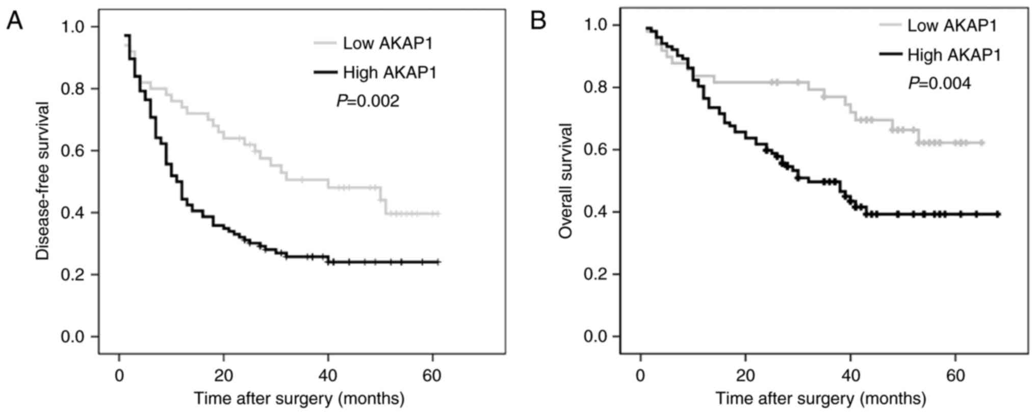
High AKAP1 expression predicts poor
overall survival (OS) and disease-free survival (DFS) rates in a
cohort of patients with HCC
To assess the association between AKAP1 expression
levels with survival of patients with HCC, Kaplan-Meier survival
analyses were performed. As depicted in Fig. 3, patients in the high expression group
exhibited poorer DFS (P=0.002) and OS (P=0.004) rates than those in
the low expression group. In addition, to determine whether AKAP1
expression level is an independent prognostic factor for DFS rate
and identify other prognostic factors for DFS rate in patients with
HCC who underwent curative resection, Cox regression analysis was
performed for 12 clinicopathological variables. Univariate analysis
demonstrated that AKAP1 expression level was associated with DFS
rate [hazard ratio (HR), 1.934; 95% confidence interval (CI),
1.243–3.01; P=0.003], and our multivariable Cox regression analyses
further confirmed that AKAP1 expression levels were an independent
risk factor of DFS rate (HR, 1.972; 95% CI, 1.177–3.306; P=0.01)
(Table II). These data indicated
that high AKAP1 expression in tumors was associated with disease
recurrence and poor survival rates for patients with HCC, and AKAP1
could also be used as a valuable prognostic factor for the DFS rate
of patients with HCC who underwent curative resection.
| Table II.Univariate and multivariate Cox
regression analysis of risk factors for disease-free survival
rate. |
Table II.
Univariate and multivariate Cox
regression analysis of risk factors for disease-free survival
rate.
|
| Univariate
analysis | Multivariable
analysis |
|---|
|
|
|
|
|---|
| Variables | Hazard ratio (95%
CI) | P-value | Hazard ratio (95%
CI) | P-value |
|---|
| Sex (male vs.
female) | 0.512
(0.249–1.055) | 0.070 |
|
|
| Age (≥50 vs. <50
years) | 0.876
(0.595–1.289) | 0.501 |
|
|
| HBV infection
(present vs. absent.) | 1.067
(0.585–1.948) | 0.832 |
|
|
| Liver cirrhosis
(present vs. absent) | 1.737
(1.083–2.787) | 0.022 | 1.681
(0.999–2.829) | 0.051 |
| AFP (≥400 ng/ml vs.
<400 ng/ml) | 1.245
(0.842–1.841) | 0.272 |
|
|
| Tumor multiplicity
(multiple vs. single) | 1.593
(1.073–2.365) | 0.021 | 0.963
(0.592–1.568) | 0.880 |
| Maximal tumor size
(≥5 vs. <5 cm) | 2.033
(1.303–3.174) | 0.002 | 2.110
(1.275–3.493) | 0.004 |
| Tumor encapsulation
(absent vs. present) | 1.707
(1.111–2.621) | 0.015 | 1.381
(0.865–2.206) | 0.176 |
| Edmondson grade
(III/IV vs. I/II) | 1.631
(0.981–2.71) | 0.059 |
|
|
| Portal vein
thrombosis (gross vs. absence) | 2.329
(1.543–3.516) | <0.001 | 1.461
(0.892–2.395) | 0.132 |
| Pathologic TNM
stage (III vs. I–II) | 1.800
(1.225–2.645) | 0.003 | 1.354
(0.861–2.130) | 0.189 |
| AKAP1 level (high
vs. low) | 1.934
(1.243–3.01) | 0.003 | 1.972
(1.177–3.306) | 0.010 |
Discussion
Recently, evidence indicates that metabolic
disorders have notable roles in HCC initiation, progression and
therapy resistance. Obesity and diabetes have long been recognized
as independent risk factors for the development of HCC (15–17).
As the energetic hub of the cell, mitochondria serve
important roles in regulating cell function and survival (18,19).
Mitochondrial dysfunction is a common trait of several human
diseases, including cancer (20–23). AKAP1
is a scaffolding protein that functions as the key regulatory
molecule responsible for controlling mitochondria function.
Therefore, it is necessary to investigate the role of AKAP1 in
cancer. In fact, the expression profiles and prognostic
implications of AKAP1 have been documented in human non-small cell
lung cancer, and AKAP1 overexpression is inversely associated with
patient survival (13). The present
study evaluated the expression level of AKAP1 in HCC for the first
time. Through studying human HCC clinical specimens, it was
revealed that AKAP1 was overexpressed in the majority of patients
with HCC, and high AKAP1 expression was associated with aggressive
clinicopathological features. These data indicated that AKAP1 may
contribute to progression of HCC.
Although early diagnosis and developments in surgery
improved the short-term survival rates of patients with HCC
(24), the long-term prognosis of
patients with HCC remains unsatisfactory, even following radical
excision. Therefore, it is necessary to reveal reliable biomarkers
for the effective identification of patients with HCC who have a
high risk of relapse following surgery. In the present study,
Kaplan-Meier survival analyses revealed that high AKAP1 expression
was associated with poor OS and DFS rates. Furthermore, univariate
and multivariate survival analyses indicated that AKAP1 expression
could serve as an independent factor for the DFS rate of patients
with HCC following hepatectomy.
In conclusion, the results of the present study
demonstrated that AKAP1 was overexpressed in the majority of
patients with HCC, and its increased expression was associated with
poor patient prognosis. AKAP1 may serve as a valuable prognostic
biomarker in predicting the survival of patients with HCC following
radical resection.
Acknowledgements
Not applicable.
Funding
This research was supported by the National Natural
Science Foundation of China (grant no. 81000971).
Availability of data and materials
The datasets used and/or analyzed during the current
study are available from the corresponding author on reasonable
request.
Authors' contributions
JY and RL designed the experiments. JY, YZ, DXZ and
JW performed the experiments, analyzed and interpreted the data,
and wrote the manuscript. RL organized and supervised the study.
All authors read and approved the final manuscript.
Ethics approval and consent to publish
All human sample collection procedures were approved
by the Biomedical Ethics Committee of Eastern Hepatobiliary Surgery
Hospital (Shanghai, China). Written informed consent for tissue
banking and future medical research was obtained from each
participant prior to surgery.
Consent for publication
Identifying information of patients were not
included in the manuscript. Informed consent for tissue banking and
future medical research was obtained from each participant prior to
surgery.
Competing interests
The authors declare that they have no competing
interests.
References
|
1
|
Ferlay J, Soerjomataram I, Dikshit R, Eser
S, Mathers C, Rebelo M, Parkin DM, Forman D and Bray F: Cancer
incidence and mortality worldwide: Sources, methods and major
patterns in GLOBOCAN 2012. Int J Cancer. 136:E359–E386. 2015.
View Article : Google Scholar : PubMed/NCBI
|
|
2
|
Bertuccio P, Turati F, Carioli G,
Rodriguez T, La Vecchia C, Malvezzi M and Negri E: Global trends
and predictions in hepatocellular carcinoma mortality. J Hepatol.
67:302–309. 2017. View Article : Google Scholar : PubMed/NCBI
|
|
3
|
Torre LA, Bray F, Siegel RL, Ferlay J,
Lortet-Tieulent J and Jemal A: Global cancer statistics, 2012. CA
Cancer J Clin. 65:87–108. 2015. View Article : Google Scholar : PubMed/NCBI
|
|
4
|
Sharma SA, Kowgier M, Hansen BE, Brouwer
WP, Maan R, Wong D, Shah H, Khalili K, Yim C, Heathcote EJ, et al:
Toronto HCC risk index: A validated scoring system to predict
10-year risk of HCC in patients with cirrhosis. J Hepatol:
S0168-8278(17)32248-1. 2017.
|
|
5
|
Chen W, Zheng R, Baade PD, Zhang S, Zeng
H, Bray F, Jemal A, Yu XQ and He J: Cancer statistics in China,
2015. CA Cancer J Clin. 66:115–132. 2016. View Article : Google Scholar : PubMed/NCBI
|
|
6
|
Fu J and Wang H: Precision diagnosis and
treatment of liver cancer in China. Cancer Lett. 412:283–288. 2018.
View Article : Google Scholar : PubMed/NCBI
|
|
7
|
Carlucci A, Adornetto A, Scorziello A,
Viggiano D, Foca M, Cuomo O, Annunziato L, Gottesman M and
Feliciello A: Proteolysis of AKAP121 regulates mitochondrial
activity during cellular hypoxia and brain ischaemia. EMBO J.
27:1073–1084. 2008. View Article : Google Scholar : PubMed/NCBI
|
|
8
|
Czachor A, Failla A, Lockey R and
Kolliputi N: Pivotal role of AKAP121 in mitochondrial physiology.
Am J Physiol Cell Physiol. 310:C625–C628. 2016. View Article : Google Scholar : PubMed/NCBI
|
|
9
|
Schiattarella GG, Cattaneo F, Pironti G,
Magliulo F, Carotenuto G, Pirozzi M, Polishchuk R, Borzacchiello D,
Paolillo R, Oliveti M, et al: Akap1 deficiency promotes
mitochondrial aberrations and exacerbates cardiac injury following
permanent coronary ligation via enhanced mitophagy and apoptosis.
PLoS One. 11:e01540762016. View Article : Google Scholar : PubMed/NCBI
|
|
10
|
Smith FD, Langeberg LK and Scott JD: The
where's and when's of kinase anchoring. Trends Biochem Sci.
31:316–323. 2006. View Article : Google Scholar : PubMed/NCBI
|
|
11
|
Cardone L, Carlucci A, Affaitati A,
Livigni A, DeCristofaro T, Garbi C, Varrone S, Ullrich A, Gottesman
ME, Avvedimento EV and Feliciello A: Mitochondrial AKAP121 binds
and targets protein tyrosine phosphatase D1, a novel positive
regulator of src signaling. Mol Cell Biol. 24:4613–4626. 2004.
View Article : Google Scholar : PubMed/NCBI
|
|
12
|
Rinaldi L, Sepe M, Delle Donne R, Conte K,
Arcella A, Borzacchiello D, Amente S, De Vita F, Porpora M, Garbi
C, et al: Mitochondrial AKAP1 supports mTOR pathway and tumor
growth. Cell Death Dis. 8:e28422017. View Article : Google Scholar : PubMed/NCBI
|
|
13
|
Bruix J and Sherman M: Practice Guidelines
Committee, American Association for the Study of Liver Diseases:
Management of hepatocellular carcinoma. Hepatology. 42:1208–1236.
2005. View Article : Google Scholar : PubMed/NCBI
|
|
14
|
Livak KJ and Schmittgen TD: Analysis of
relative gene expression data using real-time quantitative PCR and
the 2(-Delta Delta C(T)) method. Methods. 25:402–408. 2001.
View Article : Google Scholar : PubMed/NCBI
|
|
15
|
Starley BQ, Calcagno CJ and Harrison SA:
Nonalcoholic fatty liver disease and hepatocellular carcinoma: A
weighty connection. Hepatology. 51:1820–1832. 2010. View Article : Google Scholar : PubMed/NCBI
|
|
16
|
El-Serag HB, Tran T and Everhart JE:
Diabetes increases the risk of chronic liver disease and
hepatocellular carcinoma. Gastroenterology. 126:460–468. 2004.
View Article : Google Scholar : PubMed/NCBI
|
|
17
|
Yang JD, Mohamed HA, Cvinar JL, Gores GJ,
Roberts LR and Kim WR: Diabetes mellitus heightens the risk of
hepatocellular carcinoma except in patients with hepatitis C
cirrhosis. Am J Gastroenterol. 111:1573–1580. 2016. View Article : Google Scholar : PubMed/NCBI
|
|
18
|
Henze K and Martin W: Evolutionary
biology: Essence of mitochondria. Nature. 426:127–128. 2003.
View Article : Google Scholar : PubMed/NCBI
|
|
19
|
Weinberg SE and Chandel NS: Targeting
mitochondria metabolism for cancer therapy. Nat Chem Biol. 11:9–15.
2015. View Article : Google Scholar : PubMed/NCBI
|
|
20
|
Vazquez F, Lim JH, Chim H, Bhalla K,
Girnun G, Pierce K, Clish CB, Granter SR, Widlund HR, Spiegelman BM
and Puigserver P: PGC1alpha expression defines a subset of human
melanoma tumors with increased mitochondrial capacity and
resistance to oxidative stress. Cancer Cell. 23:287–301. 2013.
View Article : Google Scholar : PubMed/NCBI
|
|
21
|
Genini D, Brambilla L, Laurini E, Merulla
J, Civenni G, Pandit S, D'Antuono R, Perez L, Levy DE, Pricl S, et
al: Mitochondrial dysfunction induced by a SH2 domain-targeting
STAT3 inhibitor leads to metabolic synthetic lethality in cancer
cells. Proc Natl Acad Sci USA. 114:E4924–E4933. 2017. View Article : Google Scholar : PubMed/NCBI
|
|
22
|
Yuan D, Huang S, Berger E, Liu L, Gross N,
Heinzmann F, Ringelhan M, Connor TO, Stadler M, Meister M, et al:
Kupffer cell-derived Tnf triggers cholangiocellular tumorigenesis
through JNK due to chronic mitochondrial dysfunction and ROS.
Cancer Cell. 31:771–789.e6. 2017. View Article : Google Scholar : PubMed/NCBI
|
|
23
|
Viale A, Pettazzoni P, Lyssiotis CA, Ying
H, Sánchez N, Marchesini M, Carugo A, Green T, Seth S, Giuliani V,
et al: Oncogene ablation-resistant pancreatic cancer cells depend
on mitochondrial function. Nature. 514:628–632. 2014. View Article : Google Scholar : PubMed/NCBI
|
|
24
|
Bruix J, Reig M and Sherman M:
Evidence-based diagnosis, staging, and treatment of patients with
hepatocellular carcinoma. Gastroenterology. 150:835–853. 2016.
View Article : Google Scholar : PubMed/NCBI
|