Introduction
Thyroid nodule refers to a mass of abnormally
proliferating cells with abnormal local hardness in thyroid
(1). At present, ultrasonography of
thyroid nodules mainly relies on B-mode ultrasound diagnosis.
However, the diagnostic sensitivity and specificity by B-mode
ultrasound is not high (2,3). Ultrasound elasticity imaging, as a new
technology developed this year, can determine the mechanical
characteristics of tissues in vitro (4). The application value of ultrasound
elasticity imaging technology in breast cancer has been confirmed,
but its applications in the diagnosis of benign and malignant
thyroid nodules are relatively few. The present study compared
diagnostic values of B-mode ultrasound and elasticity imaging for
the identification of benign as well as malignant thyroid nodules.
This modality helps to improve the diagnosis rate of benign and
malignant thyroid nodules by ultrasound technology.
Materials and methods
General materials
Ninety-four confirmed cases (from January, 2011 to
January, 2015) of thyroid nodules by ultrasound in Zhengzhou
Central Hospital Affiliated to Zhengzhou University (Zhengzhou,
China) were chosen for the study. There were 46 male and 48 female
cases. The age of the patient ranged from 22–72 years and the
average age was 43.5±4.5 years.
Detection methods
Siemens Acuson Antares 5.0 Color Doppler Ultrasonic
Diagnosis Apparatus (Siemens, Erlangen, Germany) and VF13-5
frequency conversion probe were adopted as the detection
instrument. All study subjects were examined by B-mode ultrasound
and ultrasound elastography. Patients were kept in supine position
and were first given routine vertical and horizontal section
scanning of the neck. Nodule forms, sizes, boundaries, internal
echo, existence of calcification, surrounding acoustic halo, blood
flow conditions and cervical lymph nodes were observed by
two-dimensional ultrasound. Subjects were then analyzed by
elasticity imaging mode. We tried to make the probe cling to the
neck skin during the operation. It was better to make the nodule at
the center of sample frame. The size of sample frame was supposed
to be adjusted to about twice the size of the lesions. The probe
was held by hand and made to slightly vibrate at masses at middle
speed. We saved the picture, when stress figure on the device
monitor was between 3–4 grades. The color changes of elasticity
picture in the sample frame were observed (5).
Diagnostic criteria of B-mode
ultrasound
The features of benign thyroid nodules were regular
morphology, clear boundary, <1 aspect ratio, inside equal echo,
high echo, with or without an echo, coarse calcifications, no rear
echo reduction, no or a little blood flow. The resistive index of
blood flow was <0.7. The features of malignant thyroid nodules
included irregular forms, unclear boundary, the aspect ratio was
≥1, inside low echo, micro-calcification, rear echo reduction, rich
blood flow. The resistive index of blood flow was ≥0.7.
Diagnostic criteria of ultrasound
elastography
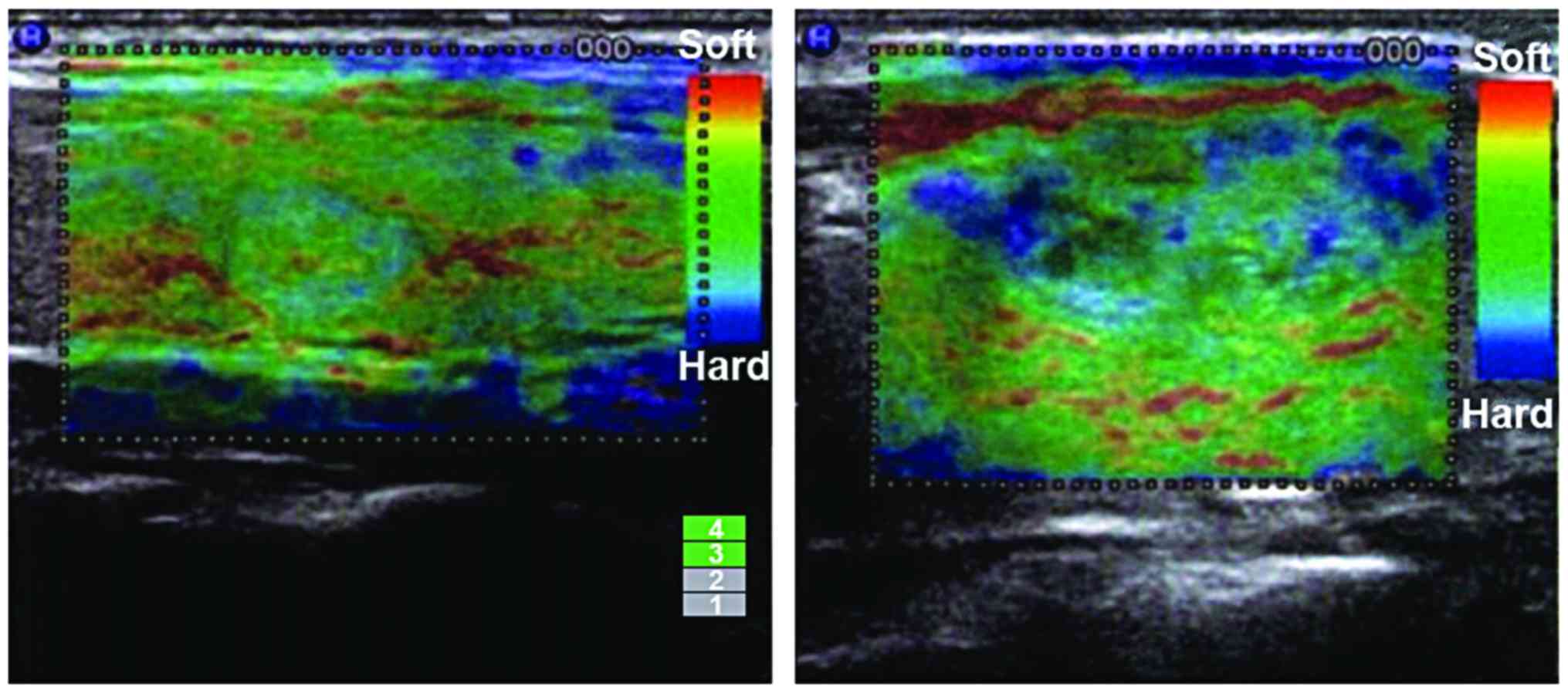
We adopted the hardness-scoring standard recommended
by the Hitachi Co. and took the 5 fractions method according to
different nodule colors. One point, the cystic components
predominated in nodules manifested by blue-green colors; 2 points,
nodules and surrounding tissues manifested by evenly green color; 3
points, >50% and <90% nodules manifested by green color; 4
points, >50% and <90% nodules manifested by blue color; 5
points, >90% nodules manifested by blue color; 1–3 points
indicated benign thyroid nodules, and 4–5 points indicated
malignant thyroid nodules (6).
Fig. 1 shows the elasticity
ultrasound imaging of the thyroid.
Statistical analysis
Data were analyzed and processed by SPSS 18.0 (SPSS,
Inc., Chicago, IL, USA). Enumeration data were examined by
χ2 test. P<0.05 was considered to be statistically
significant.
Results
Results of pathological findings
Among 94 thyroid nodules, there were 22 malignant
nodules, including 2 cases of follicular thyroid carcinoma, 9 cases
of papillary carcinoma and 1 case of metastatic carcinoma. Further,
there were 72 benign nodules, including 26 cases of goiter nodules
and 46 cases of thyroid adenoma (Table
I).
 | Table I.The classification results and
pathological diagnosis of thyroid nodule ultrasound elasticity. |
Table I.
The classification results and
pathological diagnosis of thyroid nodule ultrasound elasticity.
|
| Benign | Malignant |
|---|
|
|
|
|
|---|
| Ultrasound elasticity
grade | Nodular goiter | Thyroid adenoma | Papillary
carcinoma | Follicular thyroid
carcinoma | Non-hodgkins
lymphoma |
|---|
| Grade 0 | 7 | 6 | 0 | 0 | 0 |
| Grade I | 0 | 9 | 2 | 0 | 0 |
| Grade II | 10 | 12 | 1 | 0 | 0 |
| Grade III | 8 | 17 | 7 | 1 | 0 |
| Grade IV | 1 | 2 | 9 | 1 | 1 |
| Total | 26 | 46 | 19 | 2 | 1 |
The B-mode ultrasound evaluation of
benign and malignant thyroid nodules
The identification of benign and malignant thyroid
nodules by the B-mode ultrasound showed 84.04% (79/94) accuracy,
77.27% (17/22) sensitivity, 86.11% (62/72) specificity, 62.96%
(17/27) positive predictive value and 92.53% (62/67) negative
predictive value. On the other hand, the ultrasound elastography
showed 86.17 (81/94) accuracy, 81.82% (18/22) sensitivity, 87.50%
(63/72) specificity, 66.67% (18/27) positive predictive value and
94.02% (63/67) negative predictive value. Further the
identification of benign and malignant thyroid nodules by the
combined ultrasound diagnosis showed 96.81% (91/94) accuracy,
95.45% (21/22) sensitivity, 97.22% (70/72) specificity, 91.30%
(21/23) positive predictive value and 98.59% (70/71) negative
predictive value. The accuracy, sensitivity, specificity, positive
predictive value and negative predictive values of combined
ultrasound diagnosis were all obviously higher than those by the
B-mode ultrasound method and the ultrasound elastography method
(P<0.05). There were no significant differences between results
of the B-mode ultrasound method and the ultrasound elastography
method (P>0.05) (Table II).
| Table II.Comparisons of the accuracy,
sensitivity, specificity, positive predictive value and negative
predictive value of the benign and malignant thyroid nodule among
different methods. |
Table II.
Comparisons of the accuracy,
sensitivity, specificity, positive predictive value and negative
predictive value of the benign and malignant thyroid nodule among
different methods.
|
| Pathological |
| Pathological |
| Pathological |
|---|
| B-mode
ultrasound | Benign | Malignant | Total | Ultrasound
elastography | Benign | Malignant | Total | Combined
ultrasound | Benign | Malignant | Total |
|---|
| Benign | 62 | 5 | 67 | Benign | 63 | 4 | 67 | Benign | 70 | 1 | 71 |
| Malignant | 10 | 17 | 27 | Malignant | 9 | 18 | 27 | Malignant | 2 | 21 | 23 |
| Total | 72 | 22 | 94 | Total | 72 | 22 | 94 | Total | 72 | 22 | 94 |
| Sensitivity (%) | – | – | 77.27 | Sensitivity (%) | – | – | 81.82 | Sensitivity (%) | – | – | 95.45 |
| Specificity (%) | – | – | 86.11 | Specificity (%) | – | – | 87.50 | Specificity (%) | – | – | 97.22 |
| Positive predictive
value (%) | – | – | 62.96 | Positive predictive
value (%) | – | – | 66.67 | Positive predictive
value (%) | – | – | 91.30 |
| Negative predictive
value (%) | – | – | 92.53 | Negative predictive
value (%) | – | – | 94.02 | Negative predictive
value (%) | – | – | 98.59 |
| Accuracy (%) | – | – | 84.04 | Accuracy (%) | – | – | 86.17 | Accuracy (%) | – | – | 96.81 |
Discussion
China has listed the thyroid nodule as a health
examination item, as the detection rates are on the rise
continuously. Five to 10% of these thyroid nodules are malignant
nodules (7). The early diagnosis
would contribute to increasing the cure rate, prolonging the
survival time and improving the survival quality of patients.
Thyroid nodules are relatively occult at early stage. Therefore, it
is difficult to find the nodule completely by preliminary
diagnosis. The B-mode ultrasound diagnosis could not effectively
identify the nodule either, which is easy to be confused with the
thyroid tissue. For a long time, ultrasound has been preferred for
diagnosis of benign and malignant thyroid nodules. Thus, the B-mode
ultrasound detection is not able to identify all types of benign or
malignant thyroid nodules (2).
The prime research target in imaging currently is to
improve the detection rate and reduce the omission diagnostic rate
of malignant thyroid nodules. The real-time ultrasound elastography
is a newly developed ultrasound technology (8). This technology reflects the hardness of
monitored tissues mainly by detecting tissue distortion degree by
applying external pressure. Images are developed in accordance with
hardness differences among tissues. Ultrasound elastography could
provide information on elasticity, which is the basic mechanic
property so as to provide a new way of identifying benign and
malignant thyroid nodules. This study evaluated advantages and
disadvantages of B-mode ultrasound and ultrasound elastography in
identifying benign and malignant thyroid nodules. We combined the
B-mode ultrasound with ultrasound elastography for diagnosis, and
found that identification of benign and malignant thyroid nodules
by the combined ultrasound showed good results. The accuracy was
greatly improved which indicated that the elasticity imaging
technology could raise the accuracy of identifying benign and
malignant thyroid nodule by B-mode ultrasound.
In the present study, we found that the accuracy,
sensitivity, specificity and positive predicative value of combined
ultrasound were all better than those of B-mode ultrasound and
ultrasound elastography. Results above showed that there is no
significant difference between the diagnostic accuracy of thyroid
nodules by B-mode ultrasound and ultrasound elastography. Perhaps
it is because the elasticity imaging technology could provide
information related to tissue elasticity and is affected by many
factors. Goertz et al (9) have
compared the results of B-mode ultrasound and the elasticity
imaging technology and found the latter showed no advantages over
the former. However, the accuracy between the two technologies in
diagnosis of thyroid nodules revealed no statistical significance.
Ultrasound elasticity imaging is a technology aiming at tumor
examinations and gives relatively ideal imaging effect of tumors or
diffused diseases. At present, most clinical studies suggested that
B-mode ultrasound is better in the aspects of accuracy, sensitivity
and specificity. However, ultrasound elastography is limited in its
improvement. Therefore, in recent years, a number of literature
data have focused on the diagnosis effects of B-mode ultrasound
detection method combined with the elasticity imaging technology.
All research indicated that combined detection has greatly
increased the diagnosis sensitivity, specificity and positive
predicative values, which in turn conform to our research results
(10–12).
Ultrasound elasticity imaging technology identified
the hardness, elasticity and pathological characters of lesion
tissues. So it could serve as the basis of pathological diagnosis.
Its application in pathological diagnosis of breast diseases is
relatively good. The identification accuracy of benign and
malignant tissues reached 89.6–91.3% which is a relatively good
directive significance for clinical diagnosis (10). However, thyroid nodule tissues are
different from breast tissues as the density difference among
benign and malignant nodules is smaller than that of breast tumors.
So, the diagnosis accuracy of benign and malignant tissues is
reduced. It is still a kind of significant iconography detection
methods with wide application. There are many reliable reports on
its individual application. Also, there are some controversial
reports (11). In terms of the
diagnosis of benign and malignant thyroid nodules, some scholars
suggested that the elasticity imaging technology could not be used
as an independent diagnosis method (5). Ultrasound elasticity imaging technology
has its own advantages of identifying lesion nature. Ultrasound
elasticity imaging could locate tumors and reveal the relationships
between tumors and surrounding tissues. In recent years, the
application of real-time tissue elastography (13–15)
technology allowed recording of real-time color images. Judgment of
the nature of the tumor could be more exact based on echo signal
movement. The diagnosis effect would be further improved in the
combination of ultrasound elasticity imaging with routine
inspection. Therefore, it is more appropriate to take ultrasound
elastography as an assisting detection method for B-mode ultrasound
examination (16–18).
To conclude, ultrasound elasticity imaging
technology, as a new technology, has relatively high diagnotic
value for identifying benign and malignant thyroid nodules. The
ultrasound elasticity imaging technology could effectively
complement B-mode ultrasound diagnosis, and improve the
sensitivity, specificity and accuracy of ultrasound diagnosis of
thyroid carcinoma.
Acknowledgements
Not applicable.
Funding
No funding was received.
Availability of data and materials
The datasets used and/or analyzed during the current
study are available from the corresponding author on reasonable
request.
Authors' contributions
XZJ contributed significantly to writing the
manuscript and exam of patients by ultrasound. WWL and HFZ helped
with B-mode ultrasound. YYY and XLG analyzed and interpreted
ultrasound elastography result. All authors read and approved the
final manuscript.
Ethics approval and consent to
participate
The study was approved by the Ethics Committee of
Zhengzhou Central Hospital Affiliated tot Zhengzhou University
(Zhengzhou, China) and informed consents were signed by the
patients and/or guardians.
Consent for publication
Not applicable.
Competing interests
The authors declare that they have no competing
interests.
References
|
1
|
Wong CK and Wheeler MH: Thyroid nodules:
Rational management. World J Surg. 24:934–941. 2000. View Article : Google Scholar : PubMed/NCBI
|
|
2
|
Dudea SM and Botar-Jid C: Ultrasound
elastography in thyroid disease. Med Ultrason. 17:74–96. 2015.
View Article : Google Scholar : PubMed/NCBI
|
|
3
|
Frates MC, Benson CB, Charboneau JW, Cibas
ES, Clark OH, Coleman BG, Cronan JJ, Doubilet PM, Evans DB,
Goellner JR, et al: Society of Radiologists in Ultrasound:
Management of thyroid nodules detected at US: Society of
Radiologists in Ultrasound consensus conference statement.
Radiology. 237:794–800. 2005. View Article : Google Scholar : PubMed/NCBI
|
|
4
|
Rago T, Santini F, Scutari M, Pinchera A
and Vitti P: Elastography: New developments in ultrasound for
predicting malignancy in thyroid nodules. J Clin Endocrinol Metab.
92:2917–2922. 2007. View Article : Google Scholar : PubMed/NCBI
|
|
5
|
Jiang Y, Li GY, Qian LX, Hu XD, Liu D,
Liang S and Cao Y: Characterization of the nonlinear elastic
properties of soft tissues using the supersonic shear imaging (SSI)
technique: Inverse method, ex vivo and in vivo experiments. Med
Image Anal. 20:97–111. 2015. View Article : Google Scholar : PubMed/NCBI
|
|
6
|
Li T, Zhou P, Zhang X, Ding M, Yuchi M and
Li Y: Diagnosis of thyroid nodules using virtual touch tissue
quantification value and anteroposterior/transverse diameter ratio.
Ultrasound Med Biol. 41:384–392. 2015. View Article : Google Scholar : PubMed/NCBI
|
|
7
|
Mehrmohammadi M, Song P, Meixner DD,
Fazzio RT, Chen S, Greenleaf JF, Fatemi M and Alizad A: Comb-push
ultrasound shear elastography (CUSE) for evaluation of thyroid
nodules: Preliminary in vivo results. IEEE Trans Med Imaging.
34:97–106. 2015. View Article : Google Scholar : PubMed/NCBI
|
|
8
|
Ophir J, Céspedes I, Ponnekanti H, Yazdi Y
and Li X: Elastography: A quantitative method for imaging the
elasticity of biological tissues. Ultrason Imaging. 13:111–134.
1991. View Article : Google Scholar : PubMed/NCBI
|
|
9
|
Goertz RS: Ultrasound elastography.
Radiologe. 55:949–955. 2015.(In German). View Article : Google Scholar : PubMed/NCBI
|
|
10
|
Yoon JH, Yoo J, Kim EK, Moon HJ, Lee HS,
Seo JY, Park HY, Park WJ and Kwak JY: Real-time elastography in the
evaluation of diffuse thyroid disease: A study based on
elastography histogram parameters. Ultrasound Med Biol.
40:2012–2019. 2014. View Article : Google Scholar : PubMed/NCBI
|
|
11
|
Zhuo J, Ma Z, Fu WJ and Liu SP:
Differentiation of benign from malignant thyroid nodules with
acoustic radiation force impulse technique. Br J Radiol.
87:201302632014. View Article : Google Scholar : PubMed/NCBI
|
|
12
|
Sun J, Cai J and Wang X: Real-time
ultrasound elastography for differentiation of benign and malignant
thyroid nodules: A meta-analysis. J Ultrasound Med. 33:495–502.
2014. View Article : Google Scholar : PubMed/NCBI
|
|
13
|
Udelsman R and Zhang Y: The epidemic of
thyroid cancer in the United States: The role of endocrinologists
and ultrasounds. Thyroid. 24:472–479. 2014. View Article : Google Scholar : PubMed/NCBI
|
|
14
|
Wells PN and Liang HD: Medical ultrasound:
Imaging of soft tissue strain and elasticity. J R Soc Interface.
8:1521–1549. 2011. View Article : Google Scholar : PubMed/NCBI
|
|
15
|
Giovanella L, Clark PM, Chiovato L, Duntas
L, Elisei R, Feldt-Rasmussen U, Leenhardt L, Luster M,
Schalin-Jäntti C, Schott M, et al: Thyroglobulin measurement using
highly sensitive assays in patients with differentiated thyroid
cancer: A clinical position paper. Eur J Endocrinol. 171:R33–R46.
2014. View Article : Google Scholar : PubMed/NCBI
|
|
16
|
Remonti LR, Kramer CK, Leitão CB, Pinto LC
and Gross JL: Thyroid ultrasound features and risk of carcinoma: A
systematic review and meta-analysis of observational studies.
Thyroid. 25:538–550. 2015. View Article : Google Scholar : PubMed/NCBI
|
|
17
|
Doherty JR, Trahey GE, Nightingale KR and
Palmeri ML: Acoustic radiation force elasticity imaging in
diagnostic ultrasound. IEEE Trans Ultrason Ferroelectr Freq
Control. 60:685–701. 2013. View Article : Google Scholar : PubMed/NCBI
|
|
18
|
Andrioli M and Persani L: Elastographic
techniques of thyroid gland: Current status. Endocrine. 46:455–461.
2014. View Article : Google Scholar : PubMed/NCBI
|