Introduction
Lung cancer is the leading cause of
cancer-associated morbidity and mortality in the world (1). Lung cancer is a heterogeneous disease
with a number of histological and molecular subtypes; it is usually
classified according to the histological types associated with
tumor behavior and prognosis (2). The
majority of lung cancer types are known as non-small cell lung
cancers (NSCLCs), which are carcinoma malignancies that arise from
epithelial cells. The two most common NSCLC subtypes are
adenocarcinoma and squamous cell lung cancer (SqCC). SqCC
originates in the large airways in the central part of the lungs
and is the most common histological subtype of lung cancer among
European smokers (3,4). It is thought that the detection of lung
cancer during the early stages could reduce the mortality rates by
10- to 50-fold (5). However, SqCC is
often diagnosed at an advanced stage, by which point approximately
two-thirds of all patients have metastatic tumors when diagnosed.
Currently the low-dose computed tomography (LDCT) scan approach is
used to as a non-invasive method for detecting tumors at early
stages; however, this method has yielded conflicting results
(6,7).
Therefore, it is necessary to develop novel, minimally invasive
methods, such as molecular biomarkers, for the early detection of
lung cancer.
Lipids, which contain multiple different types of
molecules, have a number of key biological functions in cellular
energy storage, membrane structure and signaling processes. Within
the human body, lipid levels are regulated, spatially and
temporally, in various areas. Abnormal regulation of lipid
metabolism contributes to the development of a variety of human
diseases, including diabetes (8),
Alzheimer's disease (9), hypertension
(10) and many types of cancer
(11–14). Abnormal lipid metabolism in lung
cancer has also been demonstrated in a previous study, in which the
distribution of lipids in 21 pairs of resected frozen NSCLCs and
adjacent normal tissue samples were analyzed (14).
The involvement of lipid abnormalities in disease
pathogenesis increases the potential of using lipids as biomarkers
for various human diseases. However, due to technical limitations
in lipid measurement, only a few studies have investigated this
potential so far. Lipidomics is a relatively novel field that
allows for the quantitative assessment of a range (hundreds) of
fatty (lipids) species at one time; this analysis can be used to
generate lipid profiles for the majority of pathophysiological
states. Recently, lipidomics has been employed to study a number of
diseases such as diabetes (15) and
obesity (16), as well as some types
of human cancers, including colon cancer (17), thyroid papillary cancer (18) and prostate cancer (19). Direct tissue matrix-assisted laser
desorption/ionization (MALDI) mass spectrometry (MS) analysis has
been applied previously to perform lipid analysis in resected
frozen lung cancer tissue samples (14); however, large-scale studies on plasma
lipid profiling have yet to be reported in lung cancer.
In a previous study, the authors of the present
study evaluated the predictive power of this lipid panel in
diagnosis of early stage NSCLC (20).
In the present study, a lipid profiling study using tandem MS was
performed in order to measure 390 distinct lipids in the plasma
specimens obtained from patients with specifically in SqCC and
high-risk controls. The aim of the present study was to develop a
plasma lipid marker panel to use in high-risk patients as a
companion test with LDCT-based methods for the screening and early
detection of SqCC.
Patients and methods
Human research ethical statement
The present study was conducted in accordance with
the Declaration of Helsinki. The Rush University Medical Center
Institution Review Board (Chicago, IL, USA) approved the
experimental protocol, including the human rights and protection
aspects of the study, and all participants provided written
informed consent.
Patient cohorts
In the present study, ~1,250 individuals were
enrolled at the Lung Cancer Biorepository of Rush University
Medical Center between January 2004 and December 2010. Participants
were aged ≥50 years with: i) smoking histories of >30 pack-years
or ii) current smokers or a former smokers who quit within the past
15 years. Of these patients, a sub-cohort of 44 patients with SqCC
and 44 high-risk individuals were selected for this pilot study
(Table I). Patients were then further
divided into the following cohorts: i) Pathologically diagnosed
SqCC patients (n=22); and ii) matched high-risk individuals (n=22)
for the training stage; iii) independent SqCC patients (n=22); and
iv) random high-risk controls (n=22) for the validation stage. The
early-stage SqCC patient inclusion criteria included the following:
Disease was confined to the chest without evidence of distant
metastases; no preoperative chemo- or radiotherapy within 1 year of
the present study's initial blood sampling; and a minimum of 2
years of clinical follow-up data available. High risk patients
recruited to the present study were defined as individuals aged 55
to 75 years, had a smoking history of >30 years and quit smoking
<15 years prior to randomization in the present study. All
high-risk individuals were followed with annual LDCT and remained
cancer-free for a minimum of 2 years during follow-up. At the
training stage, SqCC cases and high-risk participants were matched
in terms of race, sex, age and smoking status. The demographic
information for these patients and controls is listed in Table I.
 | Table I.Patient characteristics in all of the
samples used in the training and validation stages. |
Table I.
Patient characteristics in all of the
samples used in the training and validation stages.
|
| Training stage | Validation
stage |
|---|
|
|
|
|
|---|
| Characteristic | SqCC (n=22) | High risk
(n=22) | SqCC (n=22) | High risk
(n=22) |
|---|
| Sex (n) |
|
|
|
|
|
Male | 11 | 11 | 15 | 16 |
|
Female | 11 | 11 | 7 | 6 |
| Race (n) |
|
|
|
|
|
Caucasian | 18 | 21 | 14 | 19 |
|
Noncaucasian | 4 | 1 | 8 | 3 |
| Age (years) |
|
|
|
|
|
Median | 62.95 | 64.14 | 56.75 | 73.44 |
|
Range | 49–81 | 51–82 | 69–82 | 49–73 |
| Smoking
history |
|
|
|
|
| Median
(years) | 59.17 | 47.04 | 52.90 | 32.28 |
|
Nonsmoker (n) | 2 | 0 | 0 | 0 |
| Tumor stage
(n) |
|
|
|
|
| Stage
I | 12 | − | 14 | − |
| Stage
II | 4 | − | 1 | − |
| Stage
III | 6 | − | 7 | − |
Electrospray ionization-tandem mass
spectrometry (ESI-MS) lipid profiling
In the present study, a total of 390 lipids in
plasma samples were tested using an automated ESI-MS system at the
Kansas State University Lipidomics Research Center (Manhattan, KS,
USA), as previously described (21).
Briefly, an aliquot of 3 µl plasma was used to identify plasma
lipid species at the level of the head group plus the total acyl
carbons:total double bonds in this assay; precise amounts of
internal standards were obtained and quantified, as previously
described (21). A series of spectra
were generated from the sequential precursor and neutral loss scans
of the extracts; each spectrum identified a group of lipid species
containing a common head group fragment. In total, 13 lipid classes
were measured: Phosphatidylcholine (PC), sphingomyelin (SM),
lysoPC, phosphatidylethanolamine (PE), lysoPE, phosphatidylinositol
(PI), phosphatidylserine (PS), phosphatidic acid, cholesterol
esters (CE), PS with one ether-linked (alkyl or alkenyl) chain, PC
with one ether-linked (alkyl or alkenyl) chain, PE with one
ether-linked (alkyl or alkenyl) chain and ceramide PE, as detailed
previously (19). The background of
each spectrum was removed, the data were normalized, and the
customization script and Applied Biosystems Analyst software
(version 1.5; Applied Biosystems; Thermo Fisher Scientific, Inc.,
Waltham, MA, USA) were used to integrate the peak area. The data
for the analyzed sample portion were then corrected and the sample
volume was normalized to produce data with units of nmol/µl.
Statistical and bioinformatics
analysis
In the present study, a Student's unpaired t-test
was performed using SPSS 18 software (SPSS, Inc., Chicago, IL, USA)
to compare the mean plasma concentrations of 390 lipid species
between the SqCC patient and control groups following the log
transformation of data. The GeneSpring 12.6 program (Agilent
Technologies, Inc., Santa Clara, CA, USA) was used to perform
Hierarchical Clustering Analysis (HCA), and the Weka version 3.73
software (the University of Waikato, Hamilton, New Zealand)
(22) was used in bioinformatics
analysis. The simple logistics classification algorithm and
information gain algorithm in Weka 3.7 were used to rank the
individual apparent lipid species and lipid class according to
their predictive powers in patients with SqCC; 10-fold cross
validation was employed to estimate the performance of a predictive
model. The unpaired Student's t-test in SPSS 18 software was used
to compare the mean plasma lipid concentrations in the controls and
patients. The chi-squared test was used to compare the differences
between sex and race in the patient and control groups. The
differences in age between the groups was analyzed by a Mann
Whitney U test. P<0.05 was considered to indicate a
statistically significant difference.
Results
Lipid profiling of 390 lipid species
in the training cohort
During the training stage, the plasma lipid profiles
of 390 individual lipid species from 13 classes of phospholipids
and CEs were identified using lipidomics in 44 plasma samples,
which included 22 patients with SqCC, and 22 age-, sex- and
race-matched high-risk controls. As summarized in our previous
study (20), 361 apparent lipid
species in all 13 classes were detected in all of the training
samples. Among the 361 apparent species, the highest median plasma
concentration was that of the CE species (18:2; 12.71 nmol/µl in
SqCC patients and 18.42 nmol/µl in controls). In the present study,
29 of the 390 lipid species were not detected in any of the
training cohort samples; these primarily consisted of those from
the lysoPE class (14 species) and PI class (9 species).
Identification of significantly
different lipid species between patients with SqCC and high-risk
individuals
To select individual apparent lipid biomarkers from
the hundreds of species detected, a filtration strategy was
employed to narrow down the 361 potential candidates of apparent
lipid species. During this process, the following exclusion
criteria were applied: i) Lipid species that could not be
clinically used for the diagnosis of SqCC disease due to too low a
concentration of detection; ii) there was an insignificant
difference between the patient and control groups; and iii) the
levels of plasma concentrations that were too close between the
groups to interpret (although the difference may have been
statistically significant). The inclusion criteria were as follows:
i) A significant difference in the mean plasma lipid concentration
(P≤0.05) was identified between the patient and control groups; ii)
there was a change in the mean plasma lipid concentration of ≥10%
(up or down); and iii) a mean plasma lipid concentration ≥10
nmol/µl. Using these criteria, 20 apparent lipid species were
identified that fulfilled all 3 criteria and could be selected as
potential plasma lipid biomarkers for SqCC (Table II). Among the 20 candidate lipid
markers, decreasing levels were observed in 18 lipid species in
SqCC patients; however, 2 lipid species, PC(38:6) and PI(38:4),
presented an increased level in SqCC plasma when compared with the
controls (Table II).
| Table II.Top 20 significantly differentiated
lipid species for SqCC at the training stage. |
Table II.
Top 20 significantly differentiated
lipid species for SqCC at the training stage.
|
| SqCC (n=22) | High risk
(n=22) |
|
|
|---|
|
|
|
|
|
|
|---|
| Lipids | Mean | SD | Mean | SD | Fold change | P-value |
|---|
| C16:0 CE | 0.9420 | 1.2608 | 1.0668 | 1.3492 | 1.1325 |
3.72×10² |
| C18:2 CE | 0.7879 | 1.2590 | 1.0644 | 1.3780 | 1.3509 |
1.00×10⁴ |
| C20:4 CE | 0.8406 | 1.6088 | 1.3211 | 1.5260 | 1.5717 |
4.01×10² |
| C22:6 CE | 0.6812 | 1.7158 | 1.0093 | 1.6120 | 1.4815 |
1.01×10² |
| ePC (34:2) | 0.9868 | 1.2485 | 1.0113 | 1.3051 | 1.0248 |
2.84×10² |
| ePC (34:3) | 0.9634 | 1.3843 | 1.0373 | 1.3540 | 1.0768 |
1.31×10² |
| ePC (36:2) | 0.9727 | 1.3327 | 1.1001 | 1.2649 | 1.1310 |
1.80×10³ |
| ePC (38:1) | 0.9520 | 1.3552 | 1.1049 | 1.2919 | 1.1605 |
1.07×10² |
| ePC (38:2) | 0.9768 | 1.3698 | 1.0456 | 1.2906 | 1.0705 |
1.60×10² |
| LPC (16:0) | 0.8984 | 1.6525 | 1.0497 | 1.4053 | 1.1685 |
1.88×10² |
| LPC (18:0) | 0.8089 | 1.7651 | 1.0159 | 1.3465 | 1.2559 |
3.80×10³ |
| LPC (18:2) | 0.8585 | 1.8515 | 1.3798 | 1.5443 | 1.6072 |
2.00×10⁴ |
| PC (34:2) | 1.0402 | 1.2913 | 1.1308 | 1.3081 | 1.0871 |
8.00×10⁴ |
| PC (36:2) | 0.9491 | 1.3070 | 1.1489 | 1.3165 | 1.2105 |
2.00×10⁴ |
| PC (36:4) | 0.9536 | 1.5849 | 1.2294 | 1.4101 | 1.2892 |
4.70×10² |
| PC(38:6) | 0.8903 | 1.4371 | 0.8598 | 1.5768 | 1.0355 |
1.54×10² |
| PC (40:7) | 0.8869 | 1.5719 | 0.9436 | 1.4884 | 1.0639 |
8.20×10³ |
| PI (38:4) | 0.9769 | 1.4808 | 0.9588 | 1.3437 | 0.9814 |
4.59×10² |
| SM (16:1) | 0.9819 | 1.2954 | 1.1346 | 1.2007 | 1.1556 |
2.48×10² |
| SM (22:0) | 0.5988 | 1.3069 | 0.9607 | 4.3199 | 1.6045 |
1.30×10³ |
Identification of C18:2 CE and SM 22:0
as candidate biomarkers for early-stage SqCC
A second strategy that generated an additional
differentiation of cancer and control samples was then utilized to
ensure that the selected candidates were clinically useful and
applicable, and also highly sensitive, specific and accurate in the
differentiation of SqCC from high-risk controls. Following analysis
using bioinformatics methods, any apparent lipid species of the
selected potential candidates were selected as an individual plasma
lipid biomarker for the diagnosis of SqCC cancer if the following
criteria was fulfilled: i) Sensitivity, >80%; ii) specificity,
>50%; and iii) area under the receiver operating characteristic
curve, >80%.
Using these selection strategies, 2 lipid species,
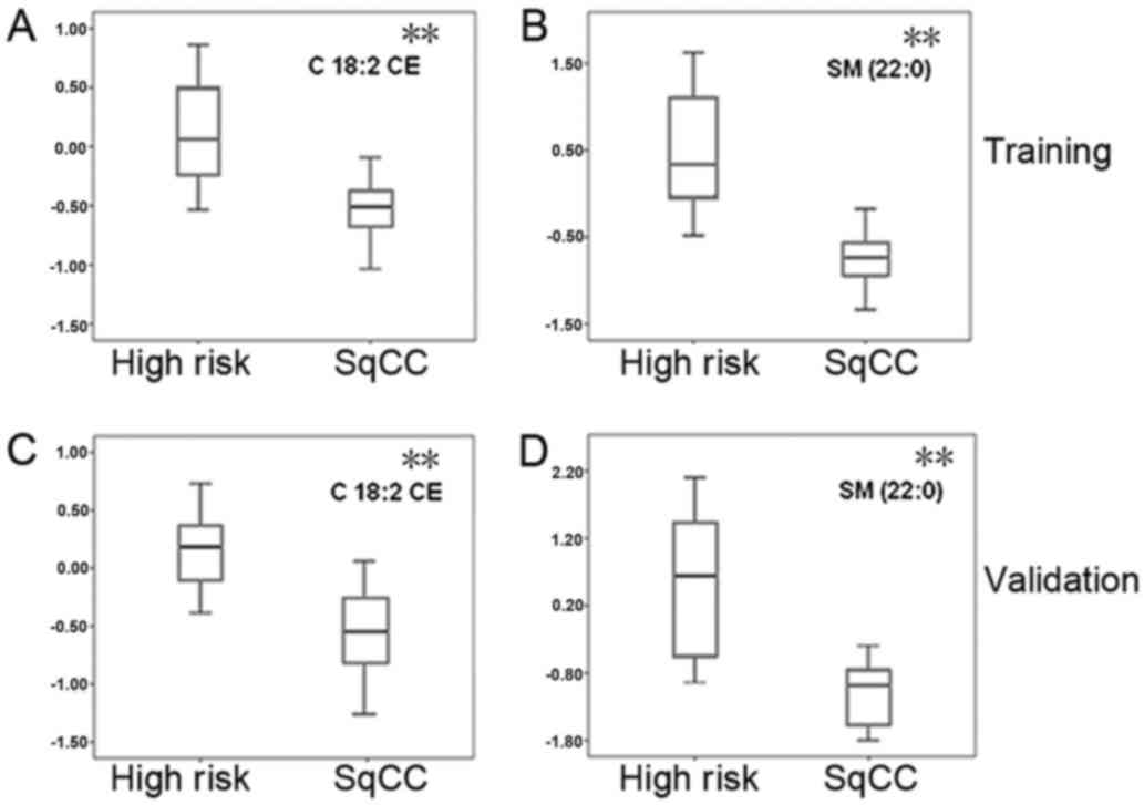
C18:2 CE and SM 22:0, were identified as candidate biomarkers for
the early detection of SqCC disease during the training stage. The
lipid C18:2 CE was detected at a mean concentration of 12.71
nmol/µl in 22 SqCC patients, with a 31.02% downregulation
(P=0.0001) when compared with the demographically matched high-risk
controls (18.42 nmol/µl; Fig. 1A).
Similarly, SM 22:0 was downregulated in SqCC plasma samples (mean
concentration, 95 pmol/µl), with a difference of 30.66% when
compared with the high-risk cohort (mean concentration, 137
pmol/µl; P=0.0013; Fig. 1B). The
lipid levels of C18:2 CE and SM 22:0 were then determined in one
high-risk individual and one SqCC case, respectively. The
predictive power of C18:2 CE in the diagnosis of early-stage SqCC
exerted a sensitivity of 86.4%, a specificity of 54.5% and an area
under the curve (AUC) value of 72.5% (Fig. 2A). In addition, SM 22:0 exerted a
sensitivity of 90.9%, a specificity of 77.3% and an AUC of 84.1%
(Table III). During the training
stages, it was also observed that the combination of these 2
markers yielded the strongest predictive power with a higher degree
of sensitivity (95.5%), specificity (90.9%) and accuracy (AUC,
95.2%), as shown in Table III and
Fig. 2A.
| Table III.Predictive values of the 2 lipid
markers at the training stage. |
Table III.
Predictive values of the 2 lipid
markers at the training stage.
| Lipid markers | Sensitivity | Specificity | PPV | NPV | OR | AUC |
|---|
| C18:2 CE | 0.864 | 0.545 | 0.800 | 0.655 | 7.6 | 0.725 |
| SM (22:0) | 0.909 | 0.773 | 0.895 | 0.800 | 34.0 | 0.841 |
| C18:2 CE + SM
(22:0) | 0.955 | 0.909 | 0.952 | 0.913 | 210.0 | 0.952 |
Validation of C18:2 CE and SM 22:0 in
independent cohorts
In order to validate C18:2 CE and SM 22:0 as
potential biomarkers for the early detection of SqCC, the 2 lipid
molecules were further investigated using the ESI-MS lipid
profiling technique in an independent cohort containing 22 SqCC
cases and 22 high-risk controls. During this validation stage, as
the biomarkers achieved in the training dataset are validated in
any SqCC dataset, the controls were randomly selected without age-,
sex- or ethnicity-matching with the SqCC patients (Table I). The box plots generated presented a
similar pattern of the 2 lipid levels among the validation cohorts
(Fig. 1C and D) and the training
samples (Fig. 1A and B). A similar
prediction power between C18:2 CE and SM 22:0 was observed in the
validation samples. The sensitivity, specificity and AUC values for
C18:2 CE were 62.5, 78.6 and 77.7%, respectively, and for SM 22:0
were 93.8, 89.3 and 91.5%, respectively. The strongest predictive
power was produced by the combination of the 2 lipids, with
markedly higher values of sensitivity (93.89%), specificity (92.9%)
and AUC (98.7%) (Table IV; Fig. 2B).
| Table IV.Predictive values of the 2 lipid
markers at the validation stage. |
Table IV.
Predictive values of the 2 lipid
markers at the validation stage.
| Lipid markers | Sensitivity | Specificity | PPV | NPV | OR | AUC |
|---|
| C18:2 CE | 0.625 | 0.786 | 0.786 | 0.625 | 6.11 | 0.777 |
| SM (22:0) | 0.938 | 0.893 | 0.962 | 0.833 | 125.00 | 0.915 |
| C18:2 CE + SM
(22:0) | 0.938 | 0.929 | 0.963 | 0.882 | 195.00 | 0.987 |
Characteristics of the 2 identified
plasma lipid biomarkers
To further reveal the characteristics of the 2
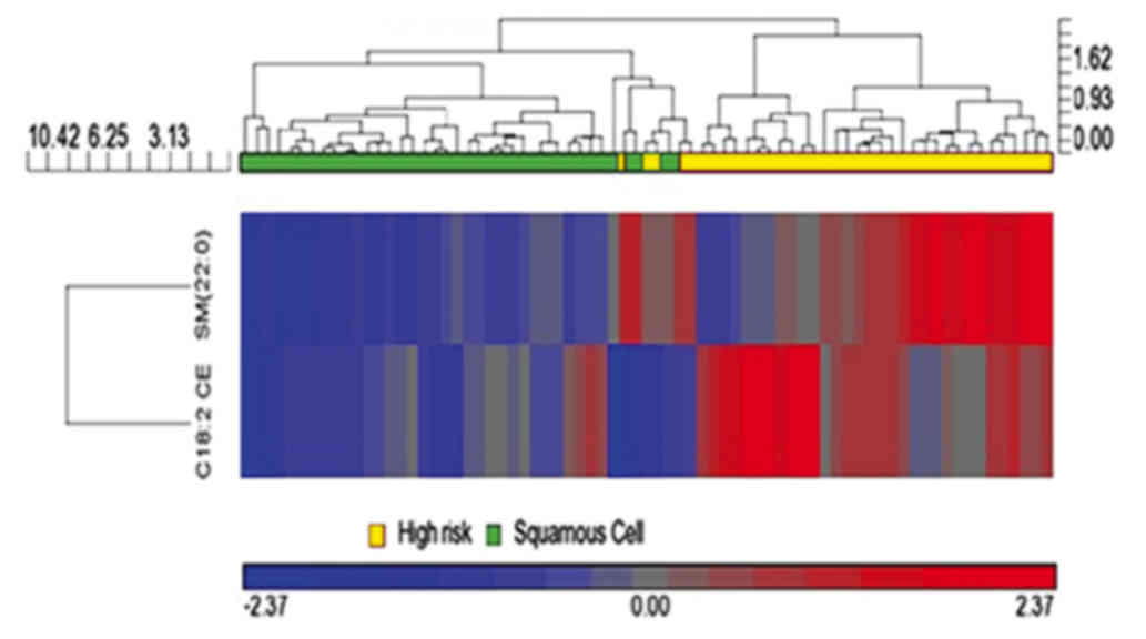
identified plasma lipid biomarkers for the diagnosis of SqCC, HCA
was conducted in order to cluster the entity (the 2 lipid
biomarkers) and condition (all 44 patients with SqCC and 44
high-risk individuals; training and validation cohorts) by
combining dendrograms and a heatmap with panels of the
characteristics at the top (Fig. 3).
HCA analysis revealed that the 2 apparent lipid species C18:2 CE
and SM 22:0 had a tendency to gradually decrease in mean plasma
lipid concentration, from right (higher concentration, shown in
red) to left (lower concentration, shown in blue; Fig. 3). This result further demonstrated
that the concentrations of C18:2 CE and SM 22:0 lipid species in
plasma samples were associated with the disease status of SqCC,
with a decreasing tendency in patients with SqCC.
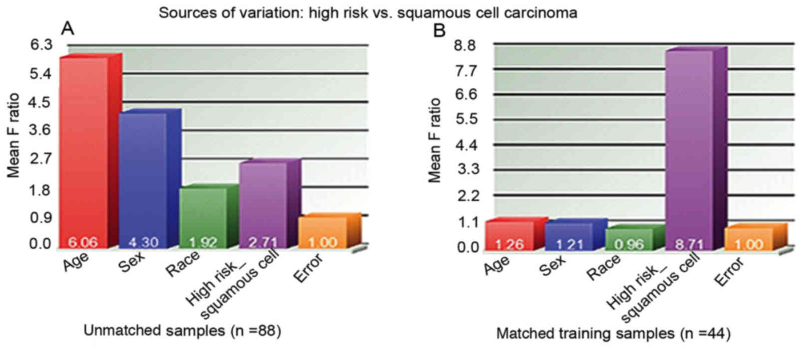
To assess the power of the sample size used during
the training and validation stages, the present study conducted the
source of variation analysis among all 88 unmatched subjects in the
training and validation stages, and among age, sex, race and
disease status (high risk vs. SqCC; Fig.
4). It was observed that the most significant difference was
shown in age, with a mean F ratio of 6.06 in all 88 unmatched
samples, which was much higher than the mean F ratio of 2.71 for
disease status (high risk vs. SqCC; Fig.
4A). However, in the 44 matched training samples, the most
significant factor was the disease status, with a mean F ratio of
8.71 (Fig. 4B). This observation
indicated that the significant difference in disease status (high
risk vs. SqCC) may contribute to the differentiated level of the 2
lipid species in plasma. As 70% of the cases (31 out of 44) who
participated in the present study were patients with stage I or II
SqCC, the panel of these 2 lipid biomarkers could also be applied
to the prediction of early-stage disease.
Discussion
In the present study, extensive plasma lipidomics
profiling of patients with SqCC was performed and a panel of 2
lipid biomarkers that were able to detect SqCC during the early
stages were identified. To the best of our knowledge, this is the
first original report on plasma lipid biomarkers for the early
detection of SqCC. The use of plasma is highly advantageous for the
development of a quick, non-invasive blood test and is easy to
obtain and use for the early diagnosis of SqCC. The results of the
present study revealed the potential of using these 2 lipid markers
in combination with LDCT-based screening methods in order to
distinguish patients with SqCC from high-risk individuals.
In the present study, it was observed that patients
with SqCC exhibited decreased plasma lipid concentrations of the 2
lipid molecules when compared with the high-risk controls. In a
previous study, the cholesteryl linoleate C18:2 CE was revealed to
be one of the three major cholesteryl esters that are present in
human low-density lipoprotein, and C18:2 CE oxidization is thought
to be associated with atherosclerosis (15). In addition, C18:2 CE can be oxidized
to generate the compounds 9-oxononanoyl secosterol-A (9-ON-secoA)
and 9-ON-secoB, which have been revealed to exhibit strong
cytotoxic activities against human leukemia HL-60 cells (16). However, it remains unknown whether
C18:2 CE oxidization is involved in lung cancer tumorigenesis.
Although smoking history is thought to be a risk factor of SqCC, in
a large-scale lipidomics analysis no associations were identified
for the plasma concentration in cholesteryl esters between smokers
and non-smokers (23). In agreement
with these results, in the present study no associations between
smoking history and the C18:2 lipid level were observed in any of
plasma samples.
In humans, SM, also known as sphingophospholipid, is
a type of sphingolipid observed in the cell membrane; it represents
~85% of all sphingolipids. SM is comprised of a phosphorylcholine
head group, a sphingosine and a fatty acid tail; the sphingosine
and fatty acid together are known as a ceramide. This composition
enables SM to serve significant roles in signaling pathways
(24) and SM degradation can produce
ceramide, which is known to be involved in the apoptotic signaling
pathway (25). In addition, ceramide
easily converts into sphingosine 1-phosphate (S1P) or ceramide
1-phosphate (C1P). When compared with ceramide, S1P and C1P produce
the opposite effect; in the regulation of cell growth and survival
they serve as pro-survival or mitogenic signals in the majority of
cell types, and they also induce different effects in controlling
tumor progression and metastasis (26). The sphingolipid, ceramide, has been
studied extensively under normal and pathological conditions,
ranging from skin development to lung cancer. A previous nested
case-control study demonstrated that higher concentrations of S1P
and total ceramide in plasma were associated with an increased risk
of lung cancer (24). A previous
study has also revealed that high-risk smokers have increased
ceramide levels; however, the underlying molecular mechanisms
associated with how smoking and ceramide accumulation cause lung
cancer remain unknown (27).
Recently, 2 signaling pathways associated with neutral
sphingomyelinase-2, an enzyme that hydrolyzes SM to ceramide, and
the EGF receptor (EGFR), respectively, have been reported to be
involved in the processes associated with cigarette smoke exposure
in the lung airways; EGFR was also revealed to be co-localized in
the plasma membrane in ceramide-enriched regions (28,29). In
addition, it is thought that these 2 signaling pathways may
converge and integrate with one another during these processes
(27,28).
In conclusion, the present study identified a panel
of 2 plasma lipid markers that may be able to distinguish patients
with SqCC from high-risk individuals with a large prediction power.
One limitation of the present study was that only relatively small
cohorts were used in the training and validation studies;
therefore, these 2 potential lipid biomarkers require further
confirmation in a greater number of samples from different
resources. Furthermore, the present study was only conducted in the
SqCC subtype of lung cancer. As 80% of lung cancers are NSCLCs, of
which ~30% are SqCC types, it is necessary to investigate plasma
lipid profiling in patients with NSCLC. As such, a global
lipidomics study is currently being performed at our laboratory in
order to identify specific lipid biomarkers for the early stages of
NSCLC.
Acknowledgements
Lipid analyses were conducted at the Kansas
Lipidomics Research Center Analytical Laboratory.
Funding
The present study was supported, in part, by The
National Institutes of Health (grant nos. R21CA164764,
5P30GM114737, P20GM103466, 2U54MD007601 and U54 MD007584) and
Hawaii Community Foundation (grant no. 17ADVC-86288).
Availability of data and materials
The datasets used and/or analysed during the current
study are available from the corresponding author on reasonable
request.
Authors' contributions
YD and WC designed and directed the research. ZY and
HC performed the experiments and wrote the manuscript. JA performed
the data analysis and prepared the tables. JB provided
bio-specimens. YL and YZ prepared the figures and assisted with the
data analysis. WG contributed to the analysis and interpretation of
the data. BJ and JZ prepared the experimental study materials and
revised the paper. All authors reviewed and approved the manuscript
prior to submission.
Ethics approval and consent to
participate
The Rush University Medical Center Institution
Review Board approved the experimental protocol, including the
human rights and protection aspects of the study, and all
participants provided written informed consent.
Consent for publication
Written informed consent was obtained from all
participants.
Competing interests
The authors declare they have no competing
interests.
Glossary
Abbreviations
Abbreviations:
|
NSCLC
|
non-small cell lung cancer
|
|
SqCC
|
squamous cell lung cancer
|
|
LDCT
|
low-dose computed tomography
|
|
MALDI-MS
|
matrix-assisted laser
desorption/ionization-mass spectrometry
|
|
ESI-MS
|
electrospray ionization-tandem mass
spectrometry
|
|
HCA
|
hierarchical clustering analysis
|
|
CE
|
cholesteryl esters
|
|
SM
|
sphingomyelin
|
References
|
1
|
Siegel R, Naishadham D and Jemal A: Cancer
statistics, 2013. CA Cancer J Clin. 63:11–30. 2013. View Article : Google Scholar : PubMed/NCBI
|
|
2
|
Beadsmoore CJ and Screaton NJ:
Classification, staging and prognosis of lung cancer. Eur J Radiol.
45:8–17. 2003. View Article : Google Scholar : PubMed/NCBI
|
|
3
|
Papi A, Casoni G, Caramori G, Guzzinati I,
Boschetto P, Ravenna F, Calia N, Petruzzelli S, Corbetta L,
Cavallesco G, et al: COPD increases the risk of squamous
histological subtype in smokers who develop non-small cell lung
carcinoma. Thorax. 59:679–681. 2004. View Article : Google Scholar : PubMed/NCBI
|
|
4
|
Janssen-Heijnen ML and Coebergh JW: Trends
in incidence and prognosis of the histological subtypes of lung
cancer in North America, Australia, New Zealand and Europe. Lung
Cancer. 31:123–137. 2001. View Article : Google Scholar : PubMed/NCBI
|
|
5
|
Edwards BK, Brown ML, Wingo PA, Howe HL,
Ward E, Ries LA, Schrag D, Jamison PM, Jemal A, Wu XC, et al:
Annual report to the nation on the status of cancer, 1975–2002,
featuring population-based trends in cancer treatment. J Natl
Cancer Inst. 97:1407–1427. 2005. View Article : Google Scholar : PubMed/NCBI
|
|
6
|
International Early Lung Cancer Action
Program Investigators1, . Henschke CI, Yankelevitz DF, Libby DM,
Pasmantier MW, Smith JP and Miettinen OS: Survival of patients with
stage I lung cancer detected on CT screening. N Engl J Med.
355:1763–1771. 2006. View Article : Google Scholar : PubMed/NCBI
|
|
7
|
Roth JA and Ramsey SD: Computed tomography
screening for lung cancer: A high-value proposition? JAMA.
315:77–78. 2016. View Article : Google Scholar : PubMed/NCBI
|
|
8
|
Sleeman MW, Wortley KE, Lai KM, Gowen LC,
Kintner J, Kline WO, Garcia K, Stitt TN, Yancopoulos GD, Wiegand SJ
and Glass DJ: Absence of the lipid phosphatase SHIP2 confers
resistance to dietary obesity. Nat Med. 11:199–205. 2005.
View Article : Google Scholar : PubMed/NCBI
|
|
9
|
Cutler RG, Kelly J, Storie K, Pedersen WA,
Tammara A, Hatanpaa K, Troncoso JC and Mattson MP: Involvement of
oxidative stress-induced abnormalities in ceramide and cholesterol
metabolism in brain aging and Alzheimer's disease. Proc Natl Acad
Sci USA. 101:2070–2075. 2004. View Article : Google Scholar : PubMed/NCBI
|
|
10
|
Nguyen NT, Magno CP, Lane KT, Hinojosa MW
and Lane JS: Association of hypertension, diabetes, dyslipidemia,
and metabolic syndrome with obesity: Findings from the National
Health and Nutrition Examination Survey, 1999 to 2004. J Am Coll
Surg. 207:928–934. 2008. View Article : Google Scholar : PubMed/NCBI
|
|
11
|
Pendaries C, Tronchère H, Plantavid M and
Payrastre B: Phosphoinositide signaling disorders in human
diseases. FEBS Lett. 546:25–31. 2003. View Article : Google Scholar : PubMed/NCBI
|
|
12
|
Ogretmen B and Hannun YA: Biologically
active sphingolipids in cancer pathogenesis and treatment. Nat Rev
Cancer. 4:604–616. 2004. View
Article : Google Scholar : PubMed/NCBI
|
|
13
|
Görke R1, Meyer-Bäse A, Wagner D, He H,
Emmett MR and Conrad CA: Determining and interpreting correlations
in lipidomic networks found in glioblastoma cells. BMC Syst Biol.
4:1262010. View Article : Google Scholar : PubMed/NCBI
|
|
14
|
Lee GK, Lee HS, Park YS, Lee JH, Lee SC,
Lee JH, Lee SJ, Shanta SR, Park HM, Kim HR, et al: Lipid MALDI
profile classifies non-small cell lung cancers according to the
histologic type. Lung Cancer. 76:197–203. 2012. View Article : Google Scholar : PubMed/NCBI
|
|
15
|
Suarna C, Dean RT, May J and Stocker R:
Human atherosclerotic plaque contains both oxidized lipids and
relatively large amounts of alpha-tocopherol and ascorbate.
Arterioscler Thromb Vasc Biol. 15:1616–1624. 1995. View Article : Google Scholar : PubMed/NCBI
|
|
16
|
Miyoshi N, Iwasaki N, Tomono S, Higashi T
and Ohshima H: Occurrence of cytotoxic 9-oxononanoyl secosterol
aldehydes in human low-density lipoprotein. Free Radic Biol Med.
60:73–79. 2013. View Article : Google Scholar : PubMed/NCBI
|
|
17
|
Fhaner CJ, Liu S, Ji H, Simpson RJ and
Reid GE: Comprehensive lipidome profiling of isogenic primary and
metastatic colon adenocarcinoma cell lines. Anal Chem.
84:8917–8926. 2012. View Article : Google Scholar : PubMed/NCBI
|
|
18
|
Ishikawa S, Tateya I, Hayasaka T, Masaki
N, Takizawa Y, Ohno S, Kojima T, Kitani Y, Kitamura M, Hirano S, et
al: Increased expression of phosphatidylcholine (16:0/18:1) and
(16:0/18:2) in thyroid papillary cancer. PLoS One. 7:e488732012.
View Article : Google Scholar : PubMed/NCBI
|
|
19
|
Zhou X, Mao J, Ai J, Deng Y, Roth MR,
Pound C, Henegar J, Welti R and Bigler SA: Identification of plasma
lipid biomarkers for prostate cancer by lipidomics and
bioinformatics. PLoS One. 7:e488892012. View Article : Google Scholar : PubMed/NCBI
|
|
20
|
Yu Z, Chen H, Ai J, Zhu Y, Li Y, Borgia
JA, Yang JS, Zhang J, Jiang B, Gu W and Deng Y: Global lipidomics
identified plasma lipids as novel biomarkers for early detection of
lung cancer. Oncotarget. 8:107899–107906. 2017. View Article : Google Scholar : PubMed/NCBI
|
|
21
|
Devaiah SP, Roth MR, Baughman E, Li M,
Tamura P, Jeannotte R, Welti R and Wang X: Quantitative profiling
of polar glycerolipid species from organs of wild-type Arabidopsis
and a phospholipase Dalpha1 knockout mutant. Phytochemistry.
67:1907–1924. 2006. View Article : Google Scholar : PubMed/NCBI
|
|
22
|
Smith TC and Frank E: Introducing machine
learning concepts with WEKA. Springer; New York, NY: pp. 353–378.
2016
|
|
23
|
Weir IM, Wong G, Barlow CK, Greeve MA,
Kowalczyk A, Almasy L, Comuzzie AG, Mahaney MC, Jowett JB, Shaw J,
et al: Plasma lipid profiling in a large population-based cohort. J
Lipid Res. 54:2898–2908. 2013. View Article : Google Scholar : PubMed/NCBI
|
|
24
|
Kolesnick R: Signal transduction through
the sphingomyelin pathway. Mol Chem Neuropathol. 21:287–297. 1994.
View Article : Google Scholar : PubMed/NCBI
|
|
25
|
Green DR: Apoptosis and sphingomyelin
hydrolysis. The flip side. J Cell Biol. 150:F5–F7. 2000. View Article : Google Scholar : PubMed/NCBI
|
|
26
|
Gangoiti P, Granado MH, Alonso A, Goñi FM
and Gomez-Munoz A: Implication of ceramide, ceramide 1-phosphate
and sphingosine 1-phosphate in tumorigenesis. Transl Oncogenomics.
3:81–98. 2008.PubMed/NCBI
|
|
27
|
Alberg AJ, Armeson K, Pierce JS, Bielawski
J, Bielawska A, Visvanathan K, Hill EG and Ogretmen B: Plasma
sphingolipids and lung cancer: A population-based, nested
case-control study. Cancer Epidemiol Biomarkers Prev. 22:1374–1382.
2013. View Article : Google Scholar : PubMed/NCBI
|
|
28
|
Goldkorn T, Chung S and Filosto S: Lung
cancer and lung injury: The dual role of ceramide. Handb Exp
Pharmacol. 93–113. 2013. View Article : Google Scholar : PubMed/NCBI
|
|
29
|
Goldkorn T and Filosto S: Lung injury and
cancer: Mechanistic insights into ceramide and EGFR signaling under
cigarette smoke. Am J Respir Cell Mol Biol. 43:259–268. 2010.
View Article : Google Scholar : PubMed/NCBI
|