Introduction
The most common type of malignant kidney cancer is
renal cell carcinoma (RCC), which accounts for 85–90% of cases
(1). Primary urothelial cancer (UC)
of the upper tract is only diagnosed in about 7–10% of all patients
with renal tumors (2,3). Macroscopic hematuria, flank pain and
palpable flank mass are three main clinical presentation of RCC but
such typical tumors are rare at present due to increasing use of
imaging (4). Microscopic or
macroscopic hematuria is the most common symptom of UC and
symptomatic hydronephrosis occurs if the tumor locates at the
ureteropelvic junction (5). Both RCC
and UC are more common in men vs. women. RCC originates from
proximal convoluted tubule including clear cell renal carcinoma,
papillary RCC, chromophobe RCC, collecting duct RCC, renal
medullary carcinoma and sarcomatoid RCC (6). While UC arises from the transitional
epithelium, including papillary and non-papillary transitional cell
carcinomas. Nephron sparing surgery (NSS) or radical nephrectomy
are the main procedures for RCC, and radical nephroureterectomy
(RNU) with en bloc excision of the periureteric bladder cuff is the
standard surgical treatment for UCs (7,8). Computed
tomography (CT) is mostly used to preoperative diagnosis of RCC and
UC. Typically, the tumors of RCC are soft tissue attenuation and
sometimes accompanied with necrosis and calcification, which have
stronger or irregular enhancement during the corticomedullary
phase. In contrast, for UC, filling defect or distortion of calyces
are shown on CT scan and the tumors have mild enhancement during
contrast CT (9,10). However, unusual infiltrative renal
masses (IRMs) are similar to intrarenal UCs (IUCs) on imaging,
which frequently results in misdiagnosis and mistreatment. The
present study retrospectively evaluated the patients who were
preoperatively diagnosed with IRM, but the subsequent pathological
results confirmed UCs in First Hospital of Jilin University. A
comprehensive review of the literature was also performed.
Case report
The study protocol was approved by the Institutional
Review Board of the First Hospital of Jilin University (Jilin,
China). A total of 22 IRM patients with pathologically confirmed
IUCs at our institution from January 2011 to December 2017 were
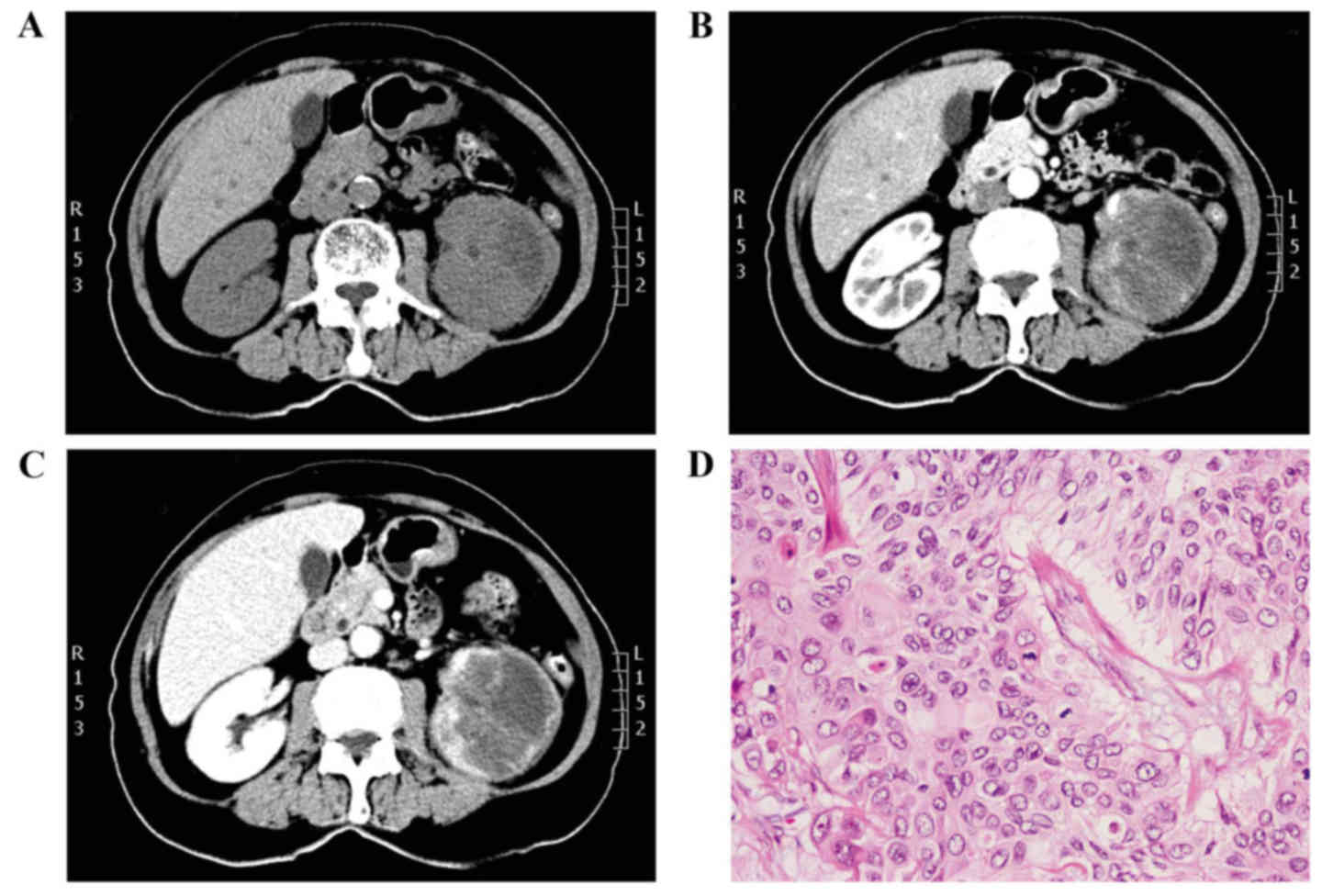
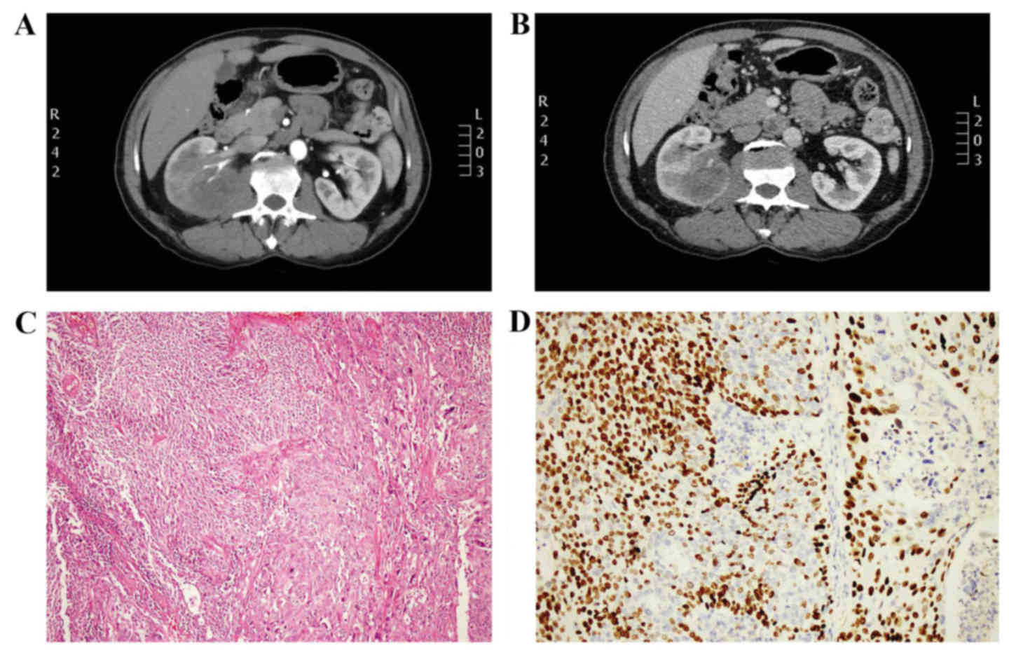
included in the present study. Representative cases are shown in
Figs. 1 and 2. Detailed clinical and pathological data
was available for all patients and the CT images were all performed
in First Hospital of Jilin University (Jilin, China).
Lists of demographic and pathological data of the
study cohort are shown in Table I.
There were 14 males and 8 females with a median age of 62 years. A
total of 11 patients were between 56–65 years (50%) and 8 patients
were >65 years old (36.4%). There was no difference between the
frequency of occurrence of tumors on the left or right side (11 vs.
10 cases, respectively). Hematuria and/or flank pain were the main
symptoms which accounted for 86.4% of patients. One patient was
diagnosed during a routine physical examination and another two
were initially admitted due to emaciation. Two patients exhibited
symptoms of lung metastasis. A total of six patients (27.3%) who
underwent laparoscopic nephroureterectomy were diagnosed by
percutaneous biopsy. The remaining 16 patients underwent
laparoscopic radical nephrectomy but the postoperative pathology
led to diagnosis of UCs, among which, 10 (62.5%) were stage T3 and
6 (37.5%) were stage T4. A total of two patients were confirmed
positive lymph node.
 | Table I.Demographic and pathological data on
22 patients. |
Table I.
Demographic and pathological data on
22 patients.
| Parameters | No. of patients
(%) |
|---|
| Age,
yearsa |
|
|
45–55 | 3 (13.6) |
|
56–65 | 11 (50) |
|
>65 | 8 (36.4) |
| Sex |
|
| Male | 14 (63.6) |
|
Female | 8 (36.4) |
| Side |
|
| Left | 11 (50) |
|
Right | 10 (45.5) |
| Both | 1 (4.5) |
| Symptoms |
|
| No | 1 (4.5) |
|
Hematuria | 6 (27.3) |
| Flank
pain | 6 (27.3) |
| Hematuria
and flank pain | 7 (31.8) |
|
Emaciation | 2 (9) |
| Suspected lung
metastasis | 2 (9) |
|
Biopsy | 6 (27.3) |
| Tumor stage |
|
| pT1 | 0 (0) |
| pT2 | 0 (0) |
| pT3 | 10 (62.5) |
| pT4 | 6 (37.5) |
| Positive lymph
node | 2 (9) |
All tumor features identified in the CT images were
shown in Table II. The mean maximum
diameter was 4.8 cm. All tumors were endophytic and solid. All
tumor boundaries were unclear. The reniform contours of the
operated kidneys were completely preserved or mildly altered (81.8
and 18.2%, respectively). In unenhanced CT images, all tumors
exhibited isoattenuation or hypoattenuation which were mildly
enhanced on enhanced CT images. A total of 2 patients demonstrated
lymph node enlargement.
| Table II.CT imaging features of tumor. |
Table II.
CT imaging features of tumor.
| Parameters | No. of patients
(%) |
|---|
| Mean maximus
diameter, cm (range) | 4.8 3.6–6.5 |
| Location |
|
|
Endophytic | 22 (100) |
|
Exophytic | 0 (0) |
| Reniform shape |
|
| No | 18 (81.8) |
| Mild | 4 (18.2) |
|
Moderate | 0 (0) |
|
Severe | 0 (0) |
| Tumor boundary |
|
|
Clear | 0 (0) |
|
Unclear | 22 (100) |
| Component |
|
|
Cystic | 0 (0) |
|
Necrosis | 0 (0) |
|
Solid | 22 (100) |
| Unenhanced CT
density |
|
| High
attenuation | 0 (0) |
|
Isoattenuation/hypoattenuation | 22 (100) |
| Degree of enhancement
on CT |
|
| Mild | 22 (100) |
|
Moderate | 0 (0) |
|
Severe | 0 (0) |
| Lymph node
enlargement | 2 (9) |
Discussion
The surgical treatment and postoperative management
of kidney cancers and renal pelvis cancers are significantly
different. Therefore, distinguishing IRMs from IUCs is critically
important but sometimes challenging (11). UC of the renal pelvis may display an
infiltrative growth pattern, which results in a close radiological
mimic of IRM.
Certain studies have attempted to differentiate IRM
from IUC but the limitations were obvious. Raza et al
(12) reviewed CT studies of 64
centrally located RCC and 34 IUC and found that the presence of a
tumor centered within the collecting system was the most valuable
characteristic identified on the CT images. Bata et al
(13), concluded that using multiple
small ROIs was valuable for distinguishing IRM from IUC. However,
based on one case and literature review, Li et al (14) reported that imaging results of
hypovascular RCC were indistinguishable from IUC. Han et al
(15), attempted to evaluate the
potential systemic inflammatory markers to differentiate between
infiltrative RCC and infiltrative UCs. Their analysis indicated
that age and lymphocyte-monocyte ratio were significantly different
between patients with IRM. To the best of our knowledge, the
present study is the first to comprehensively evaluate clinical,
imaging and pathological characteristics of IRMs that were
postoperatively confirmed as IUCs.
In the present study, 63.6% of the patients were
male and 36.4% were female. There was no obvious tendency about
sex. Zhu et al (16)
retrospectively assessed 29 patients with invasive renal
parenchymal urothelial carcinoma (IRPUC) and found that IRPUC was
more likely to occur in the right kidney (82.7%). However, in the
present study, the ratio of malignancies in left and right kidneys
was 11 vs. 10. Raza et al (12), also supported our results. It was
observed that prevalence of the disease was increased among
middle-elderly patients, over 56 years old (86.4%), which suggests
that older age may be associated with the development of IRC.
The mean maximum diameter of tumors was between 3.6
and 6.5 cm. A previous study demonstrated that if such tumors were
renal cancers, hematuria and flank pain were infrequent (17). However, in the present study, 27.3% of
patients exhibited either a symptom of hematuria or flank pain and
31.8% exhibited both hematuria and flank pain, which indicated that
hematuria and/or flank pain were the principal predictive
symptoms.
The CT images revealed that all 22 tumors were
endophytic and solid. All patients were free from cystic or
necrotic alterations. The results were similar to those previously
reported by Raza et al (12).
RCC may be associated with a moderate or severe renal shape
distortion, however, in the present study, a total of 18 patients
(81.2%) retained the renal contour and 4 patients (18.2%) underwent
mild alteration of shape. However, IRM does not necessarily alter
the reniform contour in all patients and certain patients with IUC
can sometimes exhibit a distorted renal shape (18,19). An
unclear tumor boundary was identified in each case included in the
present study, which was an important and valuable CT
characteristic. Isoattenuation or hypoattenuation occurred in all
patients and were visible in unenhanced CT images and all tumors
exhibited poor homogeneous enhancing masses in contrast-enhanced CT
images.
Although, several special CT features can help
distinguish IRM from IRC, in the majority of cases, other similar
neoplasms are present, including renal lymphoma, collecting duct
carcinoma, metastasis, xanthogranulomatous pyelonephritis and
medullary carcinoma (20,21). Biopsy is a necessary procedure in case
of misdiagnosis and mistreatment. Guarnizo et al (22), assessed the diagnostic accuracy of
ureteroscopic biopsy and found that ureteroscopic multi-biopsy can
lead to an accurate diagnosis of urothelial carcinoma in 89% of
cases and can predict the exact histopathological grade in 78% of
cases. Furukawa et al (23),
assessed a total of 40 patients and found that ureteroscopic biopsy
could lead to determination of the pathological grade of
nephroureterectomy specimens with an accuracy rate of 87.5%.
Recently, Huang et al (24)
reviewed 26 cases of upper tract lesions and found that
percutaneous biopsy was also a safe and effective method.
In conclusion, it is challenging to thoroughly make
a distinction between IRM and IUC preoperatively; however, the
elder accompanied with hematuria and/or flank pain are more likely
to indicate IRM, who with special CT features: endophytic, solid
and unclear tumor boundary on unenhanced CT and slightly
enhancement on contrast-enhanced CT. Pre-operative endoscopic or
percutaneous biopsy, is a valuable tool for complex cases.
Acknowledgements
Not applicable.
Funding
Dr. Wang was funded by the China Scholarship Council
(grant no. 201706175045).
Availability of data and materials
The datasets used and/or analyzed during the current
study are available from the corresponding author on reasonable
request.
Authors' contributions
XD, XM, YJ and HL and YW were all involved in this
conception of this study and helped to draft the manuscript. All
authors have read and approved the final manuscript.
Ethics approval and consent to
participate
Institutional Review Board of the First Hospital of
Jilin University (Jilin, China). Verbal informed consent was
obtained from the patients.
Consent for publication
Verbal informed consent was obtained from the
patients.
Competing interests
The authors declare that they have no competing
interests.
References
|
1
|
Sheir KZ, El-Azab M, Mosbah A, El-Baz M
and Shaaban AA: Differentiation of renal cell carcinoma subtypes by
multislice computerized tomography. J Urol. 174:451–455. 2005.
View Article : Google Scholar : PubMed/NCBI
|
|
2
|
Mandalapu RS and Matin SF: Contemporary
evaluation and management of upper tract urothelial cancer.
Urology. 94:17–23. 2016. View Article : Google Scholar : PubMed/NCBI
|
|
3
|
Arancibia MF, Bolenz C, Michel MS, Keeley
FX Jr and Alken P: The modern management of upper tract urothelial
cancer: Surgical treatment. BJU Int. 99:978–981. 2007. View Article : Google Scholar : PubMed/NCBI
|
|
4
|
Rossi SH, Klatte T, Usher-Smith J and
Stewart GD: Epidemiology and screening for renal cancer. World J
Urol. 2 Apr 2018 (Epub ahead of print).
|
|
5
|
Jaworski D, Szylberg Ł, Gzil A, Stawinski
P, Kasperska A and Marszałek A: Diagnostic difficulties in cases of
papillary urothelial neoplasm of low malignant potential,
urothelial proliferation of uncertain malignant potential,
urothelial dysplasia and urothelial papilloma: A review of current
literature. Ann Diagn Pathol. 17 Dec. 2017, (Epub ahead of print).
View Article : Google Scholar : PubMed/NCBI
|
|
6
|
Czarniecki M, Gautam R, Choyke PL and
Turkbey B: Imaging findings of hereditary renal tumors, a review of
what the radiologist should know. Eur J Radiol. 101:8–16. 2018.
View Article : Google Scholar : PubMed/NCBI
|
|
7
|
Pierorazio PM, Johnson MH, Patel HD, Sozio
SM, Sharma R, Iyoha E, Bass EB and Allaf ME: Management of renal
masses and localized renal cancer: Systematic review and
meta-analysis. J Urol. 196:989–999. 2016. View Article : Google Scholar : PubMed/NCBI
|
|
8
|
Soria F, Shariat SF, Lerner SP, Fritsche
HM, Rink M, Kassouf W, Spiess PE, Lotan Y, Ye D, Fernández MI, et
al: Epidemiology, diagnosis, preoperative evaluation and prognostic
assessment of upper-tract urothelial carcinoma (UTUC). World J
Urol. 35:379–387. 2017. View Article : Google Scholar : PubMed/NCBI
|
|
9
|
Nabi S, Kessler ER, Bernard B, Flaig TW
and Lam ET: Renal cell carcinoma: A review of biology and
pathophysiology. F1000Res. 7:3072018. View Article : Google Scholar : PubMed/NCBI
|
|
10
|
Froemming A, Potretzke T, Takahashi N and
Kim B: Upper tract urothelial cancer. Eur J Radiol. 98:50–60. 2018.
View Article : Google Scholar : PubMed/NCBI
|
|
11
|
Hartman DS, Davidson AJ, Davis CJ Jr and
Goldman SM: Infiltrative renal lesions: CT-sonographic-pathologic
correlation. AJR Am J Roentgenol. 150:1061–1064. 1988. View Article : Google Scholar : PubMed/NCBI
|
|
12
|
Raza SA, Sohaib SA, Sahdev A, Bharwani N,
Heenan S, Verma H and Patel U: Centrally infiltrating renal masses
on CT: differentiating intrarenal transitional cell carcinoma from
centrally located renal cell carcinoma. AJR Am J Roentgenol.
198:846–853. 2012. View Article : Google Scholar : PubMed/NCBI
|
|
13
|
Bata P, Tarnoki DL, Tarnoki AD, Novak PK,
Gyebnar J, Kekesi D, Szendroi A, Fejer B, Szasz AM, Nyirady P, et
al: Transitional cell and clear cell renal carcinoma:
Differentiation of distinct histological types with multiphase CT.
Acta Radiol. 55:1112–1119. 2014. View Article : Google Scholar : PubMed/NCBI
|
|
14
|
Li Y, Ding YU, Chen D, Yu Z, Gui Y, Yang S
and Lai Y: Renal cell carcinoma growing into the renal pelvis and
mimicking transitional cell carcinoma: A case report and literature
review. Oncol Lett. 9:1869–1872. 2015. View Article : Google Scholar : PubMed/NCBI
|
|
15
|
Han JH, Yoon YE, Kim SY, Cho YI, Rha KH,
Choi YD and Han WK: Preoperative lymphocyte-monocyte ratio
ameliorates the accuracy of differential diagnosis in
non-metastatic infiltrative renal masses. Yonsei Med J. 58:388–394.
2017. View Article : Google Scholar : PubMed/NCBI
|
|
16
|
Zhu Q, Zhu W, Wu J and Chen W:
Multidetector CT imaging features of invasive renal parenchyma
urothelial carcinoma. Br J Radiol. 89:201510682016. View Article : Google Scholar : PubMed/NCBI
|
|
17
|
Ginzburg S, Tomaszewski JJ and Kutikov A:
Focal ablation therapy for renal cancer in the era of active
surveillance and minimally invasive partial nephrectomy. Nat Rev
Urol. 14:669–682. 2017. View Article : Google Scholar : PubMed/NCBI
|
|
18
|
Prando A, Prando P and Prando D:
Urothelial cancer of the renal pelvicaliceal system: Unusual
imaging manifestations. Radiographics. 30:1553–1566. 2010.
View Article : Google Scholar : PubMed/NCBI
|
|
19
|
Yoon SK, Nam KJ, Rha SH, Kim JK, Cho KS,
Kim B, Kim KH and Kim KA: Collecting duct carcinoma of the kidney:
CT and pathologic correlation. Eur J Radiol. 57:453–460. 2006.
View Article : Google Scholar : PubMed/NCBI
|
|
20
|
Blitman NM, Berkenblit RG, Rozenblit AM
and Levin TL: Renal medullary carcinoma: CT and MRI features. AJR
Am J Roentgenol. 185:268–272. 2005. View Article : Google Scholar : PubMed/NCBI
|
|
21
|
Yan Y, Liu L, Zhou J, Li L, Li Y, Chen M,
Wang L, He W, Guan X, Zu X and Qi L: Clinicopathologic
characteristics and prognostic factors of sarcomatoid renal cell
carcinoma. J Cancer Res Clin Oncol. 141:345–352. 2015. View Article : Google Scholar : PubMed/NCBI
|
|
22
|
Guarnizo E, Pavlovich CP, Seiba M, Carlson
DL, Vaughan ED Jr and Sosa RE: Ureteroscopic biopsy of upper tract
urothelial carcinoma: Improved diagnostic accuracy and
histopathological considerations using a multi-biopsy approach. J
Urol. 163:52–55. 2000. View Article : Google Scholar : PubMed/NCBI
|
|
23
|
Furukawa J, Miyake H, Sakai I and Fujisawa
M: Significance of ureteroscopic biopsy grade in patients with
upper tract urothelial carcinoma. Curr Urol. 6:156–159. 2013.
View Article : Google Scholar : PubMed/NCBI
|
|
24
|
Huang SY, Ahrar K, Gupta S, Wallace MJ,
Ensor JE, Krishnamurthy S and Matin SF: Safety and diagnostic
accuracy of percutaneous biopsy in upper tract urothelial
carcinoma. BJU Int. 115:625–632. 2015. View Article : Google Scholar : PubMed/NCBI
|