Introduction
Endometrial cancer is one of the three major
malignant tumors in females and can occur in females of all age
groups (1). Amant et al
(2) showed that in 2016, ~1.2 million
new cases of endometrial cancer were reported worldwide, and
endometrial cancers are the third most common malignancy that
threatens the life and health of women. In recent years, incidence
of endometrial cancer showed an increasing trend. In some European
and American countries, incidence of endometrial cancer is the
highest among all gynecologic cancers (3).
Due to the high incidence, clinical treatment of
endometrial cancer has attracted increasing attention.
Breakthroughs have been made in the treatment of this disease, and
the majority of patients achieve promising prognosis after surgery,
radiotherapy and chemotherapy (4).
Plante et al (5) showed that
the 5-year survival rate of patients with endometrial cancer is
~85%. However, tumor metastasis may also occur in some patients due
to the lack of timely or improper treatment, seriously affecting
patients' prognosis (6).
At present, the pathogenesis, migration and invasion
of endometrial carcinoma are not clear. In recent years, miRNAs
have become popular research objects. As a class of non-coding
endogenous RNAs with a length of 17–23 nt, miRNAs can directly
react with the 3′-UTR region of mRNAs to regulate the expression of
target genes. miRNAs may show upregulated expression pattern to
play oncogenic roles or show downregulated expression pattern to
play a role as tumor suppressor genes (7–9). However,
Zheng et al (10) showed that
miR-106 promoted cancer cell proliferation in endometrial cancer.
Therefore, our study aimed to further investigate the regulatory
role of miR-106 in endometrial cancer with an expectation of
providing references for the targeted therapy of this disease.
Materials and methods
Main materials
Human cervical cancer cell line RL95-2 was provided
by the Department of Pathophysiology, Anhui Medical University.
RT-PCR reverse transcription kit and miRNA isolation kit were
purchased from Qiagen, Inc. (Valencia, CA, USA). Fetal bovine serum
(FBS) was purchased from Gibco (Gibco; Thermo Fisher Scientific,
Inc., Waltham, MA, USA). Tetramethyl azoline blue (MTT) and
dimethylsulfoxide (DMSO) were purchased from Sigma-Aldrich
(Sigma-Aldrich; Merck KGaA, St. Louis, MO, USA). Lipofectamine™
2000 transfection reagent was purchased from Invitrogen; Thermo
Fisher Scientific, Inc. Annexin V-FITC/PI apoptosis kit was
purchased from BestBio (Shanghai, China). The study was approved by
the Ethics Committee of Chongming Branch Hospital, Affiliated
Xinhua Hospital, School of Medicine, Shanghai Jiaotong University
(Suizhou, China). Signed informed consents were obtained from the
patients or the guardians.
Cell culture
Endometrial cancer cells RL95-2 were cultured in
RPMI-1640 medium containing 10% FBS at 37°C with 5%
CO2.
Grouping
miR-106 primers were designed and synthesized by
Shanghai GenePharma Co., Ltd. (Shanghai, China) (Table I). High-fidelity DNA polymerase was
used to amplify miR-106, and the product was subjected to
BamHI and HindIII digestion and was inserted into
pcDNA3.1(–). Cells were divided into three groups including blank
control cells (MOCK group), miR-106 transfection group (miR-106
group) and negative control group (siNC group).
 | Table I.Primer sequences. |
Table I.
Primer sequences.
| Primers | mir-106 | U6 |
|---|
| Forward |
5′-CGGCTAAAGTGCTGACAGTGC-3′ |
5′-GCTTCGGCAGCACATATACTAAAAT-3′ |
| Reverse |
5′-GTGCAGGGTCCGAGGT-3′ |
5′-CGCTTCACGAATTTGCGTGTCAT-3′ |
Methods
Cell transfection
Digestion of cells using trypsin was performed and
cells were counted. RL95-2 cells were seeded into 6-well plates
with 2×10⁵ cells/well, followed by cell culture for 24
h. Lipofectamine™ 2000 was used for transfection according to the
manufacturer's instructions when cell confluence reached 80%.
Expression of miR-106 was detected 48 h after transfection.
Reverse transcription-quantitative PCR
(RT-qPCR)
Tissues were mixed with TRIzol reagent and kept at
room temperature for 30 min to extract total RNA according to the
manufacturer's instructions. The total RNA was then reverse
transcribed into complementary deoxyribose nucleic acid (cDNA)
using cDNA synthesis kit (Biomiga Inc, San Diego, CA, USA). An
ultraviolet spectrophotometer (Hitachi, Tokyo, Japan) and 1%
denaturing agarose gel electrophoresis were used to test RNA
quality. miR-106 reverse transcription was performed according to
the manufacturer's instructions. miR-106 primers were designed and
synthesized by Shanghai GenePharma Co., Ltd. (Table I). PCR reaction systems were prepared
according to the manufacturer's instructions. PCR reaction
conditions: 95°C for 10 min, followed by 45 cycles of 95°C for 15
sec, 65°C for 30 sec and 72°C for 30 sec. U6 was as an endogenous
control, and data were processed using the 2−ΔΔCq method
(11).
MTT assay
Density of RL95-2 cells was adjusted to
1×10⁴/ml and cultured in 96-well plates (37°C, 5%
CO2) for 24 h. P-miR-106 and empty plasmid were
transfected into cells using Lipofectamine™ 2000, and three
replicate wells were set. After 72 h, 20 µl of MTT reagent (5
mg/ml) was added to each well, followed by incubation for 2 h.
Supernatant was discarded with a pipette and 100 ml of DMSO was
added and shaken for 15 min. A microplate reader (Bio-Rad,
Hercules, CA, USA) was used to measure the OD values at 450 nm and
cell growth curves were plotted.
Flow cytometry
Cells in plates were rinsed with 2 ml PBS solution,
followed by incubation with 0.5 ml 0.25% trypsin (without EDTA).
After digestion, cells were resuspended to a density of
1×10⁶ cells/ml, followed by incubation with apoptosis
detection solution Annexin V-FITC at room temperature for 15 min.
After centrifugation at a speed of 375 × g for 5 min at 4°C,
supernatant was removed, and cells were resuspended in 1X buffer.
Finally, 10 µl PI was added and flow cytometry was performed. The
experiment was repeated 3 times.
Cell scratch assay
Cells were inoculated into 6-well plates 24 h after
transfection, and three replicate wells were set. A 20 µl tip was
used to scratch cells when 90% confluence was reached. After
washing with PBS 3 times, cells were cultured in 1% FBS in DMEM.
This experiment was repeated 3 times.
Statistical analysis
SPSS 22.0 (IBM Corp., Armonk, NY, USA) statistical
software was used for data analysis. The data are expressed as mean
± SD. Comparison between groups was done using one-way ANOVA test
followed by post hoc test (Least Significant Difference). Paired
t-test was used for comparison between two groups. P<0.05 was
considered to be statistically significant.
Results
Expression of miR-106 after
transfection
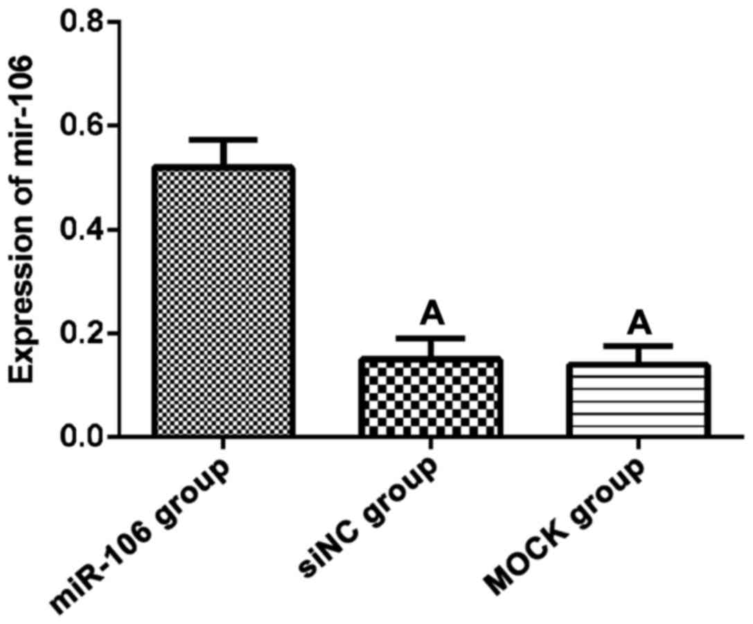
RT-qPCR results showed that the expression of
miR-106 in miR-106 group was significantly higher than that in siNC
and MOCK group (F=24.34, p<0.01). There was no significant
difference between siNC and MOCK group (t=0.19, p=0.86, Fig. 1).
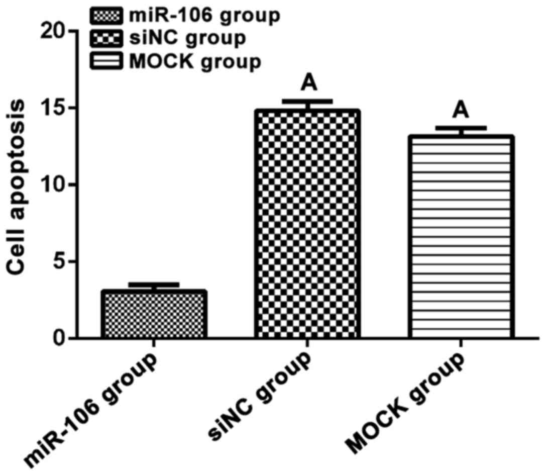
Cell apoptosis
Flow cytometry was used to detect apoptosis of
transfected cells. Apoptosis rate of miR-106 group after
transfection was 3.08±0.74%, which was significantly lower than
that of the siNC group (14.83±1.02%) and MOCK group (13.17±0.94%),
(F=147.20, p<0.01). There was no significant difference between
siNC and MOCK group (t=0.03, p=0.97, Fig.
2).
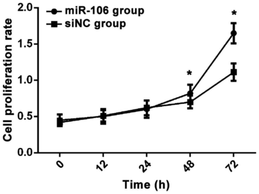
Cell proliferation
After miR-106 was transfected into RL95-2 cells,
results of MTT assay showed that there was no significant
difference in cell proliferation rate between the two groups at
12–24 h (p>0.05). After 48 h, cell proliferation rate in miR-106
group was significantly higher than that in siNC group (p<0.05,
Fig. 3).
Scratch assay results
By observing the width of scratched wounds under an
inverted light microscope, cell migration of miR-106 group was
significantly accelerated compared with siNC group (p<0.05).
Discussion
Endometrial cancer accounts for ~10% of female
malignancies, and onset age of this disease is becoming
increasingly younger (12). Early
stage of endometrial cancer has no obvious symptoms. Many patients
were diagnosed at middle or advance stages and the best treatment
timing was missed, which in turn leads to poor prognosis (13). Therefore, identification of gene
targets and development of targeted therapeutic drugs for
endometrial cancer is of great clinical significance. It has been
proved that (14–16) proliferation, apoptosis, and invasion
of cancer cells significantly affect the prognosis of patients.
Proliferation, apoptosis and invasion of cancer cells is a
multi-step and multi-factor involved biological process. In recent
years, miRNAs have been shown to inhibit the translation of mRNA in
almost all species (17,18). Jonas and Izaurralde (19) demonstrated that miRNAs are involved in
all biological processes in the body and have a strong regulatory
role in normal cellular function (20). miRNAs participate in the occurrence
and development of multiple tumors by regulating the proliferation,
migration, apoptosis, and angiogenesis of tumor cells (20). Zheng et al (10) demonstrated that miR-106 promoted
cancer cell proliferation in endometrial cancer. Therefore, we
studied the effect of miR-106 on the proliferation and apoptosis of
endometrial cancer RL95-2 cells with an expectation of providing
references for targeted gene therapy.
miR-106 is located on chromosome Xq26.2 and consists
of 23 nucleotides and is upmethyl thiazolyl tetrazoliumated in
various tumors (21). In this study,
we successfully constructed a miR-106 eukaryotic expression vector
and successfully transfected it into endometrial cancer RL95-2
cells to detect its expression and biological functions. Expression
of miR-106 in RL95-2 cells transfected with miR-106 was
significantly higher than that in siNC and MOCK group (p<0.05).
Wang et al (22) demonstrated
that miR-106 enhances the self-renewal ability of glioma cells and
the invasion ability of glioma stem cells by inhibiting the
expression of matrix metalloproteinase-2 (TIMP-2). MMT assay and
flow cytometry results also showed that miR-106 significantly
affected apoptosis of RL95-2 cells, and miR-106 can effectively
promote proliferation of endometrial cancer RL95-2 cells and
inhibit cell apoptosis.
There are still deficiencies in this study. Due to
the limited experimental conditions, in-depth investigation on the
mechanism of the function of miR-106 in patients with endometrial
cancer was not performed. Only in vitro experiments were
performed and in vivo validation is lacking. Diagnostic and
prognostic values of miR-106 for endometrial cancer were not
evaluated.
In summary, miR-106 overexpression can promote the
proliferation of endometrial cancer RL95-2 cells and inhibit cell
apoptosis. miR-106 shows promise as a new target for the treatment
of endometrial cancer.
Acknowledgements
Not applicable.
Funding
No funding was received.
Availability of data and materials
The datasets used and/or analyzed during the present
study are available from the corresponding author on reasonable
request.
Authors' contributions
XL drafted the manuscript and contributed to cell
culture. XY was responsible for cell transfection. CB and ZW helped
with RT-qPCR and MTT assay. All authors read and approved the final
manuscript.
Ethics approval and consent to
participate
The study was approved by the Ethics Committee of
Chongming Branch Hospital, Affiliated Xinhua Hospital, School of
Medicine, Shanghai Jiaotong University (Suizhou, China). Signed
informed consents were obtained from the patients or the
guardians.
Patient consent for publication
Not applicable.
Competing interests
The authors declare that they have no competing
interests.
References
|
1
|
Colombo N, Creutzberg C, Amant F, Bosse T,
González-Martín A, Ledermann J, Marth C, Nout R, Querleu D, Mirza
MR, et al: ESMO-ESGO-ESTRO Endometrial Consensus Conference Working
Group: ESMO-ESGO-ESTRO Consensus Conference on Endometrial Cancer:
Diagnosis, treatment and follow-up. Ann Oncol. 27:16–41. 2016.
View Article : Google Scholar : PubMed/NCBI
|
|
2
|
Amant F, Moerman P, Neven P, Timmerman D,
Van Limbergen E and Vergote I: Endometrial cancer. Lancet.
366:491–505. 2005. View Article : Google Scholar : PubMed/NCBI
|
|
3
|
van Gool IC, Eggink FA, Freeman-Mills L,
Stelloo E, Marchi E, de Bruyn M, Palles C, Nout RA, de Kroon CD,
Osse EM, et al: POLE proofreading mutations elicit an antitumor
immune response in endometrial cancer. Clin Cancer Res.
21:3347–3355. 2015. View Article : Google Scholar : PubMed/NCBI
|
|
4
|
Church DN, Stelloo E, Nout RA, Valtcheva
N, Depreeuw J, ter Haar N, Noske A, Amant F, Tomlinson IP, Wild PJ,
et al: Prognostic significance of POLE proofreading mutations in
endometrial cancer. J Natl Cancer Inst. 107:4022014.PubMed/NCBI
|
|
5
|
Plante M, Touhami O, Trinh XB, Renaud MC,
Sebastianelli A, Grondin K and Gregoire J: Sentinel node mapping
with indocyanine green and endoscopic near-infrared fluorescence
imaging in endometrial cancer. A pilot study and review of the
literature. Gynecol Oncol. 137:443–447. 2015. View Article : Google Scholar : PubMed/NCBI
|
|
6
|
Stelloo E, Bosse T, Nout RA, MacKay HJ,
Church DN, Nijman HW, Leary A, Edmondson RJ, Powell ME, Crosbie EJ,
et al: Refining prognosis and identifying targetable pathways for
high-risk endometrial cancer; a TransPORTEC initiative. Mod Pathol.
28:836–844. 2015. View Article : Google Scholar : PubMed/NCBI
|
|
7
|
Wilczynska A and Bushell M: The complexity
of miRNA-mediated repression. Cell Death Differ. 22:22–33. 2015.
View Article : Google Scholar : PubMed/NCBI
|
|
8
|
Yuan R, Zhi Q, Zhao H, Han Y, Gao L, Wang
B, Kou Z, Guo Z, He S, Xue X, et al: Upregulated expression of
miR-106a by DNA hypomethylation plays an oncogenic role in
hepatocellular carcinoma. Tumour Biol. 36:3093–3100. 2015.
View Article : Google Scholar : PubMed/NCBI
|
|
9
|
Yen CS, Su ZR, Lee YP, Liu IT and Yen CJ:
miR-106b promotes cancer progression in hepatitis B
virus-associated hepatocellular carcinoma. World J Gastroenterol.
22:5183–5192. 2016. View Article : Google Scholar : PubMed/NCBI
|
|
10
|
Zheng Z, Zhang Y, Zhang Z, Yang Y and Song
T: Effect of miR-106b on invasiveness of pituitary adenoma via
PTEN-PI3K/AKT. Med Sci Monit. 23:1277–1285. 2017. View Article : Google Scholar : PubMed/NCBI
|
|
11
|
Livak KJ and Schmittgen TD: Analysis of
relative gene expression data using real-time quantitative PCR and
the 2(-Delta Delta C(T)) Method. METHODS. 25:402–408. 2001.
View Article : Google Scholar : PubMed/NCBI
|
|
12
|
Billingsley CC, Cohn DE, Mutch DG,
Stephens JA, Suarez AA and Goodfellow PJ: Polymerase ε (POLE)
mutations in endometrial cancer: Clinical outcomes and implications
for Lynch syndrome testing. Cancer. 121:386–394. 2015. View Article : Google Scholar : PubMed/NCBI
|
|
13
|
Schuler KM, Rambally BS, DiFurio MJ,
Sampey BP, Gehrig PA, Makowski L and Bae-Jump VL: Antiproliferative
and metabolic effects of metformin in a preoperative window
clinical trial for endometrial cancer. Cancer Med. 4:161–173. 2015.
View Article : Google Scholar : PubMed/NCBI
|
|
14
|
Mehnert JM, Panda A, Zhong H, Hirshfield
K, Damare S, Lane K, Sokol L, Stein MN, Rodriguez-Rodriquez L,
Kaufman HL, et al: Immune activation and response to pembrolizumab
in POLE-mutant endometrial cancer. J Clin Invest. 126:2334–2340.
2016. View
Article : Google Scholar : PubMed/NCBI
|
|
15
|
Aune D, DA Rosenblatt Navarro, Chan DS,
Vingeliene S, Abar L, Vieira AR, Greenwood DC, Bandera EV and Norat
T: Anthropometric factors and endometrial cancer risk: A systematic
review and dose-response meta-analysis of prospective studies. Ann
Oncol. 26:1635–1648. 2015. View Article : Google Scholar : PubMed/NCBI
|
|
16
|
Trabert B, Wentzensen N, Felix AS, Yang
HP, Sherman ME and Brinton LA: Metabolic syndrome and risk of
endometrial cancer in the United States: A study in the
SEER-medicare linked database. Cancer Epidemiol Biomarkers Prev.
24:261–267. 2015. View Article : Google Scholar : PubMed/NCBI
|
|
17
|
Schnall-Levin M, Rissland OS, Johnston WK,
Perrimon N, Bartel DP and Berger B: Unusually effective microRNA
targeting within repeat-rich coding regions of mammalian mRNAs.
Genome Res. 21:1395–1403. 2011. View Article : Google Scholar : PubMed/NCBI
|
|
18
|
Lin S and Gregory RI: MicroRNA biogenesis
pathways in cancer. Nat Rev Cancer. 15:321–333. 2015. View Article : Google Scholar : PubMed/NCBI
|
|
19
|
Jonas S and Izaurralde E: Towards a
molecular understanding of microRNA-mediated gene silencing. Nat
Rev Genet. 16:421–433. 2015. View
Article : Google Scholar : PubMed/NCBI
|
|
20
|
Zhang J, Li S, Li L, Li M, Guo C, Yao J
and Mi S: Exosome and exosomal microRNA: Trafficking, sorting, and
function. Genomics Proteomics Bioinformatics. 13:17–24. 2015.
View Article : Google Scholar : PubMed/NCBI
|
|
21
|
Delay C, Mandemakers W and Hébert SS:
MicroRNAs in Alzheimer's disease. Neurobiol Dis. 46:285–290. 2012.
View Article : Google Scholar : PubMed/NCBI
|
|
22
|
Wang R, Li Y, Hou Y, Yang Q, Chen S, Wang
X, Wang Z, Yang Y, Chen C, Wang Z, et al: The
PDGF-D/miR-106a/Twist1 pathway orchestrates epithelial-mesenchymal
transition in gemcitabine resistance hepatoma cells. Oncotarget.
6:7000–7010. 2015.PubMed/NCBI
|