Introduction
Gastric cancer (GC) is the fifth most common type of
malignant tumor worldwide and the third most common cause of
cancer-associated mortality (1,2).
Approximately 50% of cases occur in Eastern Asia (mainly in China)
(3). Therefore, GC is a major public
health problem, particularly in China. In the majority of cases, GC
patients are diagnosed at advanced stages, at which effective
therapeutic strategies are limited and the prognosis is relatively
poor (4–6). Invasion and metastasis are hallmarks of
advanced GC progression; therefore, investigation of the molecular
pathogenesis of GC is critical to improve the survival of GC
patients.
miRNAs are evolutionary conserved small non-coding
RNAs and are involved in the regulation of gene expression and
protein translation (7). miRNAs
function in the pathogenesis of GC (8–14).
The role of miR-155-5p in various types of cancer
has been revealed in recent studies. In colorectal carcinoma (CRC),
miR-155-5p expression is upregulated and has been indicated to
promote the proliferation, invasion and metastasis of CRC cells
(15). In osteosarcoma, increased
miR-155-5p and reduced miR-148a-3p expression were demonstrated to
contribute to the suppression of tumor cell death (16). In GC, miR-155-5p was demonstrated to
be downregulated in GC tissues (17),
and miR-155-5p inhibition promoted the transformation of bone
marrow mesenchymal stem cells into GC tissue-derived MSC-like cells
via nuclear factor-κB p65 activation (18). However, the role of miR-155-5p in GC
has not been fully elucidated.
In the present study, the effect of miR-155-5p on
GC-cell proliferation and apoptosis was investigated. The data
achieved may provide a novel therapeutic target for further
investigation.
Materials and methods
Patients and tissues
A total of 14 GC tissues and corresponding adjacent
normal tissues were collected from 14 patients with GC who
underwent surgery at the Department of Gastroenterology, Center
Hospital of Nanchong City. College (Nanchong, China) from January
2013 to December 2013. The histopathological diagnoses of all
patients were confirmed by a senior pathologist at West China
Hospital (Chengdu, China). The information of the 14 GC patients
(average age: 60.2 years, age range: 36–63 years) are listed in
Table I. The sex ratio was 50:50. The
present study was approved by the ethics committee of Northern
Sichuan Medical College (Nanchong, Sichuan), and all patients
provided written informed consent.
 | Table I.The characteristics of the patients
with gastric cancer included in the present study. |
Table I.
The characteristics of the patients
with gastric cancer included in the present study.
| Patient | Age (years) | Sex | Stage | Differentiation |
|---|
| 1 | 45 | Female | III | Low |
| 2 | 63 | Female | II | Moderate |
| 3 | 56 | Female | II | Low |
| 4 | 45 | Female | III | Moderate |
| 5 | 59 | Male | III | Moderate |
| 6 | 50 | Male | IV | Moderate |
| 7 | 56 | Male | III | Moderate |
| 8 | 77 | Female | IV | Low |
| 9 | 66 | Male | II | Moderate |
| 10 | 73 | Female | II | Moderate |
| 11 | 48 | Male | III | Low |
| 12 | 67 | Male | III | Low |
| 13 | 53 | Female | IV | Moderate |
| 14 | 50 | Male | III | Moderate |
Cell lines and reagents
The GC cell lines, AGS and SGC-7901, were purchased
from the Chinese Academy of Sciences (Shanghai, China). The human
gastric epithelial mucosa cell line, GES-1, was gifted by the
Department of Gastrology, West China Hospital, Sichuan University
(Chengdu, China). Cells were cultured in RPMI-1640 medium,
supplemented with 10% fetal bovine serum (Thermo Fisher Scientific,
Inc., Waltham, MA, USA) (19).
Cisplatin was purchased from Hanson Pharma (Lianyungang, China;
http://www.hansoh.cn/). Cisplatin was added to
the cultures at a final concentration of 25 µM, as previously
described (20).
miR-155-5p mimics and oligonucleotide
transfection
The miR-155-5p mimics, miR-155-5 oligonucleotides,
miR-155-5p antisense oligonucleotides (ASO) and negative controls
were purchased from Dharmacon, Inc. (Chicago, IL, USA). Cells were
seeded at 2×10⁵ per well in 6-well plates, and
transfected with mimics (50 nM), oligonucleotides (50 nM) or
controls (50 nM) using Lipofectamine® 2000 (Thermo
Fisher Scientific, Inc.), according to the standard protocol. The
sequence of these molecules were listed as following: miR-155-5p
mimics, 5′-UUAAUGCUAAUCGUCAUAGGGGU-3′; miR-155-5p-NC,
5′-UUCUCCGAACGUGUCACGUTT-3′; miR-155-5p ASO,
5′-ACCCCUAUCACGAUUAGCAUUAA-3′; miR-155-5p ASO-NC,
5′-CAGUACUUUUGUGUAGUACAA-3′. A total of 24 h following
transfection, the miR-155-5p levels were examined by reverse
transcription-quantitative polymerase chain reaction (RT-qPCR).
Western blot analysis
The lysates were prepared with lysis buffer (Abcam,
Cambridge, UK) containing protease inhibitors and then centrifuged
(12,000 × g for 5 min in 4°C). The protein levels were determined
by BCA protein quantification kit (cat. no. ab102536; Abcam). A
total of 20 µg protein was separated by SDS-PAGE (10%) and
transferred onto PVDF membranes (EMD Millipore, Billerica, MA,
USA). The membranes were blocked in 5% skimmed milk for 15 min at
room temperature. Following washing with PBS for 15 min, the
membranes were incubated with MA3PK10 (cat. no. orb127031; 1:1,000;
BioPike, LCC, Shanghai, China), or b-actin (1:4,000; cat. no.
ab8227; Abcam) primary antibodies overnight at 4°C. The membranes
were then washed by PBS three times. Next the membranes were
incubated with a rabbit anti-mouse secondary antibody conjugated
with horseradish peroxidase (1:5,000; cat. no. ab6728; Abcam) for 2
h at room temperature. The proteins were visualized using an
enhanced chemiluminescence system (Pierce; Thermo Fisher
Scientific, Inc.).
Cell proliferation assay
Cell proliferation was assessed by MTT assay. Cells
were seeded into 96-well plates at a density of
5×10⁵/well. MTT was added to the medium at a final
concentration of 0.1 mg/ml. A total of 100 µl dimethyl sulfoxide
was used to dissolve the formazan crystals in each well. The OD was
measured using a microplate reader at a wavelength of 570 nm.
RT-qPCR
GC tissues and cell lines were incubated with
TRIzol® reagent (Thermo Fisher Scientific, Inc.),
according to the manufacturer's instructions. Total RNA was
isolated from the tissues and cell lines using a mirVana miRNA
Isolation kit (Thermo Fisher Scientific, Inc.). Reverse
transcription was performed using an All-in-one™ miRNA First-Strand
cDNA Synthesis kit (Invitrogen; Thermo Fisher Scientific, Inc.).
The primers were synthesized by the Sangon Biotech Company
(Shanghai, China, http://www.sangon.com/). PCR assay was performed as
previously described, with miR-155-5p expression normalized to U6
snRNA expression (14,21–24). The
primers were: miR-155-5p, forward, 5′-UAAUACCGUCUUAAAACCGU-3′, and
reverse, 5′-UUCUGGGAACGUGAAACCT-3′; and U6 snRNA, forward,
5′-CGCTTCGGCAGCACATATACTAAAATTGGAAC-3′, and reverse,
5′-GCTTCACGAATTTGCGTGTCATCCTTGC-3′. All the reaction mixtures were
incubated in a 96-well plate at 95°C for 10 min, followed by 40
cycles of 95°C for 15 sec and 60°C for 40 sec.
Cell apoptosis analysis
Cell apoptosis were analyzed by Annexin
V-fluorescein isothiocyanate (FITC) Apoptosis Staining Detection
kit (cat. no. ab14085; Abcam) according to the manufacturer's
protocol. In detail, 1×10⁵ cells were suspended in 1X
Annexin V Binding Buffer, then 5 µl Annexin V-FITC and 5 µl
Propidium Iodide was added for incubation at room temperature for 5
min in the dark. Samples were analyzed using a flow assisted cell
sorting analyzer instrument (BD LSR II, FACSDiva software v.1.1.0;
BD Biosciences) using the 488 nm excitation line and emission was
detected at 530 nm (green, FITC) and 575–610 nm (orange, PI)
(22).
Dual luciferase reporter assays
Cells were seeded at 2×10⁵ cells/well and
were serum-starved for 6 h pre-transfection. A mutated version of
the mitogen-activated protein kinase kinase kinase 10 (MAP3K10)
3′untranslated region (3′UTR) was generated using a Site-Directed
Mutagenesis kit (Promega Corporation), as previously described
(25). The intact 3′UTR of MAP3K10
and the mutated version were cloned into a luciferase reporter
plasmid (500 ng; cat. no. K801-200; NeoBioscience, Shanghai, China;
http://www.nbs-bio.com/). The plasmid, and a
pGL3-control (100 ng; Thermo Fisher Scientific, Inc.) were
co-transfected using Lipofectamine 2000 (Thermo Fisher Scientific,
Inc.), according to the manufacturer's instructions. A total of 24
h later, cells were harvested and the luciferase activities were
analyzed using a dual-luciferase reporter assay system (Promega
Corporation, Madison, WI, USA). The luciferase activity was
normalized to Renilla luciferase activity.
Prediction of the possible targets of
miR-155-5p
Targetscan software (http://www.targetscan.org/) (26–31) was
used to predict possible targets of miR-155-5p.
Statistical analysis
Statistical analysis was performed using SPSS 10.0
software (SPSS, Inc., Chicago, IL, USA). The difference between two
groups was analyzed using a two-tailed Student's t-test. Analysis
of variance was used to analyze differences among ≥3 groups, with
post hoc contrasts performed using Student-Newman-Keuls test.
P<0.05 was considered to indicate a statistically significant
difference.
Results
miR-155-5p expression is low in GC
tissues
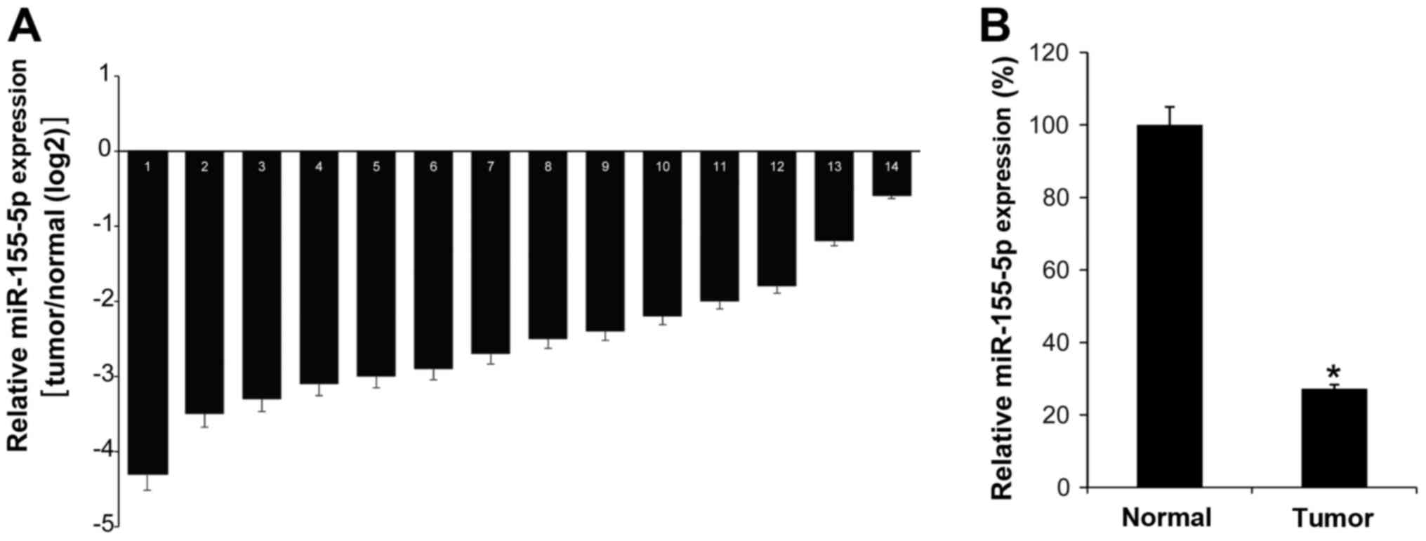
To investigate the function of miR-155-5p in the
pathogenesis of GC, the miR-155-5p expression levels in 14 GC
tissues were investigated by RT-qPCR. It was demonstrated that all
GC tissues exhibited low levels of miR-155-5p compared with their
matched adjacent normal tissues (Fig.
1A). As expected, the mean expression of miR-155-5p in GC tumor
tissues was low compared with that in normal control tissues
(Fig. 1B). Thus, we hypothesized that
miR-155-5p may serve an antitumor role in GC.
Overexpression of miR-155-5p inhibits
GC cell proliferation and promotes apoptosis
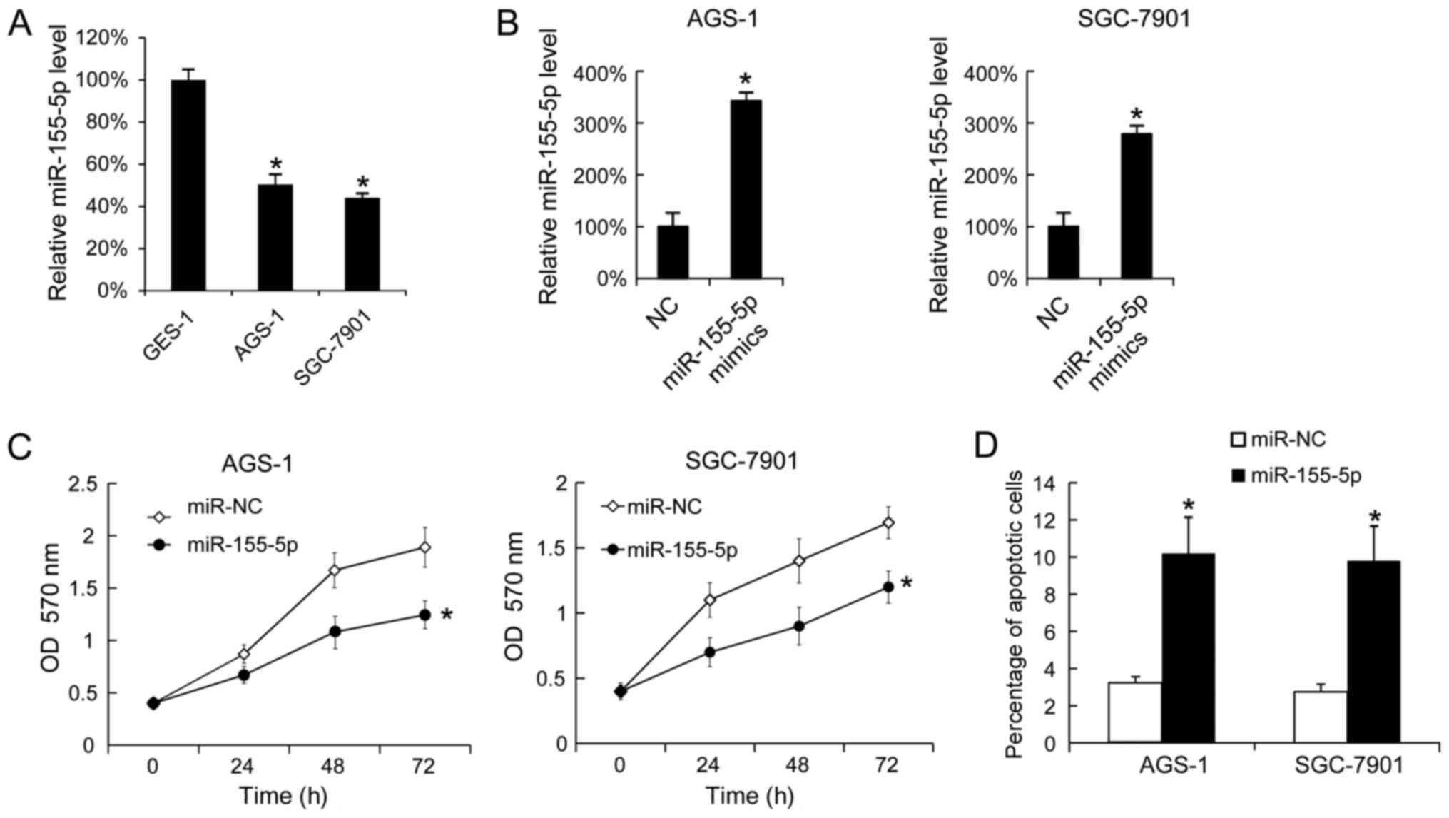
Subsequently, miR-155-5p expression levels were
analyzed in the GC cell lines, AGS-1 and SGC-7901, using the
gastric epithelial mucosa cell line, GES-1, as a negative control.
Using RT-qPCR, it was demonstrated that AGS-1 and SGC-7901 cells
expressed increased levels of miR-155-5p compared with GES-1 cells
(Fig. 2A). Overexpression of
miR-155-5p in AGS-1 and SGC-7901 was confirmed by RT-qPCR (Fig. 2B). Following transfection with
miR-155-5p mimics, proliferation was analyzed by MTT assay. This
revealed that miR-155-5p overexpression inhibited AGS-1 and
SGC-7910 cell proliferation (Fig.
2C). A total of 48 h post-transfection, apoptosis was analyzed
by Annexin V-FITC and PI staining. This indicated that miR-155-5p
overexpression increased the apoptotic rates in AGS-1 and SGC-7910
cells (Fig. 2D).
Downregulation of miR-155-5p promotes
GC cell growth and decreases the sensitivity of GC cells to
cisplatin
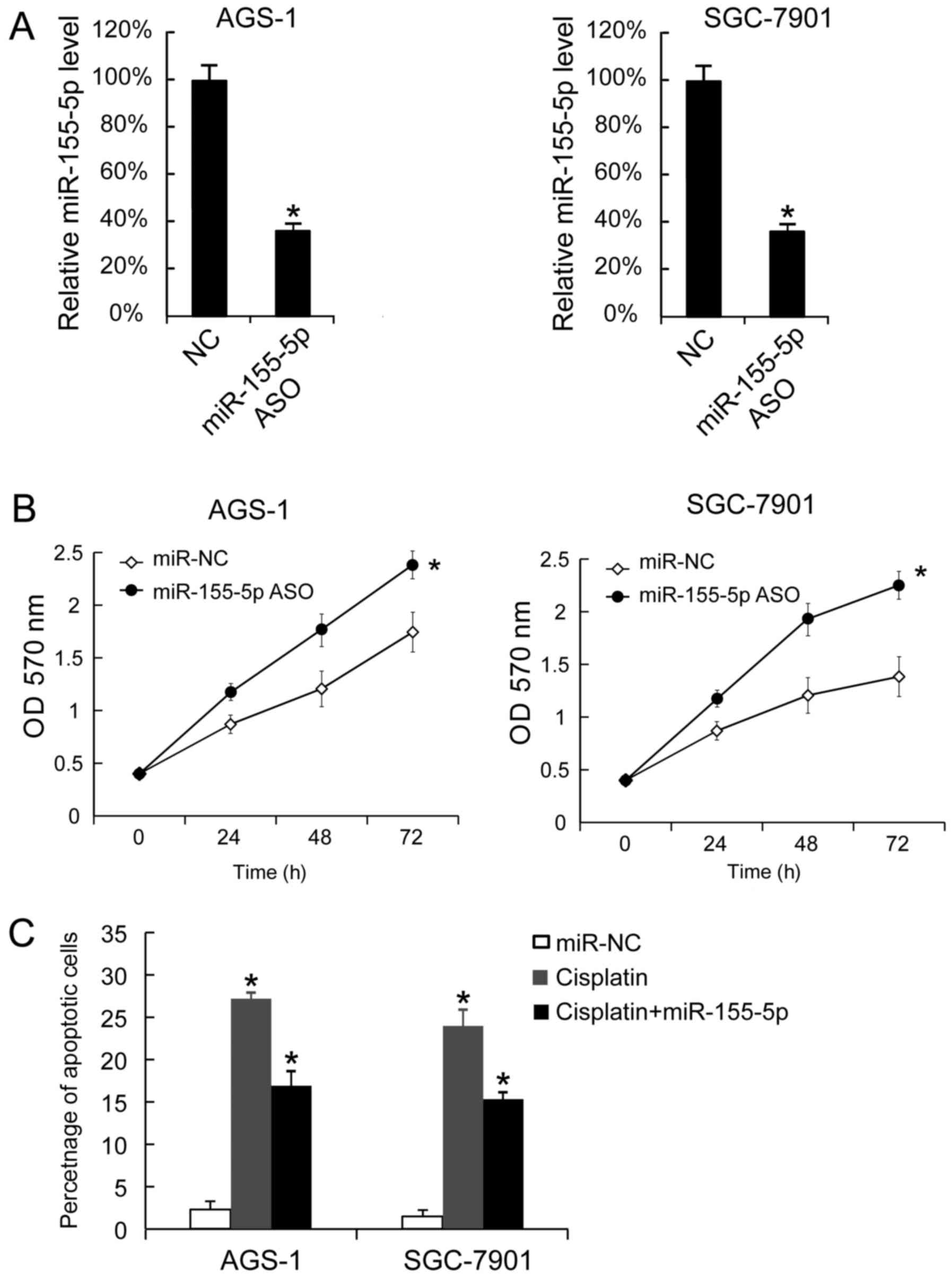
It was demonstrated that miR-155-5p ASO transfection
downregulated miR-155-5p expression levels in AGS-1 and SGC-7901
cells (Fig. 3A). miR-155-5p ASO
transfection also promoted the proliferation of AGS-1 and SGC-7901
cells (Fig. 3B). miR-155-5p ASO
transfection also decreased the apoptotic rates of AGS-1 and
SGC-7901 cells following treatment with cisplatin. Untransfected
cells treated with cisplatin exhibited an increased apoptotic rate
compared with untreated untransfected cells. Thus, miR-155-5p
reduced the effect of cisplatin-induced apoptosis (Fig. 3C).
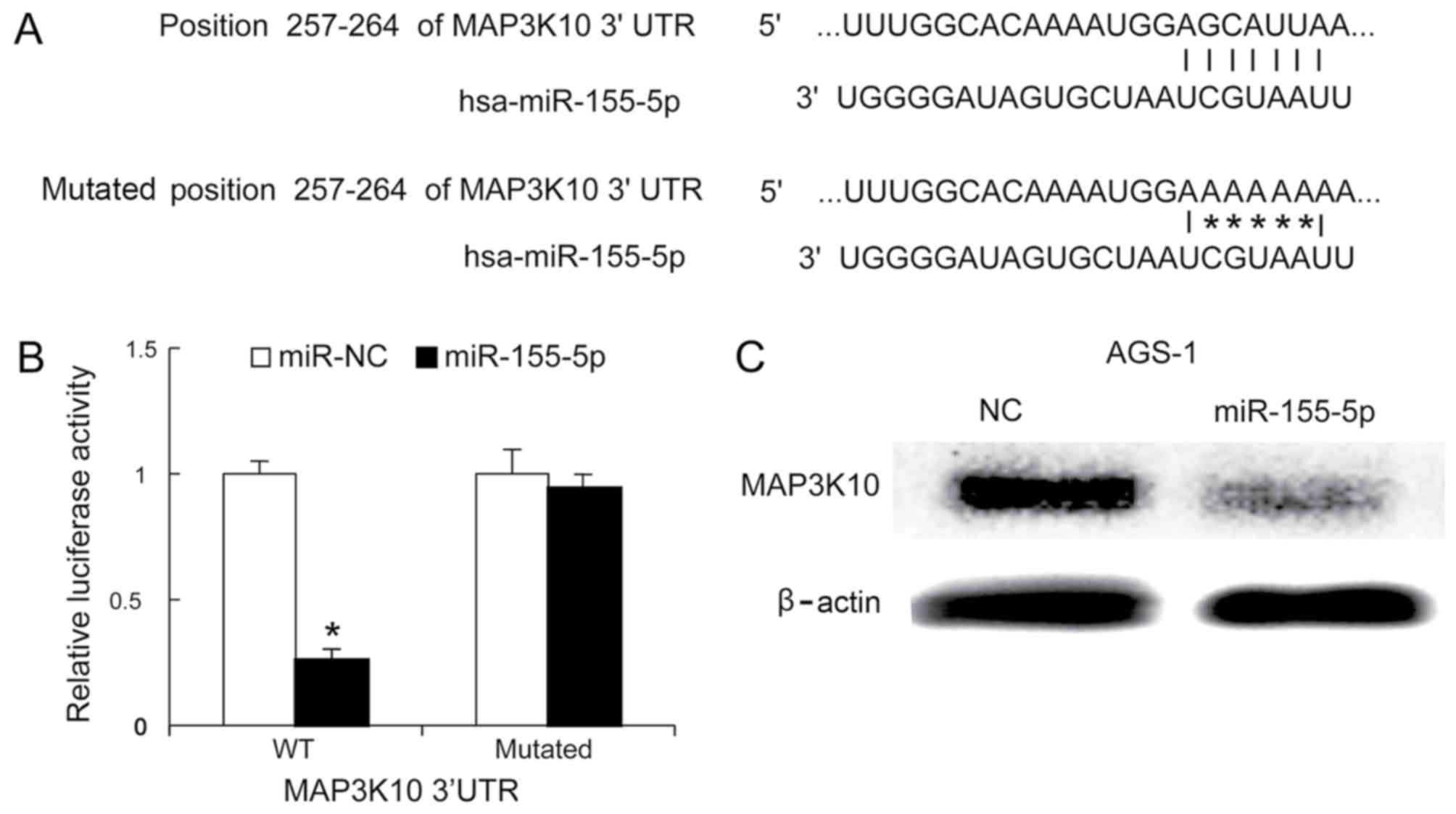
MAP3K10 is targeted by miR-155-5p
A previous study demonstrated that MAP3K10 was
overexpressed in pancreatic ductal adenocarcinoma (PDAC), and that
it promoted proliferation and decreased therapeutic impact in
pancreatic cancer cells (32).
Bioinformatics analysis suggested that MAP3K10 is a targeted gene
of miR-155-5p (Fig. 4A). The
luciferase activity of the reporter carrying the mutated 3′UTR of
MAP3K10 was not significantly different in miR-NC and
miR-155-5p-transfected cells, whereas miR-155-5p was demonstrated
to reduce the luciferase activity of the wild type 3′UTR reporter
(Fig. 4B). To investigate whether
miR-155-5p reduced MAP3K10 protein levels, western blotting was
performed 48 h following miR-155-5p mimics transfection. It was
demonstrated that miR-155-5p mimics significantly decreased the
protein expression level of MAP3K10 proteins in AGS-1 cells,
compared with negative control (Fig.
4C).
Discussion
In a previous study, patients with GC exhibiting low
expression of miR-124-3pm miR-146a-5p, miR-335-5p and miR-155-5p
were associated with increased lymph node metastasis, lymphatic
invasion, venous invasion, high Bormann stage, lymphatic invasion
and poor differentiation compared with those exhibiting high
expression (17). The present may
contribute to the elucidation of the underlying molecular mechanism
of the clinical significance of miR-155-5p. Low expression of
miR-155-5p was demonstrated to promote cellular proliferation and
decrease the cisplatin-sensitivity of GC cells. MAP3K10 was also
indicated to be a target gene of miR-155-5p. MAP3K10 is a member of
the mitogen-activated protein kinase kinase kinase (MAP3K) family,
it activates the C-Jun N-terminal kinase (JNK) signaling pathway
and the p38 mitogen-activated-protein kinase (MAPK) pathway, and
regulates apoptosis in numerous neurodegenerative diseases
(32–34). It was demonstrated in the present
study that MAP3K10 may be targeted by miR-155-5p in GC cell
lines.
MAP3K10 has been demonstrated to promote the
proliferation of pancreatic cancer cells and decrease the
sensitivity to gemcitabine by upregulating the expression of GLI
family zinc finger (Gli)-1 and Gli-2 (32). Whether Gli-1 and Gli-2 promote or
inhibit GC tumor growth requires further investigation. In
conclusion, the present study suggests that miR-155-5p serves an
antitumor role in GC.
Acknowledgements
The authors would like to thank Ms. Miao Chen
(Clinical Laboratory Department, The Affiliated Hospital of North
Sichuan Medical College, Nanchong, China) for technical her
assistance.
Funding
No funding was received.
Availability of data and materials
All data generated or analyzed during this study are
included in this published article.
Authors' contributions
SL, TZ and XZ collected patient data and performed
cell experiments. ZD and FC performed RT-qPCR, western blot
analysis and other molecular experiment. JL and QL contributed to
study design and manuscript writing.
Ethics approval and consent to
participate
The present study was approved by the ethics
committee of Northern Sichuan Medical College (Nanchong, China),
and all patients provided written informed consent.
Consent for publication
All patients gave informed consent for the use of
their tissues and publication of the data and images.
Competing interests
The authors declare that they have no competing
interests.
References
|
1
|
Ferlay J, Soerjomataram I, Dikshit R, Eser
S, Mathers C, Rebelo M, Parkin DM, Forman D and Bray F: Cance
incidence and mortality worldwide: Sources, methods and major
patterns in GLOBOCAN 2012. Int J Cancer. 136:E359–E386. 2015.
View Article : Google Scholar : PubMed/NCBI
|
|
2
|
Torre LA, Bray F, Siegel RL, Ferlay J,
Lortet-Tieulent J and Jemal A: Global cancer statistics, 2012. CA
Cancer J Clin. 65:87–108. 2015. View Article : Google Scholar : PubMed/NCBI
|
|
3
|
Ferlay J, Shin HR, Bray F, Forman D,
Mathers C and Parkin DM: Estimates of worldwide burden of cancer in
2008: GLOBOCAN 2008. Int J Cancer. 127:2893–2917. 2010. View Article : Google Scholar : PubMed/NCBI
|
|
4
|
Mocellin S, Verdi D, Pooley KA and Nitti
D: Genetic variation and gastric cancer risk: A field synopsis and
meta-analysis. Gut. 64:1209–1219. 2015. View Article : Google Scholar : PubMed/NCBI
|
|
5
|
Wadhwa R, Song S, Lee JS, Yao Y, Wei Q and
Ajani JA: Gastric cancer-molecular and clinical dimensions. Nat Rev
Clin Oncol. 10:643–655. 2013. View Article : Google Scholar : PubMed/NCBI
|
|
6
|
Riquelme I, Saavedra K, Espinoza JA, Weber
H, García P, Nervi B, Garrido M, Corvalán AH, Roa JC and Bizama C:
Molecular classification of gastric cancer: Towards a
pathway-driven targeted therapy. Oncotarget. 6:24750–24779. 2015.
View Article : Google Scholar : PubMed/NCBI
|
|
7
|
Giordano S and Columbano A: MicroRNAs: New
tools for diagnosis, prognosis, and therapy in hepatocellular
carcinoma? Hepatology. 57:840–847. 2013. View Article : Google Scholar : PubMed/NCBI
|
|
8
|
Chen DL, Zhang DS, Lu YX, Chen LZ, Zeng
ZL, He MM, Wang FH, Li YH, Zhang HZ, Pelicano H, et al:
microRNA-217 inhibits tumor progression and metastasis by
downregulating EZH2 and predicts favorable prognosis in gastric
cancer. Oncotarget. 6:10868–10879. 2015.PubMed/NCBI
|
|
9
|
Zheng B, Liang L, Wang C, Huang S, Cao X,
Zha R, Liu L, Jia D, Tian Q, Wu J, et al: MicroRNA-148a suppresses
tumor cell invasion and metastasis by downregulating ROCK1 in
gastric cancer. Clin Cancer Res. 17:7574–7583. 2011. View Article : Google Scholar : PubMed/NCBI
|
|
10
|
Li J, Dong G, Wang B, Gao W and Yang Q:
miR-543 promotes gastric cancer cell proliferation by targeting
SIRT1. Biochem Biophys Res Commun. 469:15–21. 2016. View Article : Google Scholar : PubMed/NCBI
|
|
11
|
Zhou X, Zhu W, Li H, Wen W, Cheng W, Wang
F, Wu Y, Qi L, Fan Y, Chen Y, et al: Diagnostic value of a plasma
microRNA signature in gastric cancer: A microRNA expression
analysis. Sci Rep. 5:112512015. View Article : Google Scholar : PubMed/NCBI
|
|
12
|
Ibarrola-Villava M, Llorca-Cardeñosa MJ,
Tarazona N, Mongort C, Fleitas T, Perez-Fidalgo JA, Roselló S,
Navarro S, Ribas G and Cervantes A: Deregulation of ARID1A, CDH1,
cMET and PIK3CA and target-related microRNA expression in gastric
cancer. Oncotarget. 6:26935–26945. 2015. View Article : Google Scholar : PubMed/NCBI
|
|
13
|
Ge X, Liu X, Lin F, Li P, Liu K, Geng R,
Dai C, Lin Y, Tang W, Wu Z, et al: MicroRNA-421 regulated by HIF-1α
promotes metastasis, inhibits apoptosis, and induces cisplatin
resistance by targeting E-cadherin and caspase-3 in gastric cancer.
Oncotarget. 7:24466–24482. 2016. View Article : Google Scholar : PubMed/NCBI
|
|
14
|
Du Y, Wang L, Wu H, Zhang Y, Wang K and Wu
D: MicroRNA-141 inhibits migration of gastric cancer by targeting
zinc finger E-box-binding homeobox 2. Mol Med Rep. 12:3416–3422.
2015. View Article : Google Scholar : PubMed/NCBI
|
|
15
|
Qu YL, Wang HF, Sun ZQ, Tang Y, Han XN, Yu
XB and Liu K: Up-regulated miR-155-5p promotes cell proliferation,
invasion and metastasis in colorectal carcinoma. Int J Clin Exp
Pathol. 8:6988–6994. 2015.PubMed/NCBI
|
|
16
|
Bhattacharya S, Chalk AM, Ng AJ, Martin
TJ, Zannettino AC, Purton LE, Lu J, Baker EK and Walkley CR:
Increased miR-155-5p and reduced miR-148a-3p contribute to the
suppression of osteosarcoma cell death. Oncogene. 35:5282–5294.
2016. View Article : Google Scholar : PubMed/NCBI
|
|
17
|
Li H, Xie S, Liu M, Chen Z, Liu X, Wang L,
Li D and Zhou Y: The clinical significance of downregulation of
mir-124-3p, mir-146a-5p, mir-155-5p and mir-335-5p in gastric
cancer tumorigenesis. Int J Oncol. 45:197–208. 2014. View Article : Google Scholar : PubMed/NCBI
|
|
18
|
Zhu M, Wang M, Yang F, Tian Y, Cai J, Yang
H, Fu H, Mao F, Zhu W, Qian H and Xu W: miR-155-5p inhibition
promotes the transition of bone marrow mesenchymal stem cells to
gastric cancer tissue derived MSC-like cells via NF-κB p65
activation. Oncotarget. 7:16567–16580. 2016.PubMed/NCBI
|
|
19
|
Zhu Y, Zhang C, Gu C, Li Q and Wu N:
Function of deubiquitinating enzyme USP14 as oncogene in different
types of cancer. Cell Physiol Biochem. 38:993–1002. 2016.
View Article : Google Scholar : PubMed/NCBI
|
|
20
|
Price PM, Yu F, Kaldis P, Aleem E, Nowak
G, Safirstein RL and Megyesi J: Dependence of cisplatin-induced
cell death in vitro and in vivo on cyclin-dependent kinase 2. J Am
Soc Nephrol. 17:2434–2442. 2006. View Article : Google Scholar : PubMed/NCBI
|
|
21
|
Li D, Liu X, Lin L, Hou J, Li N, Wang C,
Wang P, Zhang Q, Zhang P, Zhou W, et al: MicroRNA-99a inhibits
hepatocellular carcinoma growth and correlates with prognosis of
patients with hepatocellular carcinoma. J Biol Chem.
286:36677–36685. 2011. View Article : Google Scholar : PubMed/NCBI
|
|
22
|
Song B, Zhang C, Li G, Jin G and Liu C:
MiR-940 inhibited pancreatic ductal adenocarcinoma growth by
targeting MyD88. Cell Physiol Biochem. 35:1167–1177. 2015.
View Article : Google Scholar : PubMed/NCBI
|
|
23
|
Zhou Q and Yu Y: Upregulated CDK16
expression in serous epithelial ovarian cancer cells. Med Sci
Monit. 21:3409–3414. 2015. View Article : Google Scholar : PubMed/NCBI
|
|
24
|
Li H, Xu Y, Qiu W, Zhao D and Zhang Y:
Tissue miR-193b as a novel biomarker for patients with ovarian
cancer. Med Sci Monit. 21:3929–3934. 2015. View Article : Google Scholar : PubMed/NCBI
|
|
25
|
Yuan B, Liang Y, Wang D and Luo F: MiR-940
inhibits hepatocellular carcinoma growth and correlates with
prognosis of hepatocellular carcinoma patients. Cancer Sci.
106:819–824. 2015. View Article : Google Scholar : PubMed/NCBI
|
|
26
|
Lewis BP, Burge CB and Bartel DP:
Conserved seed pairing, often flanked by adenosines, indicates that
thousands of human genes are microRNA targets. Cell. 120:15–20.
2005. View Article : Google Scholar : PubMed/NCBI
|
|
27
|
Friedman RC, Farh KK, Burge CB and Bartel
DP: Most mammalian mRNAs are conserved targets of microRNAs. Genome
Res. 19:92–105. 2009. View Article : Google Scholar : PubMed/NCBI
|
|
28
|
Grimson A, Farh KK, Johnston WK,
Garrett-Engele P, Lim LP and Bartel DP: MicroRNA targeting
specificity in mammals: Determinants beyond seed pairing. Mol Cell.
27:91–105. 2007. View Article : Google Scholar : PubMed/NCBI
|
|
29
|
Garcia DM, Baek D, Shin C, Bell GW,
Grimson A and Bartel DP: Weak seed-pairing stability and high
target-site abundance decrease the proficiency of lsy-6 and other
microRNAs. Nat Struct Mol Biol. 18:1139–1146. 2011. View Article : Google Scholar : PubMed/NCBI
|
|
30
|
John B, Sander C and Marks DS: Prediction
of human microRNA targets. Methods Mol Biol. 342:101–113.
2006.PubMed/NCBI
|
|
31
|
Lee S, Paulson KG, Murchison EP, Afanasiev
OK, Alkan C, Leonard JH, Byrd DR, Hannon GJ and Nghiem P:
Identification and validation of a novel mature microRNA encoded by
the Merkel cell polyomavirus in human Merkel cell carcinomas. J
Clin Virol. 52:272–275. 2011. View Article : Google Scholar : PubMed/NCBI
|
|
32
|
An Y, Cai B, Chen J, Lv N, Yao J, Xue X,
Tu M, Tang D, Wei J, Jiang K, et al: MAP3K10 promotes the
proliferation and decreases the sensitivity of pancreatic cancer
cells to gemcitabine by upregulating Gli-1 and Gli-2. Cancer Lett.
329:228–235. 2013. View Article : Google Scholar : PubMed/NCBI
|
|
33
|
Gallo KA and Johnson GL: Mixed-lineage
kinase control of JNK and p38 MAPK pathways. Nat Rev Mol Cell Biol.
3:663–672. 2002. View
Article : Google Scholar : PubMed/NCBI
|
|
34
|
Xu Z, Maroney AC, Dobrzanski P, Kukekov NV
and Greene LA: The MLK family mediates c-Jun N-terminal kinase
activation in neuronal apoptosis. Mol Cell Biol. 21:4713–4724.
2001. View Article : Google Scholar : PubMed/NCBI
|