Introduction
The growth rate and metastasis of non-small cell
lung cancer (NSCLC) have been reported to be slower and later than
that of small cell carcinoma (1).
Moreover, NSCLC accounts for 80–85% of the total lung cancer, and
now chemotherapy is the main treatment for NSCLC (2). Moreover, low overall 5-year survival and
high recurrence rate of NSCLC patients (3) make it urgent to develop novel treatments
for NSCLC. Therefore, future direction is to further analyze
potential therapeutic targets in the process of NSCLC
occurrence.
Recently, microRNA has been paid increasing
attention in cancer research in regulation of many biological
processes including growth, proliferation, migration, invasion, and
apoptosis (4–6). Especially in NSCLC, some downregulated
miRNAs such as miR-134, miR-204, miR-34c, and miR-200b were
reported to repress the development of NSCLC (7–10). In
addition, many upregulated miRNAs such as miR-21, miR-205, miR-211,
and miR-484 were reported to promote cell growth, cell
proliferation, cell cycle and invasion (11–14). These
previous studies elucidated that the alteration of miRNA expression
could affect tumorigenesis of NSCLC.
Among these miRNAs, miR-486-5p has been identified
in various human cancers and diseases. It has been reported that
miR-486-5p was downregulated in hepatocellular carcinoma (15), breast cancer (16), duchenne muscular dystrophy (17) and chronic kidney disease (18). However, studies are rare on miR-486-5p
in NSCLC. Based on the above research, we speculated that
miR-486-5p expression level might be declined in regulation of the
progression of NSCLC. Additionally, targeted genes such as NOB1
(19), RUNX3 (20) and FOXM1 (21) have been found to influence the
tumorigenesis of NSCLC. Nonetheless, the role of GAB2 in NSCLC is
rarely reported.
In the present study, we explored the effect of
miR-486-5p on the NSCLC development. As predicted, miR-486-5p
expression was decreased in NSCLC. Moreover, miR-486-5p repressed
cell proliferation and invasion in NSCLC through suppressing GAB2.
These findings provide a new way for treatment of NSCLC.
Materials and methods
Clinical tissues
Forty-six NSCLC and adjacent tissues were obtained
from The Central Hospital of Wuhan, Tongji Medical College,
Huazhong University of Science and Technology (Wuhan, China) after
receiving written informed consent. None of the patients received
treatment prior to the operation. Then the tissues were frozen in
liquid nitrogen and stored at −80°C in a refrigerator for further
experiment. This study was approved by the Central Hospital of
Wuhan, Tongji Medical College, Huazhong University of Science and
Technology institutional Ethics Committee.
Cell cultures and cell
transfection
The human NSCLC cell lines A549, SPC-A1 and BEAS-2B
(control) were used for this study. All the cell lines were
obtained from the Shanghai Cell Bank, China Academy of Sciences
(Shanghai, China). Then these cell lines were seeded in DMEM medium
(Cellgro; Corning Incorporated, Corning, NY, USA) with 10% fetal
bovine serum (FBS) and cultured at 37°C with 5% CO2.
The miR-486 mimic/mimic control
(miR10004762-1-5/miR01201-1-5), the miR-486 inhibitor/inhibitor
control (miR20004762-1-5/miR02201-1-5) and the GAB2 siRNA (si-GAB2,
5′-AAACGCUGGUUUAUACUGCGG-3′), purchased from RiboBio Co., Ltd.
(Guangzhou, China), were transferred into NSCLC cells by
Lipofectamine 2000 (Thermo Fisher Scientific, Inc., Waltham, MA,
USA) based on the manufactures' instructions.
Quantitative RT-qPCR
TRIzol reagent (Invitrogen; Thermo Fisher
Scientific, Inc.) was applied for extracting total RNA containing
miRNA to quantitate miR-486-5p expression in NSCLC. To obtain the
cDNA templates, 1 µg total RNA of each sample was used for reverse
transcription using a miScript Reverse Transcription kit (Qiagen
GmbH, Hilden, Germany). This reaction was performed at 37°C for 60
min, then 95°C for 5 min. Quantitative RT-qPCR was carried out
through the SYBR-Green PCR kit (Takara Bio, Inc., Otsu, Japan) on
ABI 7500 Fast Real-Time PCR system (ABI; Thermo Fisher Scientifc,
Inc.). The cycling conditions for RT-qPCR were as follows: 5 min at
95°C, followed by 40 cycles of 95°C for 30 sec and 60°C for 45 sec.
U6 and GAPDH were used as control for miR-486-5p and GAB2. The
primers were: GAPDH forward, 5′-TGTTCGTCATGGGTGTGAAC-3′ and
reverse, 5′-ATGGCATGGACTGTGGTCAT-3′; GAB2 forward,
5′-CGAAGAGAACTATGTCCCTATGC-3′; reverse, 5′-AGGGGCAGGACTGTTCGT-3′
miR-486-5p forward, 5′-ACACTCCAGCTGGGTCCTGTACTGAGCTGCCC-3′ and
reverse, 5′-CTCAACTGGTGTCGTGGAGTCGGCAATTCAGTTGAGCCCCGAG-3′; U6
forward, 5′-CTCGCTTCGGCAGCACA-3′ and reverse,
5′-AACGCTTCACGAATTTGCGT-3′. The expression was calculated using the
2−ΔΔcq method (22).
Luciferase activity assay
The wild or mutant type of 3′-UTR of GAB2 was
inserted into the pGL3 promoter vector (Invitrogen; Thermo Fisher
Scientific, Inc.) for luciferase reporter experiments. GAB2 vector
(pCDNA3.1-GAB2) was purchased from RiboBio Co., Ltd. Then, we
transfected GAB2 vector and miR-486-5p mimic into A549 cells.
Subsequently, the dual luciferase reporter assay (Promega
Corporation, Madison, WI, USA) was applied to perform luciferase
assays.
MTT assay
The MTT
(3-(4,5-dimethyl-2-thiazolyl)-2,5-diphenyl-2H-tetrazolium bromide)
assay was applied to measure cell proliferation. The cells
(4×10³ cells/well) were added onto 96-well plates in
medium with 10% FBS. The cells with miR-486-5p mimic or inhibitor
were incubated for 0–72 h. After incubation, the cells added with
MTT (Sigma-Aldrich; Merck KGaA, Darmstadt, Germany) and were
cultured at 37°C for 4 h. The absorbance at 490 nm (OD=490 nm) was
detected with a spectrophotometer (Model 680 microplate reader;
Bio-Rad Laboratories, Inc., Hercules, CA, USA).
Cell invasion assay
The cells were plated into the upper chambers (8 µm
pore size; Corning Incorporated) and medium with 10% FBS was put
into the lower chamber. Then these cells (5×10³) were
cultured for 24 h at 37°C in atmosphere with 5% CO2.
Then the invasive cells in the lower chamber were fixed with 70%
ethanol and stained using crystal violet. Finally, light microscope
(DM IRB; Leica Microsystems GmbH, Wetzlar, Germany) was used to
measure the cell number.
Western blot analysis
The protein samples were obtained using RIPA lysis
buffer. 10% SDS-PAGE was employed to separate proteins which were
incubated with 5% non-fat milk in polyvinylidene difluoride
membranes (EMD Millipore, Billerica, MA, USA) at room temperature.
Next, we incubated the membranes overnight at 4°C with rabbit
monoclonal anti-GAB2 (1:1,000; catalog no. ab203478; Abcam,
Shanghai, China), rabbit polyclonal anti-GAPDH (1:1,000; catalog
no. ab70699; Abcam) and subsequently incubated with goat
anti-rabbit IgG-H&L secondary antibody (1:1,000; catalog no.
ab150077; Abcam). Then, protein expression levels were measured by
an Enhanced Chemiluminescence Immunoblot Detection system (Pierce;
Thermo Fisher Scientific, Inc.) and analyzed using Quantity One
software (version 4.62; Bio-Rad Laboratories, Inc., Hercules, CA,
USA).
Statistical analysis
The obtained data are shown as the mean ± standard
deviation. The difference between the groups was calculated through
Chi-square test or one-way ANOVA with Tukey's as a post hoc test.
Statistical analysis was analyzed with GraphPad Prism 6.0 (GraphPad
Software, Inc., La Jolla, CA, USA) and SPSS 19.0 (IBM Corp.,
Armonk, NY, USA). P<0.05 was considered to indicate a
statistically significant difference.
Results
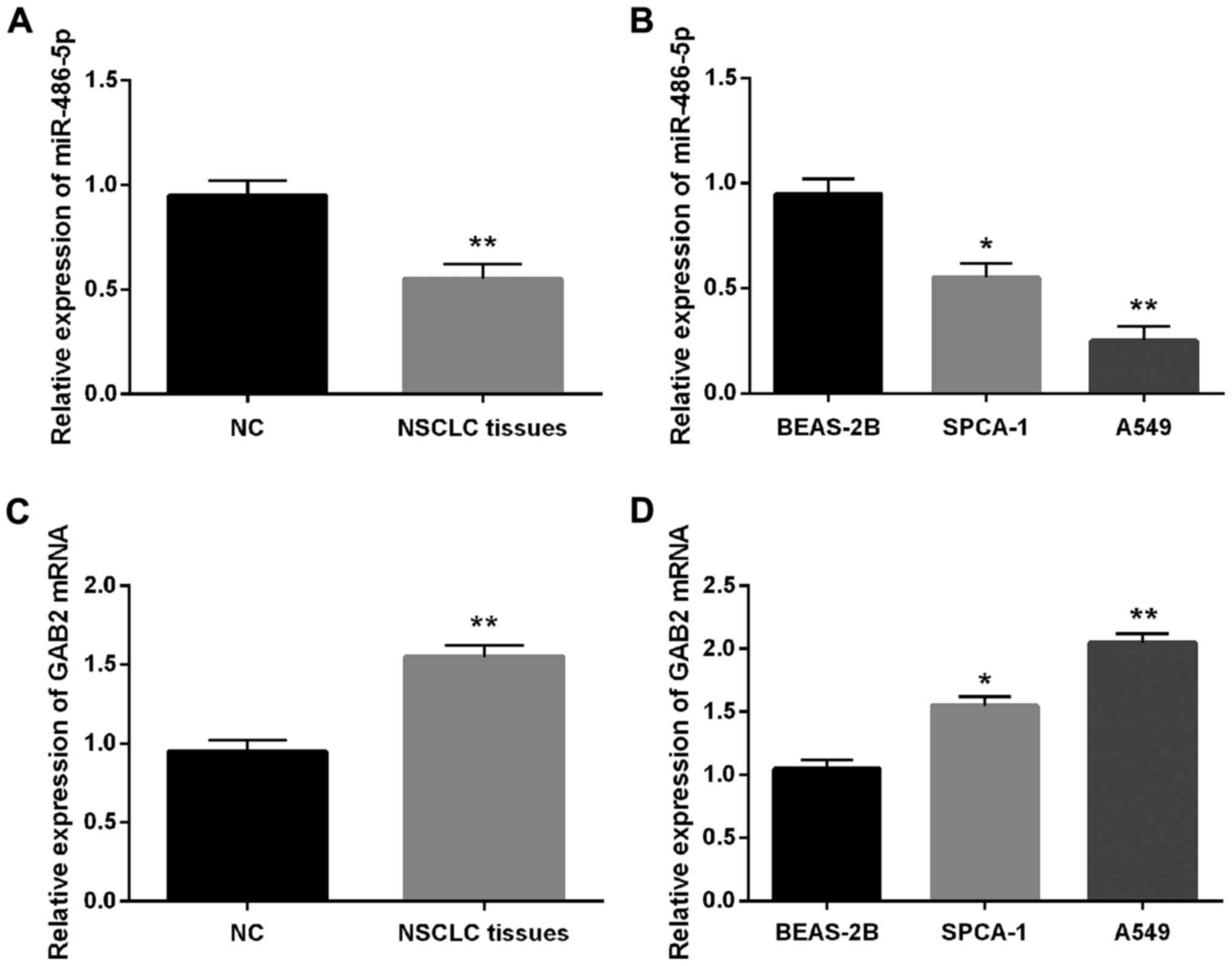
Expression of miR-486-5p and GAB2 mRNA
was detected in NSCLC
First, we detected miR-486-5p expressions in NSCLC
via quantitative RT-qPCR. The results suggested that miR-486-5p
expression was reduced in NSCLC tissues compared with normal
tissues (Fig. 1A). Moreover, the
downregulation of miR-486-5p was also identified in SPC-A1 and A549
cell lines (Fig. 1B). Subsequently,
the expression of GAB2 was analyzed in NSCLC as well.
Interestingly, GAB2 mRNA expression was increased in NSCLC tissues
and cell lines (Fig. 1C and D) which
was contrary to miR-486-5p expression.
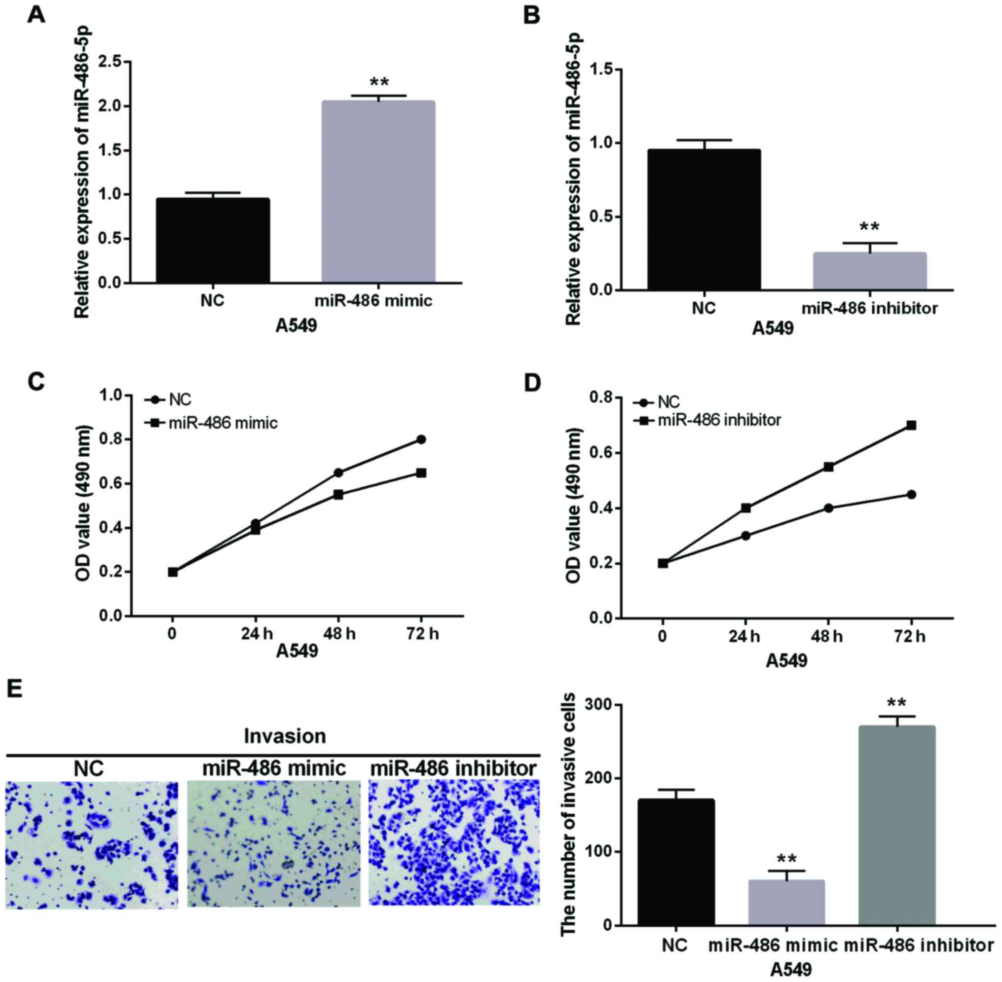
miR-486-5p suppresses cell
proliferation and invasion in NSCLC
The function of miR-486-5p in NSCLC was verified by
performing MTT and Transwell assay in A549 cells containing
miR-486-5p mimic or inhibitor. Primarily, the miR-486-5p expression
level was detected in cells with miR-486-5p mimic or inhibitor as
shown in Fig. 2A and B. Importantly,
MTT assay showed that miR-486-5p overexpression suppressed cell
proliferation, while miR-486-5p downregulation exhibited the
opposite result in NSCLC (Fig. 2C and
D). Transwell assay suggested that the cell invasion was
significantly repressed by miR-486-5p overexpression. However, it
was promoted by miR-486-5p inhibitor in NSCLC cells (Fig. 2E). These findings indicated that cell
proliferation and invasion were inhibited in NSCLC by miR-486-5p
overexpression.
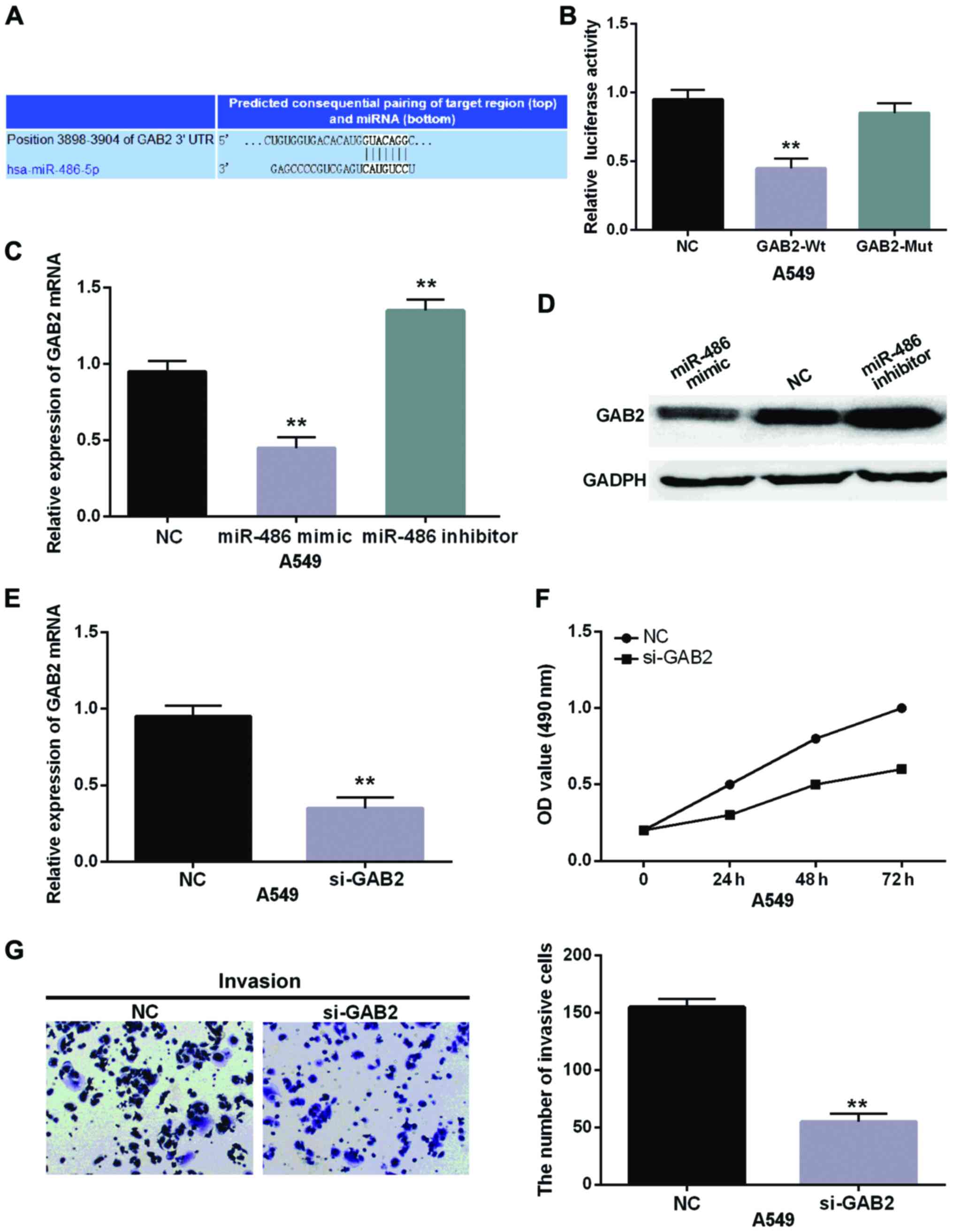
GAB2 is a direct target gene of
miR-486-5p in NSCLC
The target genes of miR-486-5p were searched through
the database of TargetScan Human (http://www.targetscan.org/vert_71/), which indicated
that it binds with the 3′-UTR of GAB2 (Fig. 3A). Moreover, we confirmed that
miR-486-5p directly targeted GAB2 and had a binding site with the
wild-type of GAB2 by dual luciferase reporter assay (Fig. 3B). Additionally, mRNA and protein
expression of GAB2 were reduced by miR-486-5p mimics whereas
increased by miR-486-5p inhibitor (Fig.
3C and D). Furthermore, si-GAB2 was transfected into A549 cells
to further explore its role in NSCLC (Fig. 3E). Additionally, we found that si-GAB2
had the similar effect as the upregulation of miR-486-5p which
suppressed cell proliferation and invasion in NSCLC (Fig. 3F and G). Therefore, it was inferred
that miR-486-5p directly targeted GAB2 and knockout of GAB2
inhibited NSCLC cell proliferation and invasion.
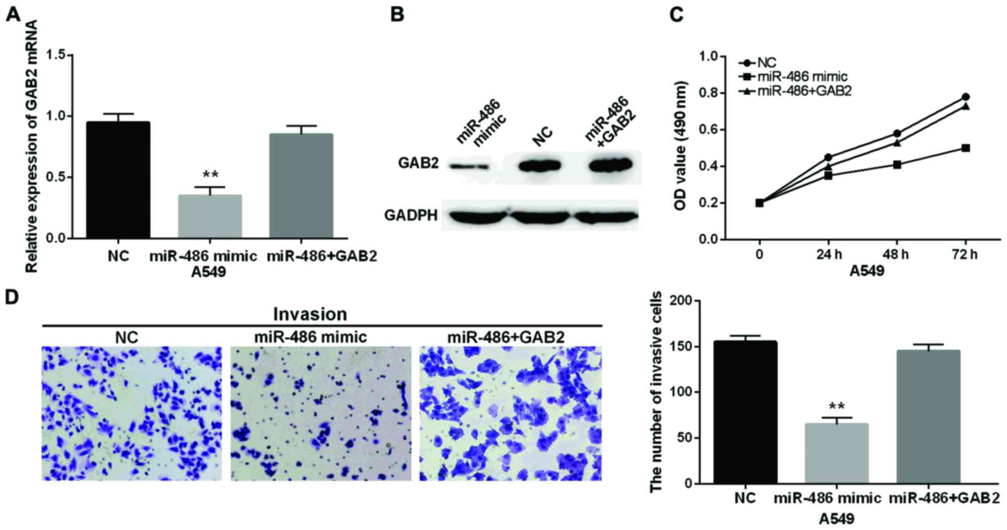
miR-486-5p regulates cell
proliferation and invasion through suppressing GAB2 in NSCLC
Finally, we transfected GAB2 vector and miR-486-5p
mimics into NSCLC cells to verify their relationship, and we found
that there was almost no change in expression of mRNA and protein
of GAB2 in cells with miR-486-5p mimic and GAB2 vector in
comparison with the control (Fig. 4A and
B). Importantly, the suppression of miR-486-5p for
proliferation of NSCLC cells was impaired by GAB2 vector (Fig. 4C). Moreover, the inhibitory action of
miR-486-5p for cell invasion almost disappeared in GAB2
overexpression group (Fig. 4D). All
these findings indicated that miR-486-5p inhibited cell
proliferation and invasion to a certain extent through the
regulation of GAB2 in NSCLC.
Discussion
A full understanding of the occurrence and
development mechanisms of NSCLC is necessary for opening a new
pathway for NSCLC patients to improve survival. We conducted this
research to explore the alteration of miR-486-5p expression and its
effect on the progression of NSCLC. Moreover, miR-486-5p expression
levels were declined in NSCLC which promoted cell proliferation and
invasion in NSCLC. Additionally, GAB2 was confirmed as a direct
target gene of miR-486-5p in NSCLC. We considered that miR-486-5p
would repress NSCLC development by affecting GAB2 expression,
indicating that miR-486-5p had a suppressive effect on NSCLC
progression.
In various human cancers, miR-486 is usually
expressed aberrantly and has inhibitory action. Previous studies
demonstrated that miR-486 could be used as a biomarker for early
diagnosis and recurrence of NSCLC (23), and had an inhibitory action for the
growth and development of NSCLC (24). Other researchers also found the
downregulation of miR-486 in prostatic carcinoma (25) and esophageal cancer (26) which was similar to our results.
Additionally, low miR-486-5p expression was identified in NSCLC and
promoted tumor metastasis and development by regulating ARHGAP5
(27). Our findings also suggested
miR-486-5p repressed cell proliferation and invasion by suppressing
GAB2 in NSCLC. Although miR-486 was confirmed to associate with
tumorigenesis and progression of NSCLC, its relationship with GAB2
in NSCLC is still not clear.
Grb2-associated binding protein 2 (GAB2) was
frequently detected in many human malignancies (28). As an oncogene, GAB2 has been
identified in glioblastoma (29),
gastric cancer (30) and renal cell
carcinoma (31). Previously, GAB2 was
identified to express highly in lung cancer (32). We found the upregulation of GAB2 in
NSCLC as well. Functionally, GAB2 promoted cell proliferation and
invasion in NSCLC. Similar to our results, GAB2 was also reported
to have a positive impact on cell migration in NSCLC via Akt
signaling pathway (33). Based on
these results, we considered that GAB2 obviously promoted growth
and metastasis of NSCLC. Hence, the low expression of GAB2 induced
by miR-486-5p could help repress NSCLC development. However, due to
the poor current laboratory conditions, there are still several
limitations in this research such as the deficiency of in
vivo experiments. We will further perform the experiments to
validate our conclusion in future research.
In conclusion, we found that miR-486-5p was
downregulated in NSCLC. Moreover, miR-486-5p inhibited cell
proliferation and invasion through repressing GAB2 in NSCLC. This
new pathway may help us better understand NSCLC pathogenesis and
provide beneficial clues for the diagnosis and treatment of
NSCLC.
Acknowledgements
Not applicable.
Funding
No funding was received.
Availability of data and materials
The datasets used and/or analyzed during the present
study are available from the corresponding author on reasonable
request.
Authors' contributions
SY contributed significantly to the study design and
data acquisition; SG performed the data analyses and wrote the
manuscript. YH contributed to the conception of the study. All
authors read and approved the final study.
Ethics approval and consent to
participate
The study was approved by the Ethics Committee of
The Central Hospital of Wuhan, Tongji Medical College, Huazhong
University of Science and Technology (Wuhan, China). Signed
informed consents were obtained from the patients or the
guardians.
Patient consent for publication
Not applicable.
Competing interests
The authors declare that they have no competing
interests.
References
|
1
|
Chen W, Zheng R, Baade PD, Zhang S, Zeng
H, Bray F, Jemal A, Yu XQ and He J: Cancer statistics in China,
2015. CA Cancer J Clin. 66:115–132. 2016. View Article : Google Scholar : PubMed/NCBI
|
|
2
|
Torre LA, Bray F, Siegel RL, Ferlay J,
Lortet-Tieulent J and Jemal A: Global cancer statistics, 2012. CA
Cancer J Clin. 65:87–108. 2015. View Article : Google Scholar : PubMed/NCBI
|
|
3
|
Fassina A, Cappellesso R and Fassan M:
Classification of non-small cell lung carcinoma in transthoracic
needle specimens using microRNA expression profiling. Chest.
140:1305–1311. 2011. View Article : Google Scholar : PubMed/NCBI
|
|
4
|
Garzon R, Calin GA and Croce CM: MicroRNAs
in cancer. Annu Rev Med. 60:167–179. 2009. View Article : Google Scholar : PubMed/NCBI
|
|
5
|
Bartel DP: MicroRNAs: Target recognition
and regulatory functions. Cell. 136:215–233. 2009. View Article : Google Scholar : PubMed/NCBI
|
|
6
|
Oom AL, Humphries BA and Yang C:
MicroRNAs: Novel players in cancer diagnosis and therapies. BioMed
Res Int. 2014:9594612014. View Article : Google Scholar : PubMed/NCBI
|
|
7
|
Li J, Wang Y, Luo J, Fu Z, Ying J, Yu Y
and Yu W: miR-134 inhibits epithelial to mesenchymal transition by
targeting FOXM1 in non-small cell lung cancer cells. FEBS Lett.
586:3761–3765. 2012. View Article : Google Scholar : PubMed/NCBI
|
|
8
|
Wang P, Lv HY, Zhou DM and Zhang EN:
miR-204 suppresses non-small-cell lung carcinoma (NSCLC) invasion
and migration by targeting JAK2. Genet Mol Res. 15:gmr64152016.
|
|
9
|
Zhou YL, Xu YJ and Qiao CW: MiR-34c-3p
suppresses the proliferation and invasion of non-small cell lung
cancer (NSCLC) by inhibiting PAC1/MAPK pathway. Int J Clin Exp
Pathol. 8:6312–6322. 2015.PubMed/NCBI
|
|
10
|
Xiao P, Liu W and Zhou H: miR-200b
inhibits migration and invasion in non-small cell lung cancer cells
via targeting FSCN1. Mol Med Rep. 14:1835–1840. 2016. View Article : Google Scholar : PubMed/NCBI
|
|
11
|
Zhang JG, Wang JJ, Zhao F, Liu Q, Jiang K
and Yang GH: MicroRNA-21 (miR-21) represses tumor suppressor PTEN
and promotes growth and invasion in non-small cell lung cancer
(NSCLC). Clin Chim Acta. 411:846–852. 2010. View Article : Google Scholar : PubMed/NCBI
|
|
12
|
Duan B, Guo T, Sun H, Cai R, Rui Q and Xi
Z: miR-205 as a biological marker in non-small cell lung cancer.
Biomed Pharmacother. 91:823–830. 2017. View Article : Google Scholar : PubMed/NCBI
|
|
13
|
Ye L, Wang H and Liu B: miR-211 promotes
non-small cell lung cancer proliferation by targeting SRCIN1.
Tumour Biol. 37:1151–1157. 2016. View Article : Google Scholar : PubMed/NCBI
|
|
14
|
Li T, Ding ZL, Zheng YL and Wang W:
MiR-484 promotes non-small-cell lung cancer (NSCLC) progression
through inhibiting Apaf-1 associated with the suppression of
apoptosis. Biomed Pharmacother. 96:153–164. 2017. View Article : Google Scholar : PubMed/NCBI
|
|
15
|
Sun H, Cui C, Xiao F, Wang H, Xu J, Shi X,
Yang Y, Zhang Q, Zheng X, Yang X, et al: miR-486 regulates
metastasis and chemosensitivity in hepatocellular carcinoma by
targeting CLDN10 and CITRON. Hepatol Res. 45:1312–1322. 2015.
View Article : Google Scholar : PubMed/NCBI
|
|
16
|
Tan K, Huang G and Fang Q: MiR-486-5p
prevents migration, invasion and EMT by regulating smad2 in breast
cancer. Int J Clin Exp Med. 10:8942–8949. 2017.
|
|
17
|
Alexander MS, Casar JC, Motohashi N,
Vieira NM, Eisenberg I, Marshall JL, Gasperini MJ, Lek A, Myers JA,
Estrella EA, et al: MicroRNA-486-dependent modulation of
DOCK3/PTEN/AKT signaling pathways improves muscular
dystrophy-associated symptoms. J Clin Invest. 124:2651–2667. 2014.
View Article : Google Scholar : PubMed/NCBI
|
|
18
|
Xu J, Li R, Workeneh B, Dong Y, Wang X and
Hu Z: Transcription factor FoxO1, the dominant mediator of muscle
wasting in chronic kidney disease, is inhibited by microRNA-486.
Kidney Int. 82:401–411. 2012. View Article : Google Scholar : PubMed/NCBI
|
|
19
|
Li Y, Ma C, Qian M, Wen Z, Jing H and Qian
D: Downregulation of NOB1 suppresses the proliferation and tumor
growth of non-small cell lung cancer in vitro and in
vivo. Oncol Rep. 31:1271–1276. 2014. View Article : Google Scholar : PubMed/NCBI
|
|
20
|
Wang Y, Li Y, Wu B, Shi C and Li C:
MicroRNA-661 promotes non-small cell lung cancer progression by
directly targeting RUNX3. Mol Med Rep. 16:2113–2120. 2017.
View Article : Google Scholar : PubMed/NCBI
|
|
21
|
Ke Y, Zhao W, Xiong J and Cao R: MiR-149
inhibits non-small-cell lung cancer cells emt by targeting FOXM1.
Biochem Res Int. 2013:5067312013. View Article : Google Scholar : PubMed/NCBI
|
|
22
|
Livak KJ and Schmittgen TD: Analysis of
relative gene expression data using real-time quantitative PCR and
the 2(-Delta Delta C(T)) method. Methods. 25:402–408. 2001.
View Article : Google Scholar : PubMed/NCBI
|
|
23
|
Li W, Wang Y, Zhang Q, Tang L, Liu X, Dai
Y, Xiao L, Huang S, Chen L, Guo Z, et al: Correction: MicroRNA-486
as a biomarker for early diagnosis and recurrence of non-small cell
lung cancer. PLoS One. 11:e01485892016. View Article : Google Scholar : PubMed/NCBI
|
|
24
|
Shao Y, Shen YQ, Li YL, Liang C, Zhang BJ,
Lu SD, He YY, Wang P, Sun QL, Jin YX, et al: Direct repression of
the oncogene CDK4 by the tumor suppressor miR-486-5p in non-small
cell lung cancer. Oncotarget. 7:34011–34021. 2016. View Article : Google Scholar : PubMed/NCBI
|
|
25
|
Zhang X, Zhang T, Yang K, Zhang M and Wang
K: miR-486-5p suppresses prostate cancer metastasis by targeting
Snail and regulating epithelial-mesenchymal transition. OncoTargets
Ther. 9:6909–6914. 2016. View Article : Google Scholar
|
|
26
|
Yi Y, Lu X, Chen J, Jiao C, Zhong J, Song
Z, Yu X and Lin B: Downregulated miR-486-5p acts as a tumor
suppressor in esophageal squamous cell carcinoma. Exp Ther Med.
12:3411–3416. 2016. View Article : Google Scholar : PubMed/NCBI
|
|
27
|
Wang J, Tian X, Han R, Zhang X, Wang X,
Shen H, Xue L, Liu Y, Yan X, Shen J, et al: Downregulation of
miR-486-5p contributes to tumor progression and metastasis by
targeting protumorigenic ARHGAP5 in lung cancer. Oncogene.
33:1181–1189. 2014. View Article : Google Scholar : PubMed/NCBI
|
|
28
|
Ding CB, Yu WN, Feng JH and Luo JM:
Structure and function of Gab2 and its role in cancer (Review). Mol
Med Rep. 12:4007–4014. 2015. View Article : Google Scholar : PubMed/NCBI
|
|
29
|
Tian LQ, Liu EQ, Zhu XD, Wang XG, Li J and
Xu GM: MicroRNA-197 inhibits cell proliferation by targeting GAB2
in glioblastoma. Mol Med Rep. 13:4279–4288. 2016. View Article : Google Scholar : PubMed/NCBI
|
|
30
|
Lee SH, Jeong EG, Nam SW, Lee JY, Yoo NJ
and Lee SH: Increased expression of Gab2, a scaffolding adaptor of
the tyrosine kinase signalling, in gastric carcinomas. Pathology.
39:326–329. 2007. View Article : Google Scholar : PubMed/NCBI
|
|
31
|
Gu DH, Mao JH, Pan XD, Zhu H, Chen X,
Zheng B and Shan Y: microRNA-302c-3p inhibits renal cell carcinoma
cell proliferation by targeting Grb2-associated binding 2 (Gab2).
Oncotarget. 8:26334–26343. 2017.PubMed/NCBI
|
|
32
|
Xu XL, Wang X, Chen ZL, Jin M, Yang W,
Zhao GF and Li JW: Overexpression of Grb2-associated binder 2 in
human lung cancer. Int J Biol Sci. 7:496–504. 2011. View Article : Google Scholar : PubMed/NCBI
|
|
33
|
Xu LJ, Wang YC, Lan HW, Li J and Xia T:
Grb2-associated binder-2 gene promotes migration of non-small cell
lung cancer cells via Akt signaling pathway. Am J Transl Res.
8:1208–1217. 2016.PubMed/NCBI
|