Introduction
Extrahepatic bile duct cancer (EHBDCA) and
gallbladder cancer (GBCA) are rare diseases worldwide, but they are
the sixth leading causes of cancer death in Japan, with nearly
17,000 deaths annually (compiled by the Statistics and Information
Department, Minister's Secretariat, Ministry of Health, Labour and
Welfare). Surgical resection is the only curative treatment, but
the 5-year survival rate even after R0 resection is poor, ranging
from 20 to 40% (1). The incidence of
EHBDCA and GBCA is increasing throughout the world with a high
fatality rate; therefore, new prognostic markers and treatments for
EHBDCA and GBCA patients are required.
Recently, cancer stem cells (CSCs) were reported to
play important roles in various kinds of cancer (2). CD133, which is also known as prominin-1,
is a cell surface marker of hematopoietic cells and, originally, of
progenitor cells. Although the physiological function of CD133
remains to be elucidated, it has been widely used as a marker for
CSCs (3–5). Immunohistochemistry has been used to
show the clinical significance of CD133 expression in various types
of solid tumors including brain tumors (6), breast cancer (7), lung cancer (8), pancreatic cancer (9), hepatocellular carcinoma (10), and colorectal cancer (11), in which positive CD133 expression is
significantly associated with poor prognosis. However, the
clinicopathological significance of CD133 in intrahepatic
cholangiocarcinoma remains controversial (12–14), and
there are no reports on EHBDCA and GBCA. Therefore, in this study,
we investigated the potential clinical role of CD133 expression,
using immunohistochemistry, in a large series of EHBDCA and GBCA
patients.
Patients and methods
Patient demographics
The participants in this study were 82 patients who
underwent radical surgery for EHBDCA and GBCA between 2000 and 2010
at Hokkaido University Hospital, in the Department of
Gastroenterological Surgery I, Hokkaido University, Graduate School
of Medicine (Sapporo, Japan). The patients' clinicopathological
characteristics are summarized in Table
I.
 | Table I.Clinicopathological characteristics of
82 patients with extrahepatic bile duct cancer and gallbladder
cancer. |
Table I.
Clinicopathological characteristics of
82 patients with extrahepatic bile duct cancer and gallbladder
cancer.
| Parameter | No. of cases |
|---|
| Age, years (mean ±
standard deviation) | 66.4±8.7 |
| Sex |
|
|
Male/female | 62/20 |
| Location |
|
|
Bp/Bd/G+C | 32/27/23 |
| Tumor diameter,
cm |
|
|
<5/≥5 | 70/12 |
| Histopathological
grade |
|
| G1,
2/G3 | 60/22 |
| Lymphatic
permeation |
|
|
Negative/positive | 32/50 |
| Blood vessel
permeation |
|
|
Negative/positive | 33/49 |
| Perineural
invasion |
|
|
Negative/positive | 17/65 |
| pT factor |
|
|
pT1-2/pT3-4 | 47/35 |
| pN factor |
|
|
Negative/positive | 36/46 |
| Staging |
|
| I,
II/III, IV | 44/38 |
| Resection status |
|
|
R0/R1 | 32/50 |
| Median survival
(months) | 40.7 |
The patients' mean age (± standard deviation) was
66.4±8.7 years, and 62 patients (75.6%) were male and 20 patients
(24.4%) were female. The predominant sites of the cancer were the
proximal bile duct in 32 patients (39.0%), distal bile duct in 27
patients (33.0%), and cystic duct and gallbladder in 23 patients
(28.0%). The pathological T-factor, N-factor, and stage were
assigned according to the TNM classification of the Union
Internationale Contre le Cancer (UICC). Intraoperative diagnosis of
the ductal resection margins was performed using frozen sections.
When a positive margin was found, additional resection of the
marginal bile duct was performed to the maximum extent possible. R0
curative resection was achieved in 32 patients (39.0%), and R1
resection was achieved in 50 patients (61.0%). The patients' median
survival time was 40.7 months.
Recurrence was diagnosed based on
clinical examinations and imaging studies
Time to death, final follow-up examination, and the
diagnosis of recurrence was measured from the date of surgery.
Surviving patients were followed up until May 2014.
Written informed consent was obtained from all 82
patients before enrollment into the study, and this study design
and protocol were approved by the institutional Review Board of
Hokkaido University Hospital (clinical research approval no.
014-0134).
Pathological specimens
Formalin-fixed and paraffin-embedded specimens were
retrieved from the surgical pathology files of the Pathology
Department at Hokkaido University Hospital. Sections were cut and
stained with hematoxylin and eosin (H&E) for routine
histopathologic examination. All specimens were diagnosed as EHBDCA
and GBCA. A representative tissue block was selected from each
patient case to perform immunohistochemical studies.
Immunohistochemistry
The resected tissues were fixed in 10% formalin and
embedded in paraffin blocks, and the most representative block was
chosen for each patient. Each block was cut into serial 4-µm-thick
sections for staining with H&E and immunohistochemistry for
CD133. Immunohistochemistry was performed using the EnVision+
System-HRP (Dako; Agilent Technologies, Inc., Santa Clara, CA,
USA).
Briefly, the sections were mounted on charged glass
slides, deparaffinized, and rehydrated through a graded ethanol
series. Antigens were retrieved in Dako EnVision FLEX Target
Retrieval Solution low pH using Dako PT Link for 20 min at 97°C, in
accordance with the manufacturer's instructions (Dako; Agilent
Technologies, Inc.). After blocking endogenous peroxidase activity
using 0.03% hydrogen peroxide, the tissue sections were incubated
with a mouse monoclonal antibody against CD133 (clone W6B3C1
diluted 1:100; Miltenyi Biotec, Tokyo, Japan) at room temperature
for 30 min and then reacted with a dextran polymer reagent combined
with secondary antibodies and peroxidase for 30 min at room
temperature. Specific antigen-antibody reactions were visualized
with diaminobenzidine chromogen, which was applied for 10 min.
Slides were counterstained with hematoxylin, dehydrated, and
mounted.
Pancreatic acinar cells were defined as the positive
control, and the negative control tissue sections were prepared by
omitting the primary antibody.
Immunohistochemical evaluation
All assessments were performed on the tumor region
in the whole section (×200). Each slide was evaluated independently
by two observers (TaM and ToM) who did not know the clinical
outcomes. CD133 expression was defined as positive if any cells
stained in the cytoplasm were observed in the tumor, in accordance
with previous reports (10,12).
Statistical analysis
We used the Chi-squared or Fisher's exact test to
evaluate the relationships between clinicopathological data and
CD133 expression. Overall survival (OS) and relapse-free survival
(RFS) rates were calculated using the Kaplan-Meier method, and
comparisons between groups were performed using the log-rank test.
A Cox proportional hazards model was used for multivariate
analysis. P<0.05 was considered to indicate a statistically
significant difference. All statistical analyses were performed
using JMP Pro 10 (SAS Institute, Inc., Cary, NC, USA).
Results
CD133 expression in EHBDCA and
GBCA
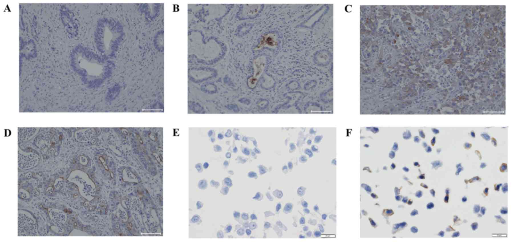
Representative stained images of EHBDCA and GBCA are
shown in Fig. 1.
Proportion of cells with cytoplasmic
CD133-positive expression
We found two types of CD133 expression as follows:
C-type is the cytoplasmic staining pattern, and M-type is the
luminal membranous staining pattern with or without intraluminal
stains. Overall, C-type was observed in 20 (24.4%) patients whereas
M-type was observed in 47 (57.3%) patients, and overlap between the
two types was observed in 9 (11.0%) patients. Among C-type
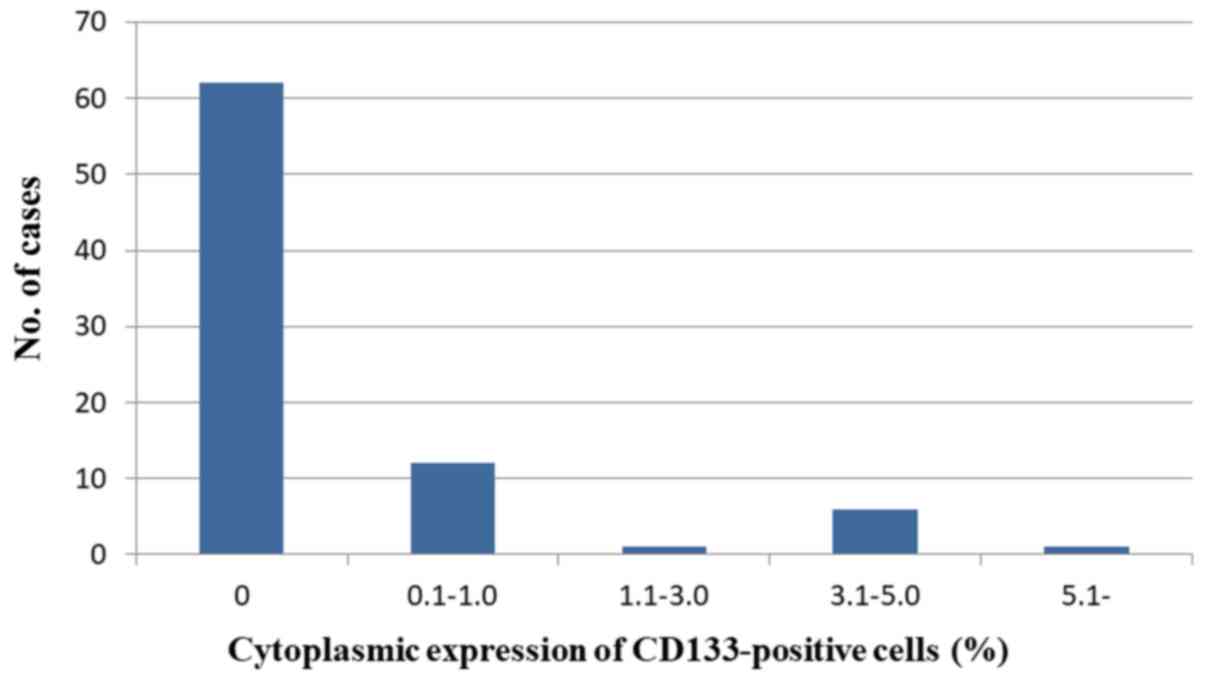
patients, the frequency of cytoplasm-stained cells in the tumor was
<5% in most cases (Fig. 2).
Correlation between cytoplasmic CD133
expression and clinicopathological features
Statistical analysis revealed that the incidence of
CD133-positive expression was correlated with the histopathological
grade (P=0.035), pT factor (P=0.020), and recurrence (P=0.048).
However, there was no significant association between CD133
immunoreactivity and tumor diameter, lymphatic permeation,
perineural invasion, pN factor, staging, or resection status
(Table II).
| Table II.Association between cytoplasmic CD133
expression and clinicopathological features. |
Table II.
Association between cytoplasmic CD133
expression and clinicopathological features.
|
| Cytoplasmic
expression of CD133 |
|---|
|
|
|
|---|
| Clinicopathological
feature | Positive (n=20) | Negative (n=62) | P-value |
|---|
| Tumor diameter, cm:
<5/≥5 | 16/4 | 54/8 | 0.474 |
| Histopathological
grade: G1, 2/G3 | 11/9 | 49/13 | 0.035a |
| Lymphatic permeation:
Negative/positive | 8/12 | 24/38 | 0.918 |
| Blood vessel
permeation: Negative/positive | 10/10 | 23/39 | 0.306 |
| Perineural invasion:
Negative/positive | 3/17 | 14/48 | 0.545 |
| pT factor:
pT1-2/pT3-4 | 7/13 | 40/22 | 0.020a |
| pN factor:
Negative/positive | 6/14 | 30/32 | 0.150 |
| Staging: I, II/III,
IV | 8/12 | 36/26 | 0.159 |
| Resection status:
R0/R1 | 4/16 | 28/34 | 0.065 |
| Recurrence:
No/yes | 2/18 | 22/40 | 0.046a |
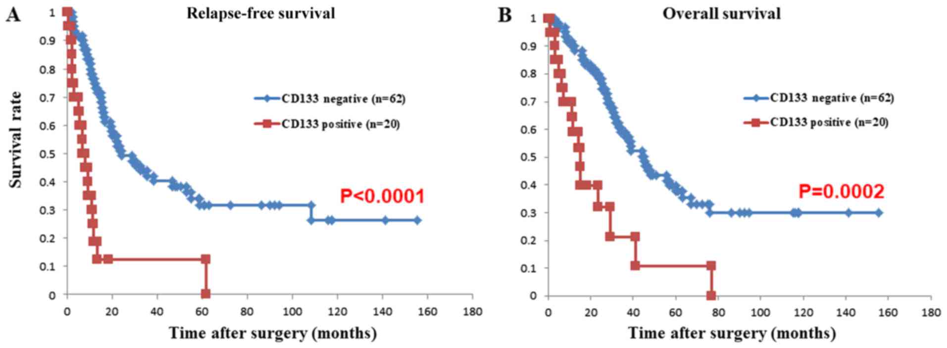
Correlation of cytoplasmic CD133
expression with OS and RFS
There was a significant difference in OS between the
patients with and without cytoplasmic CD133 expression (P=0.0002).
Additionally, patients with cytoplasmic CD133 expression showed
significantly worse RFS compared with those without cytoplasmic
CD133 expression (P<0.0001; Fig.
3).
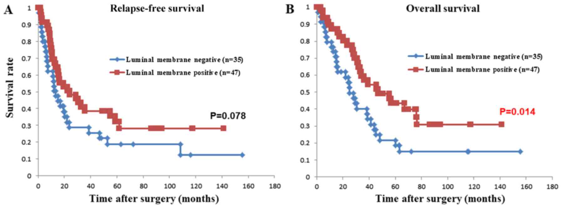
Correlation of luminal membrane CD133
expression with OS and RFS
There was a significant difference in OS between the
patients with and without luminal membrane CD133 expression
(P=0.014). Additionally, patients with luminal membrane CD133
expression tended to show better RFS compared with those without
luminal membrane expression (P=0.078; Fig. 4).
Univariate and multivariate analyses
of OS
Univariate analysis showed that OS was significantly
correlated with histological grade (G3) and CD133-positive
expression. Although perineural invasion also tended to be
correlated with OS, the difference was not significant (P=0.067).
Multivariate analysis showed that G3 (P=0.018), perineural invasion
(P=0.040), and CD133-positive expression (P=0.0036) were
independent prognostic factors for OS (Table III).
| Table III.Univariate and multivariate analyses
of prognostic parameters for overall survival in extrahepatic bile
duct cancer and gallbladder cancer. |
Table III.
Univariate and multivariate analyses
of prognostic parameters for overall survival in extrahepatic bile
duct cancer and gallbladder cancer.
|
| Univariate
analysis | Multivariate
analysis |
|---|
|
|
|
|
|---|
| Parameters | No. of
patients | 5-year survival
rate (%) | P-value | Hazard ratio (95%
CI) | P-value |
|---|
| Tumor diameter,
cm |
|
<5 | 70 | 35.5 | 0.215 | ND |
|
| ≥5 | 12 | 22.9 |
|
|
|
| Histopathological
grade |
| G1,
2 | 60 | 40.4 | 0.001b | 1 | 0.018b |
| G3 | 22 | 12.4 |
| 2.227
(1.152–4.122) |
|
| Lymphatic
permeation |
|
Negative | 32 | 44.5 | 0.214 | ND |
|
|
Positive | 50 | 26.6 |
|
|
|
| Blood vessel
permeation |
|
Negative | 33 | 46.0 | 0.303 | ND |
|
|
Positive | 49 | 25.0 |
|
|
|
| Perineural
invasion |
|
Negative | 17 | 56.7 | 0.067a | 1 | 0.040b |
|
Positive | 65 | 27.2 |
| 2.101
(1.031–4.879) |
|
| pT factor |
|
pT1-2 | 47 | 40.4 | 0.160 | ND |
|
|
pT3-4 | 35 | 22.3 |
|
|
|
| pN factor |
|
Negative | 36 | 45.8 | 0.142 | ND |
|
|
Positive | 46 | 22.4 |
|
|
|
| Staging |
| I,
II | 44 | 39.8 | 0.738 | ND |
|
| III,
IV | 38 | 25.6 |
|
|
|
| Resection
status |
| R0 | 32 | 41.3 | 0.167 | ND |
|
| R1 | 50 | 28.9 |
|
|
|
| Cytoplasmic
expression of CD133 |
|
Negative | 20 | 39.1 |
<0.001b | 1b |
|
|
Positive | 62 | 11.6 |
| 2.947
(1.447–5.718) | 0.004b |
Univariate and multivariate analyses
of RFS
Univariate analysis showed that RFS was
significantly correlated with G3, perineural invasion, and CD133
positive expression. Although pN factor and staging also tended to
be correlated with RFS, the difference was not significant (P=0.063
and P=0.079, respectively). Multivariate analysis showed that
perineural invasion (P=0.045) and CD133-positive expression
(P<0.0001) were independent prognostic factors for RFS (Table IV).
| Table IV.Univariate and multivariate analyses
of prognostic parameters for recurrence-free survival in
extrahepatic bile duct cancer and gallbladder cancer. |
Table IV.
Univariate and multivariate analyses
of prognostic parameters for recurrence-free survival in
extrahepatic bile duct cancer and gallbladder cancer.
|
| Univariate
analysis | Multivariate
analysis |
|---|
|
|
|
|
|---|
| Parameters | No. of
patients | 5-year survival
rate (%) | P-value | Hazard ratio (95%
confidence interval) | P-value |
|---|
| Tumor diameter,
cm |
|
<5 | 70 | 36.6 | 0.320 | ND |
|
| ≥5 | 12 | 22.9 |
|
|
|
| Histopathological
grade |
|
G1,2 | 60 | 41.5 | 0.005b | 1 | 0.143 |
| G3 | 22 | 12.0 |
| 1.624
(0.843–2.992) |
|
| Lymphatic
permeation |
|
Negative | 2 | 40.6 | 0.202 | ND |
|
|
Positive | 50 | 29.8 |
|
|
|
| Blood vessel
permeation |
|
Negative | 33 | 44.6 | 0.274 | ND |
|
|
Positive | 49 | 27.2 |
|
|
|
| Perineural
invasion |
|
Negative | 17 | 52.9 | 0.036 | 1 | 0.045b |
|
Positive | 65 | 28.8 |
| 2.132
(1.016–4.941) |
|
| pT factor |
|
pT1-2 | 47 | 38.7 | 0.216 | ND |
|
|
pT3-4 | 35 | 27.8 |
|
|
|
| pN factor |
|
Negative | 36 | 44.5 | 0.063a | 1 | 0.896 |
|
Positive | 46 | 26.0 |
| 1.043
(0.559–1.978) |
|
| Staging |
| I,
II | 44 | 42.2 | 0.079a | 1 | 0.191 |
| III,
IV | 38 | 25.0 |
| 1.484
(0.821–2.700) |
|
| Resection
status |
| R0 | 32 | 41.5 | 0.135 | ND |
|
| R1 | 50 | 29.4 |
|
|
|
| Cytoplasmic
expression of CD133 |
|
Negative | 20 | 42.0 | <0.001b | 1 |
<0.001b |
|
Positive | 62 | 12.5 |
| 2.947
(1.447–5.718) |
|
Discussion
In our study, we used immunohistochemistry to
demonstrate the clinical role of CD133 expression in EHBDCA and
GBCA. Our major findings are as follows: (1) Cytoplasmic CD133 expression was closely
correlated with poor prognosis and was an independent prognostic
factor; (2) cytoplasmic CD133
expression was related to histological differentiation, which is
also an independent prognostic factor in EHBDCA and GBCA. There
have been three reports to date on the prognostic value of CD133 in
intrahepatic cholangiocarcinoma, but the prognostic significance of
CD133 remains controversial (12–14).
Shimada et al (12) maintained
that cytoplasmic CD133 expression was independently related to a
worse prognosis, but Leelawat et al (14) demonstrated that, although CD133
expression significantly correlates with lymph node metastasis and
surgical margin status, it does not correlate with poor prognosis.
Moreover, Fan et al (13)
stressed that CD133 expression was correlated with prolonged
patient survival. These results seem to be contradictory.
One of the reasons why these results, including
ours, are different from each other despite the same type of cancer
may be because of the antibodies used. In our study, we selected
the clone W6B3C1 from Miltenyi Biotec because this clone is
difficult to stain nonspecifically (15), so we suggest that we can obtain more
accurate information about site of CD133 expression using the
W6B3C1 clone. To assess CD133 expression using
immunohistochemistry, we isolated breast cancer cell lines (MCF-7
and MDA-MB-468) using fluorescence activated cell sorting (FACS),
as previously reported by Croker et al (16), We confirmed that MCF-7 cells did not
show CD133 expression, but MDA-MB-468 cells did consistently
express CD133 only on their surface using this clone (Fig. 1).
The opposing results may also be explained by
confusing the cytoplasmic expression and luminal membrane CD133
expression. Leelawat et al (14) and Fan et al (13) defined CD133 positivity based on the
percentage of stained cells without distinguishing between the
staining sites in their papers. Based on a similar definition,
CD133 expression on the luminal membrane with or without
intraluminal contents resulted in better OS compared with no CD133
expression in our study (Fig. 4).
These results suggested that cytoplasmic, not
luminal membrane, CD133 expression may be an important risk factor
in EHBDCA and GBCA, which is similar to a report by Shimada et
al (12).
Several studies also showed a significant
correlation between cytoplasmic CD133 expression and poor prognosis
in gastric cancer (17), colorectal
cancer (11), hepatocellular
carcinoma (10), and ovarian cancer
(18) patients. Among these papers,
some authors suggested that cells with CD133 expression in the
cytoplasm may have CSC-like characteristics. They suggested that
cytoplasmic CD133 expression is found in only a small population of
cells. To be more precise, the percentage of CD133-positive cells
was 16.2% in hepatocellular carcinoma, 48.3% in cholangiocarcinoma,
and 31.2% in ovarian cancer, but the proportion of the cells with
CD133 expression in the cytoplasm was <10%. These results are in
agreement with our data (Fig. 2).
Cytoplasmic CD133 expression is likely to be a
powerful prognostic factor and it may have CSCs-like
characteristics, but localization of the CD133 antigen in the
cytoplasm remains unclear, even though its expression was
originally on the cell membrane.
What is the significance of cell membrane and
cytoplasmic CD133 expression? The significance of cell membrane
CD133 expression was described previously (19). It was reported that CD133 was
exclusively expressed on the luminal membrane of gland-forming
cells and it was never found on poorly differentiated diffuse-type
cells in human gastric cancers. Considering that poorly
differentiated tumors often develop from well-differentiated tumors
during tumor progression and they are detected using FACS analysis,
it was suggested that loss of CD133 expression on the luminal
membrane may be related to tumor progression.
However, for the significance of cytoplasmic CD133
expression, Bauer et al revealed the process of
differentiation involved in CD133 in hematopoietic stem cells
(20). In their report, they
explained that during the process of differentiation, hematopoietic
progenitors release small membrane vesicles containing the CD133
protein by exportation via exosomes and upon release, CD133
protein-containing membrane vesicles are internalized by feeder
cells. It is widely known that some types of proteins gain
biological function based on their site of expression. CD133 may be
one of these types of proteins and we speculated that a shift from
membranous localization to cytoplasmic localization reflects the
transition of epithelial cells to a more invasive phenotype, even
in EHBDCA and GBCA.
This hypothesis may be supported by the significant
association between cytoplasmic CD133 expression and histologic
differentiation, and because CD133 is an independent prognostic
factor in our study. Thus, understanding its mechanism might allow
us to manipulate the fate of these cells for novel molecular target
therapies.
In conclusion, we first demonstrated that
cytoplasmic CD133 expression was correlated with histologic
differentiation and was a significant prognostic factor in EHBDCA
and GBCA. Our study suggests that cytoplasmic CD133 expression
might be a useful marker for the clinical prognosis of EHBDCA and
GBCA patients and that CD133 might be a powerful molecular target
for cancer treatment. However, our results were obtained using a
relatively small cohort (n=82) of EHBDCA and GBCA patients, and
additional studies in larger cohorts are required to clarify the
predictive significance.
Acknowledgements
The authors would like to thank Mrs. Y. Hirano, Mr.
T. Shimizu and Mr. K. Marukawa for their technical assistance. The
authors would like to thank Dr Jodi Smith for editing a draft of
this manuscript.
Funding
The present study was supported in part by a
grant-in-aid from the foundation for the Department of
Gastroenterological Surgery I, Hokkaido University Alumni
Association.
Availability of data and materials
The datasets used and analyzed during the current
study are available from the corresponding author on reasonable
request.
Authors' contributions
TaM performed the immunohistochemistry, evaluated
the immunostaining, performed the statistical analysis and drafted
the manuscript. HK designed the study, analyzed the data and helped
to revise the manuscript. ToM confirmed the diagnosis of the
samples, evaluated the immunostaining and helped to revise the
manuscript. TE participated in the follow-up study. YH performed
immunohistochemistry and participated in designing the study. TK
participated designing the study and analyzed the data. AT
supervised the research, analyzed the data and edited the
manuscript. All authors read and approved the final manuscript.
Ethics approval and consent to
participate
Ethics approval was requested and obtained from the
Institutional Review Board of Hokkaido University Hospital
(clinical research approval no. 014-0134). Written informed consent
was obtained from all participants.
Patient consent for publication
Written informed consent was obtained from all
participants.
Competing interests
The authors declare that they have no competing
interests.
Glossary
Abbreviations
Abbreviations:
|
EHBDCA
|
extrahepatic bile duct cancer
|
|
GBCA
|
gallbladder cancer
|
|
CSCs
|
cancer stem cells
|
|
UICC
|
Union Internationale Contre le
Cancer
|
|
DFS
|
disease-free survival
|
|
OS
|
overall survival
|
References
|
1
|
Ruzzenente A, Iacono C, Conci S, Bertuzzo
F, Salvagno G, Ruzzenente O, Campagnaro T, Valdegamberi A, Pachera
S, Bagante F and Guglielmi A: A novel serum marker for biliary
tract cancer: Diagnostic and prognostic values of quantitative
evaluation of serum mucin 5AC (MUC5AC). Surgery. 155:633–639. 2014.
View Article : Google Scholar : PubMed/NCBI
|
|
2
|
Visvader JE and Lindeman GJ: Cancer stem
cells in solid tumours: Accumulating evidence and unresolved
questions. Nat Rev Cancer. 8:755–768. 2008. View Article : Google Scholar : PubMed/NCBI
|
|
3
|
Wu Y and Wu PY: CD133 as a marker for
cancer stem cells: Progresses and concerns. Stem Cells Dev.
18:1127–1134. 2009. View Article : Google Scholar : PubMed/NCBI
|
|
4
|
Irollo E and Pirozzi G: CD133: To be or
not to be, is this the real question? Am J Transl Res. 5:563–581.
2013.PubMed/NCBI
|
|
5
|
Li Z: CD133: A stem cell biomarker and
beyond. Exp Hematol Oncol. 2:172013. View Article : Google Scholar : PubMed/NCBI
|
|
6
|
Pallini R, Ricci-Vitiani L, Montano N,
Mollinari C, Biffoni M, Cenci T, Pierconti F, Martini M, De Maria R
and Larocca LM: Expression of the stem cell marker CD133 in
recurrent glioblastoma and its value for prognosis. Cancer.
117:162–174. 2011. View Article : Google Scholar : PubMed/NCBI
|
|
7
|
Zhao P, Lu Y, Jiang X and Li X:
Clinicopathological significance and prognostic value of CD133
expression in triple-negative breast carcinoma. Cancer Sci.
102:1107–1111. 2011. View Article : Google Scholar : PubMed/NCBI
|
|
8
|
Wu S, Yu L, Wang D, Zhou L, Cheng Z, Chai
D, Ma L and Tao Y: Aberrant expression of CD133 in non-small cell
lung cancer and its relationship to vasculogenic mimicry. BMC
Cancer. 12:5352012. View Article : Google Scholar : PubMed/NCBI
|
|
9
|
Maeda S, Shinchi H, Kurahara H, Mataki Y,
Maemura K, Sato M, Natsugoe S, Aikou T and Takao S: CD133
expression is correlated with lymph node metastasis and vascular
endothelial growth factor-C expression in pancreatic cancer. Br J
Cancer. 98:1389–1397. 2008. View Article : Google Scholar : PubMed/NCBI
|
|
10
|
Sasaki A, Kamiyama T, Yokoo H, Nakanishi
K, Kubota K, Haga H, Matsushita M, Ozaki M, Matsuno Y and Todo S:
Cytoplasmic expression of CD133 is an important risk factor for
overall survival in hepatocellular carcinoma. Oncol Rep.
24:537–546. 2010. View Article : Google Scholar : PubMed/NCBI
|
|
11
|
Takahashi S, Kamiyama T, Tomaru U, Ishizu
A, Shida T, Osaka M, Sato Y, Saji Y, Ozaki M and Todo S: Frequency
and pattern of expression of the stem cell marker CD133 have strong
prognostic effect on the surgical outcome of colorectal cancer
patients. Oncol Rep. 24:1201–1212. 2010. View Article : Google Scholar : PubMed/NCBI
|
|
12
|
Shimada M, Sugimoto K, Iwahashi S,
Utsunomiya T, Morine Y, Imura S and Ikemoto T: CD133 expression is
a potential prognostic indicator in intrahepatic
cholangiocarcinoma. J Gastroenterol. 45:896–902. 2010. View Article : Google Scholar : PubMed/NCBI
|
|
13
|
Fan L, He F, Liu H, Zhu J, Liu Y, Yin Z,
Wang L, Guo Y, Wang Z, Yan Q and Huang G: CD133: A potential
indicator for differentiation and prognosis of human
cholangiocarcinoma. BMC Cancer. 11:3202011. View Article : Google Scholar : PubMed/NCBI
|
|
14
|
Leelawat K, Thongtawee T, Narong S,
Subwongcharoen S and Treepongkaruna SA: Strong expression of CD133
is associated with increased cholangiocarcinoma progression. World
J Gastroenterol. 17:1192–1198. 2011. View Article : Google Scholar : PubMed/NCBI
|
|
15
|
Hermansen SK, Christensen KG, Jensen SS
and Kristensen BW: Inconsistent immunohistochemical expression
patterns of four different CD133 antibody clones in glioblastoma. J
Histochem Cytochem. 59:391–407. 2011. View Article : Google Scholar : PubMed/NCBI
|
|
16
|
Croker AK, Goodale D, Chu J, Postenka C,
Hedley BD, Hess DA and Allan AL: High aldehyde dehydrogenase and
expression of cancer stem cell markers selects for breast cancer
cells with enhanced malignant and metastatic ability. J Cell Mol
Med. 13:2236–2252. 2009. View Article : Google Scholar : PubMed/NCBI
|
|
17
|
Hashimoto K, Aoyagi K, Isobe T, Kouhuji K
and Shirouzu K: Expression of CD133 in the cytoplasm is associated
with cancer progression and poor prognosis in gastric cancer.
Gastric Cancer. 17:97–106. 2014. View Article : Google Scholar : PubMed/NCBI
|
|
18
|
Ferrandina G, Martinelli E, Petrillo M,
Prisco MG, Zannoni G, Sioletic S and Scambia G: CD133 antigen
expression in ovarian cancer. BMC Cancer. 9:2212009. View Article : Google Scholar : PubMed/NCBI
|
|
19
|
Fukamachi H, Shimada S, Ito K, Ito Y and
Yuasa Y: CD133 is a marker of gland-forming cells in gastric tumors
and Sox17 is involved in its regulation. Cancer Sci. 102:1313–1321.
2011. View Article : Google Scholar : PubMed/NCBI
|
|
20
|
Bauer N, Wilsch-Bräuninger M, Karbanová J,
Fonseca AV, Strauss D, Freund D, Thiele C, Huttner WB, Bornhäuser M
and Corbeil D: Haematopoietic stem cell differentiation promotes
the release of prominin-1/CD133-containing membrane vesicles-a role
of the endocytic-exocytic pathway. EMBO Mol Med. 3:398–409. 2011.
View Article : Google Scholar : PubMed/NCBI
|