Introduction
Myelodysplastic syndrome (MDS) constitutes a
heterogeneous group of clonal myeloid disorders characterized by
refractory cytopenias and dysplastic changes in ≥2 hematopoietic
cell lineages, frequently representing an intermediate disease
stage prior to progression to acute myeloid leukemia (AML)
(1). MDS progresses to AML in ~30% of
patients following multiple intervals of time from diagnosis.
Therefore, the early diagnosis and treatment of MDS may aid in
improving the survival rate of patients with MDS and AML. The
pathogenesis of MDS remains unclear with no validated biomarkers
for early diagnosis of MDS.
Numerous genetic and epigenetic alterations occur
during the pathogenesis of MDS (2,3). Among
these alterations, methylation of the tumor suppressor gene
promoter results in gene silencing, which often take place during
the early stages of tumor development (4). These aberrant DNA methylations of tumor
suppressor genes may be used as diagnostic markers for MDS. Thus,
defining altered gene expression and understanding the underlying
molecular mechanism of MDS are required.
RAP1GTPase activating protein 1 (Rap1GAP) is one of
the genes thought to be involved in hematopoietic regulation. The
authors' previous study confirmed that the mRNA and protein
expression of Rap1GAP in the majority of patients with MDS were
significantly higher compared with those with AML and non-malignant
blood diseases (NM) (5,6). However, the mechanistic basis of Rap1GAP
upregulation in patients with MDS remains unclear.
In the present study, the methylation levels and
pattern in the transcriptional regulation region (TRR) of the
Rap1GAP gene were detected in the bone marrow mononuclear cells
(BMNCs) cells of patients with MDS, NM and AML to assess the
altered methylation levels of the Rap1GAP gene, and to analyze its
possible association with clinicopathological characteristics of
patients with MDS.
Materials and methods
Cells and patient specimens
The present study included patients with MDS, AML
and NM. In total, 5 human leukemia cell lines (SKM-1, K562, SHI-1,
U937 and HL-60) were used as the control of Rap1GAP methylation
level, including SKM-1, a cell line established from a patient with
MDS-refractory anemia with excess blasts-II with progression to
AML. All cell lines were stored at and supplied by the Cell Bank of
Jiangsu Institute of Hematology (The First Affiliated Hospital of
Soochow University, Collaborative Innovation Center of Hematology,
Soochow University, Key Laboratory of Thrombosis and Hemostasis of
Ministry of Health, Suzhou, China). Cells were cultured in
RPMI-1640 (Hyclone; GE Healthcare Life Sciences, Logan, UT, USA) or
Iscove's modified Dulbecco's medium (Hyclone; GE Healthcare Life
Sciences) with 10% fetal bovine serum (Bovogen Biologicals, Keilor
East, Victoria, Australia) at 37°C in a humidified atmosphere with
5% CO2. Cells were passaged every 3 days for 24 days.
BMNC samples from 86 patients were obtained from The First
Affiliated Hospital of Soochow University (Suzhou, China) between
August 2010 and December 2011. The samples were stored at −80°C
following separation. These samples were collected from 29
pathologically proven cases of MDS, 31 cases of AML and 26 cases of
NM. Clinicopathological data, including age, gender, clinical
classification and International Prognostic Scoring System (IPSS)
(7), were collected from the medical
records of patients (Table I). The
present study was approved by the Ethics Committee of The First
Affiliated Hospital of Soochow University and written informed
consent was obtained from all participants.
 | Table I.Clinicopathological characteristics of
patients with MDS. |
Table I.
Clinicopathological characteristics of
patients with MDS.
| Case | Sex | Age | Karyotype | WHO
classification | IPSS |
|---|
| 1 | M | 77 | Normal | MDS-RCMD | 0 |
| 2 | F | 66 | Normal | MDS-RCMD | 0 |
| 3 | M | 39 | 46,XY,
3q+,inv(9)(p12q13) | MDS-RAEB1 | 1.5 |
| 4 | M | 35 | 47,XY, +8 | MDS-RCMD | 0.5 |
| 5 | F | 75 | Normal | MDS-RCMD | 0.5 |
| 6 | F | 59 | Normal | MDS-RCMD | 0.5 |
| 7 | M | 61 | Normal | MDS-RCMD | 0.5 |
| 8 | M | 59 | Normal | MDS-RCMD | 0.5 |
| 9 | F | 60 | Normal | MDS-RCUD | 0.5 |
| 10 | M | 25 | Normal | MDS-RAEB1 | 1 |
| 11 | M | 79 | Normal | MDS-RARS | 0.5 |
| 12 | F | 64 | Normal | MDS-RAEB2 | 2 |
| 13 | M | 84 | Normal | MDS-RAEB2 | 2 |
| 14 | F | 62 | 46,XX,
del(2)(p15),?der(5)del(5)(q31)t(2;5)(p15;q31)[8] | MDS-RCMD | 1.5 |
| 15 | M | 78 | Normal | MDS-RAEB1 | 0.5 |
| 16 | F | 65 | 46,XX,
5q-,20q-[9]/46,XX[1] | MDS-U | 1 |
| 17 | M | 73 | 47,XY,
+8[8]/46,XY[2] | MDS-RAEB2 | 2 |
| 18 | F | 79 | 46,XX,
del(5),del(13),del(20) | MDS-U | 1 |
| 19 | M | 66 | Normal | MDS-RCMD | 1 |
| 20 | M | 21 | 47,XY,
+8[9]/46,XY[1] | MDS-RAEB1 | 1 |
| 21 | M | 85 |
45,X-Y[3]/46,XY[17] | MDS-RCMD | 1 |
| 22 | F | 50 | Normal | MDS-RCMD | 0.5 |
| 23 | F | 53 | Normal | MDS-RAEB1 | 0 |
| 24 | M | 77 | 46,XY,
der(18)[8]/46,XY[4] | MDS-RCMD | 1 |
| 25 | F | 64 | Normal | MDS-RARS | 1 |
| 26 | M | 17 | 46,XY,
20q-[9]/46,XY[4] | MDS-RCMD | 0 |
| 27 | M | 20 | 46,XY,
der(7)t(1;7)[4]/46,XY[6] | MDS-RCMD | 1.5 |
| 28 | F | 38 | Normal | MDS-RAEB2 | 1.5 |
| 29 | M | 65 | 47,XY,
+8[2]/45,X,-Y/46,XY[12] | MDS-RCMD | 0.5 |
DNA extraction and bisulfite
modification of DNA
BMNCs were separated from the bone marrow of
patients using Ficoll-Hypaque (Invitrogen; Thermo Fisher
Scientific, Inc., Waltham, MA, USA) at 800 × g for 20 min at room
temperature. Genomic DNA from MNCs was extracted using EpiTectFast
DNA Bisulfite kit™ (Qiagen, Inc., Valencia, CA, USA). The
concentration of DNA extraction was measured using SkanIt software
(version 3.2; Thermo Fisher Scientific, Inc.) and the mass of DNA
was measured, for which the A260/A280was 1.8.
Genomic DNA (1 µg) was treated using the EpiTectFast DNA Bisulfite
kit according to the manufacturer's protocol. The bisulfite
modified DNA was subsequently suspended in 20 µl deionized water
and used immediately or stored at −80°C.
Bisulfite-specific polymerase chain
reaction (BSP) and DNA sequencing
The primers used to detect the methylation of the
RAP1GAP gene promoter TRR were designed to specifically amplify
bisulfite-converted RAP1GAP DNA. The primers were custom
synthesized by Sangon Biotech Co., Ltd. (Shanghai, China) with the
following sequences: Forward, 5′-AGTTGTTTAGTTTAGAGATAAAGTTTAAGAG-3′
and Reverse, 5′-ACAACCCAACTATCCAAACA-3′. A total of 2 µl
bisulfite-converted DNA (0.1 µg) from each sample was subjected to
PCR analysis in a 25 µl volume containing 1X PCR buffer, 2 mmol/l
MgCl2, 2.5 mmol/l dNTP, 1 mmol/l primer, and 800 U/l EX
TaqHS DNA polymerase (Invitrogen; Thermo Fisher Scientific, Inc.).
The reaction mixture was preheated at 95°C and then amplified (35
cycles of 95°C for 30 sec, 52°C for 30 sec, 72°C for 45 sec), with
a final extension of 10 min at 72°C. The PCR products were treated
using 2% agarose gel electrophoresis and a DNA fragment
purification kit (Axygen; Corning Life Sciences, Hangzhou, China)
according to the manufacturer's protocol, then subjected to cloning
into the pMD-18-T vector (Takara Biotechnology Co., Ltd., Dalian,
China). The purified DNA (4.5 µl) was incubated with the pMD-18-T
vector in a water bath overnight at 16°C, then cultured in lysogeny
broth medium (Jiangsu Institute of Hematology) at 37°C with
Escherichia coli H5a (Jiangsu Institute of Hematology), with
colonies forming after 24 h. Following the cloning, 10–18 clones
from each sample were randomly selected for DNA sequencing.
Sequencing data analysis
Sequencing analysis was performed by Shanghai
BioSune Co., Ltd. (Shanghai, China). Based on the data from BSP
PCR-based sequencing analysis, the methylation level of each CpG
site in a given sample was calculated as follows: Cpeak height/(C
height + T height). The percentage of methylation of each CpG site
in a given sample was calculated as follows: Number of methylated
CpG sites/total number of observed sequenced clones. The percentage
of the region methylation in a given sample was the average of each
CpG sites methylation percentage in the DNA region.
Statistical analysis
Statistical analyses were performed using SPSS
software (version 17.0; SPSS, Inc., Chicago, IL, USA) and GraphPad
Prism software (version 6.0; GraphPad Software, Inc., La Jolla, CA,
USA). The median ± 2nd and 3rd quartiles were assessed to analyze
differences in the percentage of the region methylation among MDS,
AML and NM samples. Variance of the variables among groups was
calculated using nonparametric tests (Wilcoxon-Mann-Whitney U-test
and Kruskal-Wallis test) in SPSS 17.0. P≤0.05 was considered to
indicate a statistically significant difference.
Results
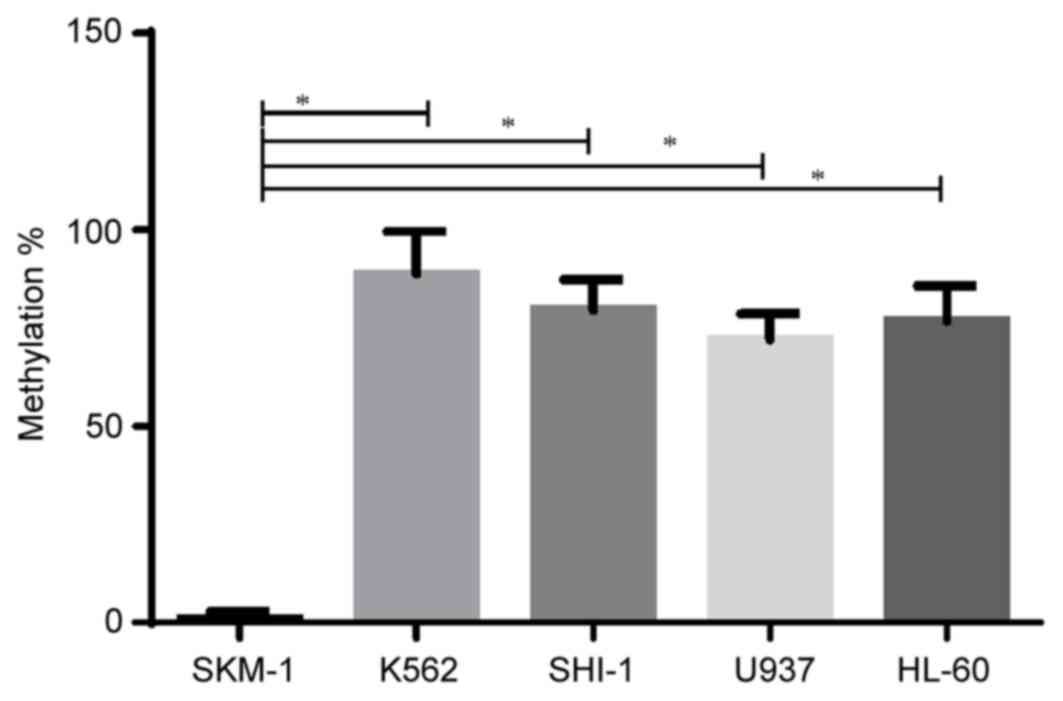
Methylation of the RAP1GAP gene in
cells and MNCs of patients with MDS
Firstly, the methylation status of 20 CpG sites in
the promoter region of RAP1GAP gene in 5 human leukemia cell
lines was examined. The methylation level of RAP1GAP in SKM-1 cells
was significantly lower compared with that of the other 4 leukemia
cell lines, which was consistent with the clinical data of the
present study (Fig. 1). Furthermore,
a statistically significant difference in the methylation level of
RAP1GAP was detected between SKM-1 cells and all other cell lines
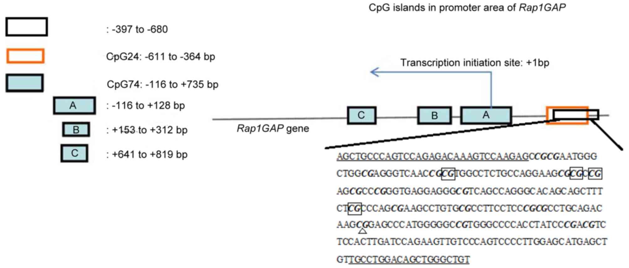
(Fig. 1). According to the National
Center for Biotechnology Information Gene Expression Omnibus
(https://www.ncbi.nlm.nih.gov/geo/)
database, the RAP1GAP gene was analyzed in 29 MDS, 31 AML
and 26 NM samples using Methyl Primer Express analysis software
(version 1.0; Thermo Fisher Scientific, Inc.). CpG islands were
found upstream of the transcriptional start site (designated as
‘0’) between −680 and −398 bp. The structure of the RAP1GAP gene is
presented in Fig. 2, indicating the
position of the CpG island containing 20 CpG sites.
In these 20 CpG sites, the majority of the CpG sites
were slightly methylated or unmethylated in all samples, including
8 CpG sites, which exhibited statistically significant differences
among MDS, AML and NM samples. The results of the current study
confirmed that the methylation of Rap1GAP in patients with MDS was
100%, distributing over various CpG sites in different patients.
The overall methylation level in patients with MDS was decreased
compared with that in patients with NM. The methylation pattern in
20 CpG sites differed among NM, MDS and AML. A significant
difference in 4 CpG sites was identified in the patients with NM
compared with those with MDS. These preliminary results suggest
that the methylation state of the whole region, rather than the
methylation state of a single CpG site, is associated with the
sample groups. Statistical analysis of the data was subsequently
performed.
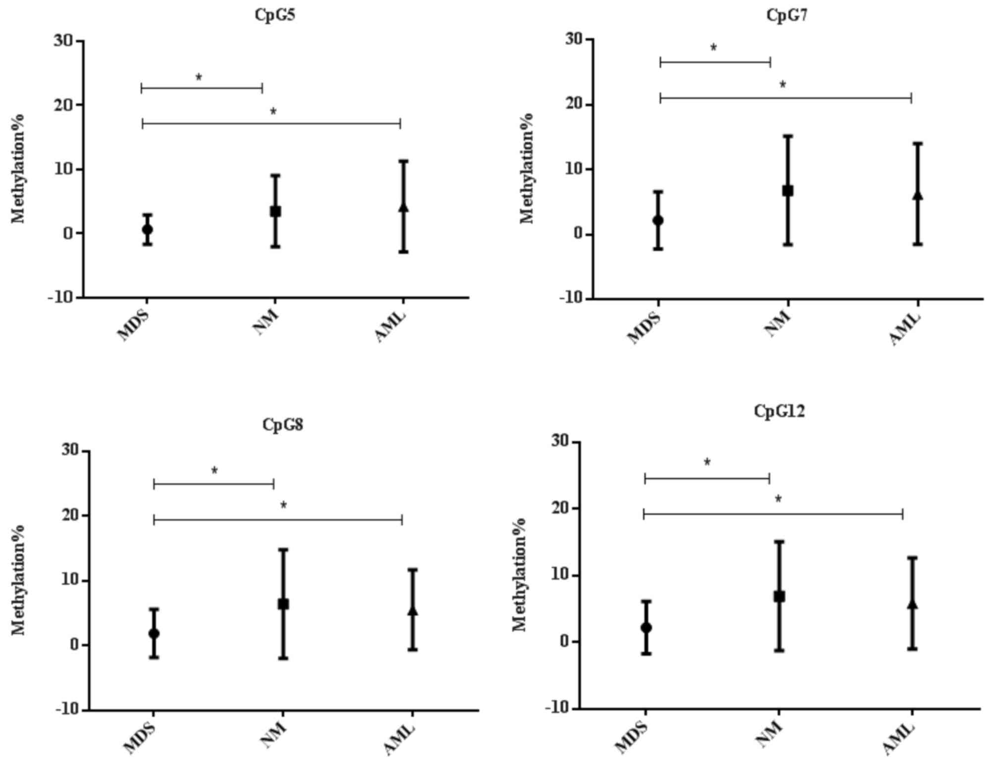
Statistical analysis results
The associations between the methylation degree of
each of the 6 CpG sites and tumor types were analyzed. Firstly, the
hypomethylated level in 4 CpG sites (CpG5, CpG7, CpG8 and CpG12)
were identified to be significantly different between patients with
MDS and those with NM (P<0.05; Fig.
4), and was significantly different in one site (CpG17) between
patients with AML and those with NM (P=0.023; data not shown).
Compared with AML, the methylation level of patients with MDS is
lower in four CpG sites (CpG5, CpG7, CpG8 and CpG12) (P<0.05;
Fig. 4). Based on these results, it
was demonstrated that the methylation level at four recurring sites
(CpG5, CpG7, CpG8 and CpG12) was significantly different among the
groups, which may correlate with the progression of MDS to AML.
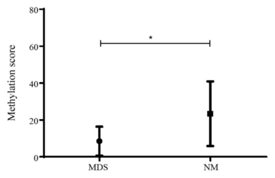
A previous study reported the methylation score as a
novel variance to measure and compare the methylation status of the
CpG sites in the differentially methylated region of each patient
(8). As the methylation score was
more stable and reproducible for the evaluation of DNA methylation
status (8), methylation scoring was
performed on 4 CpG sites (5, 7, 8 and 12) using GraphPad Prism 6. A
marked difference in methylation scores was identified among
patient groups (Fig. 5). It is
hypothesized that the methylation score reflects the methylation
status for more CpG sites, and could be more helpful for diagnosis
and risk stratification.
Clinical association analysis
The mechanism underlying the hypomethylation in
promoter of Rap1GAP remains unclear. To access the clinical
significance of these findings, the association between the
methylation status in promoter of Rap1GAP in patients with MDS, and
clinical parameters, including age, gender, clinical classification
and IPSS was further analyzed (Table
I). As older patients with MDS possess poorer prognoses,
reduced disease-free survival (DFS) and reduced overall survival
(OS) times (3 and 8.5 months, respectively) (9) and the majority of patients with
high-risk MDS and AML are >60 years (10), the age threshold was set at 60. Based
on the present clinical data, no significant differences in age,
gender and IPSS were identified between patient groups with
different methylation statuses (Table
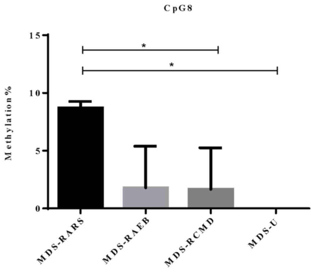
II). However, according to the 2008 World Health Organization
classification of MDS (11), only the
difference in methylation level at CpG site 8 of Rap1GAP promoter
was identified to be significantly increased in patients with
MDS-refractory anemia with ring sideroblasts (RARS) compared with
that in the MDS-refractory cytopenia with multilineage dysplasia
(RCMD; P=0.033) or MDS-unclassified (U; P=0.034) groups (Fig. 6). A previous study reported that
40–60% of all patients with MDS possess identifiable karyotypic
abnormalities (12) and that
cytogenetic subgroups of outcome are associated with prognosis
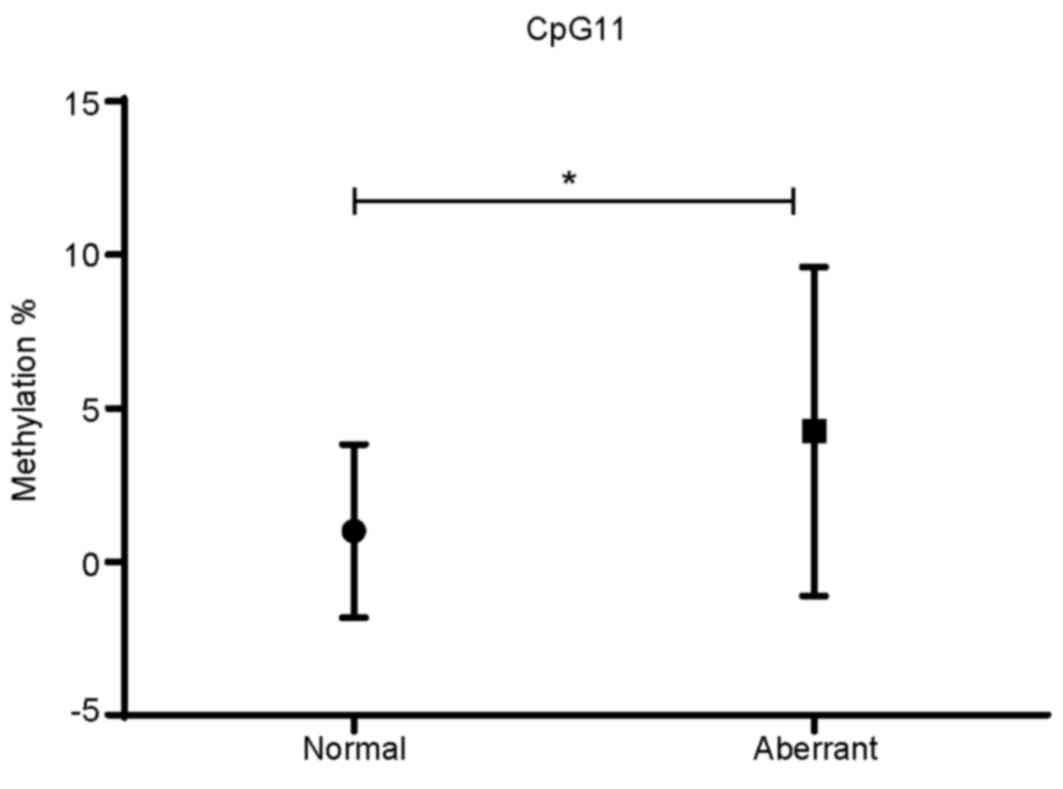
(13). In the present cohort, 13
patients exhibited abnormal karyotypes, accounting for 45% of all
patients in the sample, which is consistent with the previous study
described. However, the majority of the karyotypes identified in
these patients were not beneficial as prognostic indicators in MDS
according to IPSS (14). Furthermore,
it was revealed that the methylation level of CpG11 was
significantly different between patients with the normal and
abnormal karyotypes, which may be associated with prognosis in MDS
(P=0.0429; Fig. 7).
| Table II.Clinical data analysis of MDS. |
Table II.
Clinical data analysis of MDS.
|
| CpG5 | CpG7 | CpG8 | CpG12 |
|---|
|
|
|
|
|
|
|---|
|
| Methylation, %
(OR) | P-value | Methylation, %
(OR) | P-value | Methylation, %
(OR) | P-value | Methylation, %
(OR) | P-value |
|---|
| Age, years |
| 0.1643 |
| 0.6817 |
| 0.5659 |
| 0.3163 |
|
<60 | 0.0 (0.0) |
| 0.0 (0.0) |
| 0.0 (0.0) |
| 0.0 (0.0) |
|
|
≥60 | 0.0 (0.0) |
| 0.0 (8.3) |
| 0.0 (8.3) |
| 0.0 (8.3) |
|
| Sex |
| 0.4592 |
| 0.2562 |
| 0.2540 |
| 0.2726 |
|
Male | 0.0 (0.0) |
| 0.0 (4.15) |
| 0.0 (8.3) |
| 0.0 (8.3) |
|
|
Female | 0.0 (0.0) |
| 0.0 (8.3) |
| 0.0 (0.0) |
| 0.0 (0.0) |
|
| IPSS |
| 0.7727 |
| 0.8219 |
| 0.2988 |
| 0.2502 |
|
Low | 0.0 (12.5) |
| 0.0 (0.0) |
| 0.0 (14.6) |
| 0.0 (0.0) |
|
|
Int-1 | 0.0 (0.0) |
| 0.0 (8.3) |
| 0.0 (8.3) |
| 0.0 (8.3) |
|
|
Int-2 | 0.0 (0.0) |
| 0.0 (8.3) |
| 0.0 (0.0) |
| 0.0 (0.0) |
|
|
High | – |
| – |
| – |
| – |
|
Discussion
Rap1GAP codes for an enzyme that catalyzes the
hydrolytic switch from ATP to ADP bound to Rap1, a member of small
G proteins. The intrinsic GTPase activity enhanced by Rap1GAP
inactivates Rap1GTP, influencing a variety of essential biological
processes in eukaryotic cells, including cell proliferation,
adhesion and migration via specific signal molecules, including
BRAF, and ERK (15). A previous study
on hematopoietic cells reported multiple functions for Rap1,
including its involvement in the maturation of megakaryocytes, and
the adhesion of leukocytes and T lymphocytes (15).
Rap1GAP has been identified as a putative tumor
suppressor gene in pancreatic cancer. The downregulation of Rap1GAP
expression is associated with pancreatic cancer progression, which
may function through the modulation of integrin activity (16). Rap1GAP-knockdown in human colon cancer
cells results in more aggressive migratory and invasive properties
compared with normal colon cancer cells (17). Similar results were demonstrated in
melanoma whereby the overexpression of Rap1GAP in melanoma cells
inhibited cell proliferation and survival (18). In thyroid cancer Rap1GAP expression is
decreased due to promoter hypermethylation, and progressively
higher frequencies of promoter hypermethylation are identified in
the more aggressive types of thyroid cancer, thus promoting thyroid
cancer cell invasion (19). In
addition, upregulation of Rap1GAP expression was demonstrated to
inhibit focal adhesion formation and decrease melanoma cell
migration (18). Altered expression
of Rap1GAP in multiple types of cell may also be associated with
the pathology of multiple diseases. For example, the monocytes of
patients with chronic lymphocytic leukemia exhibiting increased
Rap1GAP expression exerted impaired phagocytosis compared with
those exhibiting decreased Rap1GAP expression (20). Furthermore, the proliferation of T
cells in contact with these monocytes was inhibited (20). The increased expression of Rap1GAP in
podocytes contributes to their dysfunction and the injury
underlying the pathogenesis of all proteinuric kidney diseases
(21). However, certain studies have
reported contradictory results regarding the expression and
function of Rap1GAP in different cells (6,22). A
previous study demonstrated that patients with MDS express Rap1GAP
at a significantly increased level compared with that in patients
with NM or de novo AML (6).
Furthermore, Rap1GAP-transfected leukemia cells exhibited an
increased apoptosis rate in response to arsenic trioxide and
elevated invasion compared with non-transfected leukemia cells
(22).
Kim et al (23)
demonstrated that promoter hypermethylation of Rap1GAP in renal
cell carcinoma may lead to decreased Rap1GAP expression. Zheng
et al (19) confirmed these
results in multiple melanoma tumors and cell lines using
demethylating agent treatment. Although aberrant DNA methylation
has been considered the dominant mechanism underlying the
progression of MDS to AML, whether promoter hypomethylation is the
cause of the elevated expression of Rap1GAP previously observed in
the cluster of differentiation 34+ cells of patients
with MDS remains unclear (6).
To qualitatively or quantitatively analyze DNA
methylation levels, various techniques can be used, including
methylation-specific PCR, sequencing of certain regions of DNA
fragments and high throughput genome-wide sequencing. As the
distribution of CpG sites and the methylation pattern in Rap1GAP
promoter is presently unknown and whole genome sequencing is
unnecessary, the DNA fragments of 20 CpG sites within the Rap1GAP
promoter were amplified, cloned and sequenced in the present study.
The frequency of subclones in which methylation could be detected
at a certain CpG site may estimate the level of methylation for
that CpG site. The results of the current study demonstrated a
relatively lower overall methylation level within Rap1GAP promoter
in patients with MDS compared with that in patients with NM or AML.
However, only 4 CpG sites were confirmed to be methylated at a
significantly lower level in MDS compared with that in NM or AML.
Furthermore, based on the clinical records of the patient cohort in
the present study, no significant associations were identified
between the methylation status of Rap1GAP promoter and the
clinicopathological characteristics of patients with MDS, which
included age, gender, and IPSS.
Together, the results of the current study suggest
that the upregulation of Rap1GAP expression in patients with MDS is
associated with a lower methylation status in the promoter region
of this gene. However, further studies are required to elucidate
the association between the methylation of Rap1GAP promoter and the
clinical course, and prognosis of patients with MDS. Currently,
demethylating agents, including decitabine, have been used for the
treatment of patients with MDS based on a putative hypothesis that
certain tumor suppressor genes are inactivated by the
hypermethylation in their promoter leading to the initiation and
progression of MDS (24). Therefore,
demethylating the promoter may reactivate these tumor suppressor
genes to inhibit or reverse the progression of MDS towards AML.
However, in the present study, although Rap1GAP can be considered
as a tumor suppressor gene in multiple tumor types, inconsistently
the expression of this gene in MDS is upregulated in parallel with
a lower level of methylation in promoter of this gene. This
phenomenon appears to contradict the usage of demethylation agents
for the treatment of MDS. As MDS is recognized as a dynamic
pre-leukemic process, the epigenetic mechanism in addition to
genetic alterations may serve essential roles in the initiation of
MDS and its progression towards AML. The detailed function of
Rap1GAP and its signaling pathway during this process remains
unclear. Further studies are warranted to identify other epigenetic
mechanisms in addition to promoter methylation that underlie MDS
progression.
In conclusion, the present study has demonstrated
that the methylation level in the 4 CpG sites distinguished MDS
from the NM. Based on the clinical results of the present study, no
significant differences in age, gender and IPSS were identified
between patient groups with different methylation statuses. The
difference in methylation level of Rap1GAP promoter was identified
to be statistically significant between patients with MDS-RARS and
MDS-RCMD or MDS-U, and between the normal with abnormal karyotypes.
This suggests that methylation level is associated with prognosis
in MDS. Methylation within Rap1GAP promoters in patients with MDS
was decreased compared with that in patients with NM or AML.
Acknowledgements
Not applicable.
Funding
The present study was supported by the Jiangsu
Provincial Special Program of Medical Science (grant no. BL2012005,
the National key scientific projects of China (grant no.
2011CB933501), the National Scientific Foundation of China (grant
no. 81070402), the Jiangsu Province's Key Medical Center (grant no.
ZX201102), the National Public Health Grand Research Foundation
(grant no. 201202017) and the Priority Academic Program Development
of Jiangsu Higher Education Institutions.
Availability of data and materials
All data generated or analyzed during this study are
included in this published article.
Authors' contributions
ZXC, YYW and SNC designed the study and gave final
approval of the manuscript to be published. WJD and YY performed
the experiments and aquistion of data. WJD, YY and FJ analyzed the
experimental data. XFQ, JNC and WLD analyzed and interpreted the
patient data regarding the hematological disease. WJD, YY and YYW
wrote the manuscript. All authors read and approved the final
manuscript.
Ethics approval and consent to
participate
Patient's data and sample usage protocol was
approved by the Ethics Committee of The First Affiliated Hospital
of Soochow University with documented consent.
Patient consent for publication
All patients and their families consent to the
publication.
Competing interests
The authors declare that they have no competing
interests.
References
|
1
|
Jiang Y, Dunbar A, Gondek LP, Mohan S,
Rataul M, O'Keefe C, Sekeres M, Saunthararajah Y and Maciejewski
JP: Aberrant DNA methylation is a dominant mechanism in MDS
progression to AML. Blood. 113:1315–1325. 2009. View Article : Google Scholar : PubMed/NCBI
|
|
2
|
Issa JP: The myelodysplastic syndrome as a
prototypical epigenetic disease. Blood. 121:3811–3817. 2013.
View Article : Google Scholar : PubMed/NCBI
|
|
3
|
Lindsley RC and Ebert BL: The biology and
clinical impact of genetic lesions in myeloid malignancies. Blood.
122:3741–3748. 2013. View Article : Google Scholar : PubMed/NCBI
|
|
4
|
Ushijima T: Detection and interpretation
of altered methylation patterns in cancer cells. Nat Rev Cancer.
5:223–231. 2005. View
Article : Google Scholar : PubMed/NCBI
|
|
5
|
Ika SA, Qi XF and Chen ZX: Protein RAP1GAP
in human myelodysplastic syndrome detected by flow cytometry and
its clinical relevance. Zhongguo Shi Yan Xue Ye Xue Za Zhi.
17:612–617. 2009.PubMed/NCBI
|
|
6
|
Qi X, Chen Z, Qian J, Cen J and Gu M:
Expression of Rap1GAP in human myeloid disease following microarray
selection. Genet Mol Res. 7:379–87. 2008. View Article : Google Scholar : PubMed/NCBI
|
|
7
|
Greenberg P, Cox C, Lebeau MM, Fenaux P,
Morel P, Sanz G, Sanz M, Vallespi T, Hamblin T, Oscier D, et al:
International scoring system for evaluating prognosis in
myelodysplastic syndromes. Blood. 89:2079–88. 1997.PubMed/NCBI
|
|
8
|
Keller S, Angrisano T, Florio E, Pero R,
Decaussin-Petrucci M, Troncone G, Capasso M, Lembo F, Fusco A and
Chiariotti L: DNA methylation state of the galectin-3 gene
represents a potential new marker of thyroid malignancy. Oncol
Lett. 6:86–90. 2013. View Article : Google Scholar : PubMed/NCBI
|
|
9
|
Lübbert M, Suciu S, Baila L, Rüter BH,
Platzbecker U, Giagounidis A, Selleslag D, Labar B, Germing U and
Salih HR: Low-dose decitabine versus best supportive care in
elderly patients with intermediate-or high-risk Myelodysplastic
Syndrome (MDS) ineligible for intensive chemotherapy: Final results
of the randomized phase III study of the European Organisation for
Research and Treatment of Cancer Leukemia Group and the German MDS
Study Group. J Clin Oncol. 29:1987–1996. 2011. View Article : Google Scholar : PubMed/NCBI
|
|
10
|
Deschler B, de Witte T, Mertelsmann R and
Lübbert M: Treatment decision-making for older patients with
high-risk myelodysplastic syndrome or acute myeloid leukemia:
Problems and approaches. Haematologica. 91:1513–1522.
2006.PubMed/NCBI
|
|
11
|
Breccia M: From FAB to 2008 WHO
classification: The wide heterogeneity of refractory anemia subtype
among different countries. Leuk Res. 34:967–968. 2010. View Article : Google Scholar : PubMed/NCBI
|
|
12
|
Nilsson L, Astrand-Grundström I, Arvidsson
I, Jacobsson B, Hellström-Lindberg E, Hast R and Jacobsen SE:
Isolation and characterization of hematopoietic progenitor/stem
cells in 5q-deleted myelodysplastic syndromes: Evidence for
involvement at the hematopoietic stem cell level. Blood.
96:2012–2021. 2000.PubMed/NCBI
|
|
13
|
Greenberg P, Cox C, LeBeau MM, Fenaux P,
Morel P, Sanz G, Sanz M, Vallespi T, Hamblin T, Oscier D, et al:
International scoring system for evaluating prognosis in
myelodysplastic syndromes. Blood. 89:2079–2088. 1997.PubMed/NCBI
|
|
14
|
Hasle H, Baumann I, Bergsträsser E, Fenu
S, Fischer A, Kardos G, Kerndrup G, Locatelli F, Rogge T, Schultz
KR, et al: The international prognostic scoring system (IPSS) for
childhood myelodysplastic syndrome (MDS) and juvenile
myelomonocytic leukemia (JMML). Leukemia. 18:2008–2014. 2004.
View Article : Google Scholar : PubMed/NCBI
|
|
15
|
Stork PJ and Dillon TJ: Multiple roles of
Rap1 in hematopoietic cells: Complementary versus antagonistic
functions. Blood. 106:2952–2961. 2005. View Article : Google Scholar : PubMed/NCBI
|
|
16
|
Zhang L, Chenwei L, Mahmood R, van Golen
K, Greenson J, Li G, D'Silva NJ, Li X, Burant CF, Logsdon CD and
Simeone DM: Identification of a putative tumor suppressor gene
Rap1GAP in pancreatic cancer. Canc Res. 66:898–906. 2006.
View Article : Google Scholar
|
|
17
|
Tsygankova OM, Wang H and Meinkoth JL:
Tumor cell migration and invasion are enhanced by depletion of Rap1
GTPase-activating protein (Rap1GAP). J Biol Chem. 288:24636–24646.
2013. View Article : Google Scholar : PubMed/NCBI
|
|
18
|
Zuo H, Gandhi M, Edreira MM, Hochbaum D,
Nimgaonkar VL, Zhang P, Dipaola J, Evdokimova V, Altschuler DL and
Nikiforov YE: Downregulation of Rap1GAP through epigenetic
silencing and loss of heterozygosity promotes invasion and
progression of thyroid tumors. Cancer Res. 70:1389–1397. 2010.
View Article : Google Scholar : PubMed/NCBI
|
|
19
|
Zheng H, Gao L, Feng Y, Yuan L, Zhao H and
Cornelius LA: Down-regulation of Rap1GAP via promoter
hypermethylation promotes melanoma cell proliferation, survival,
and migration. Cancer Res. 69:449–457. 2009. View Article : Google Scholar : PubMed/NCBI
|
|
20
|
Maffei R, Bulgarelli J, Fiocari S,
Bertoncelli L, Martinelli S, Guarnotta C, Castelli I, Deaglio S,
Debbia G, De Biasi S, et al: The monocytic population in chronic
lymphocytic leukemia shows altered composition and deregulation of
genes involved in phagocytosis and inflammation. Haematologica.
98:1115–1123. 2013. View Article : Google Scholar : PubMed/NCBI
|
|
21
|
Potla U, Ni J, Vadaparampil J, Yang G,
Leventhal JS, Campbell KN, Chuang PY, Morozov A, He JC, D'Agati VD,
et al: Podocyte-specific Rap1GAP expression contributes to focal
segmental glomerulosclerosis-associated glomerular injury. J Clin
Invest. 124:1757–1769. 2014. View
Article : Google Scholar : PubMed/NCBI
|
|
22
|
Qiu T, Qi X, Cen J and Chen Z: Rap1GAP
alters leukemia cell differentiation, apoptosis and invasion in
vitro. Oncol Rep. 28:622–628. 2012. View Article : Google Scholar : PubMed/NCBI
|
|
23
|
Kim WJ, Gersey Z and Daaka Y: Rap1GAP
regulates renal cell carcinoma invasion. Cancer Lett. 320:65–71.
2012. View Article : Google Scholar : PubMed/NCBI
|
|
24
|
Tsujioka T, Yokoi A, Uesugi M, Kishimoto
M, Tochigi A, Suemori S, Tohyama Y and Tohyama K: Effects of DNA
methyltransferase inhibitors (DNMTIs) on MDS-derived cell lines.
Exp Hematol. 41:189–197. 2013. View Article : Google Scholar : PubMed/NCBI
|