Introduction
Ependymoma represents one of the most common
pediatric intracranial malignancies constituting approximately 9%
of all pediatric brain tumors (1,2). Despite
their relatively high incidence, there is a lack of appropriate
prognostic factors besides extent of surgical resection (3).
Previous studies have shown that it is hard to
predict patient outcome based on histology and/or genetics of the
tumor. One of the underlying reason is a heterogeneous nature of
the disease (4,5).
The traditional histopathological grading also
remains controversial and fails to predict clinical behavior and
outcome of the disease (6). The
extent of surgical resection still remains the most important
predictor of overall survival and time-to-relapse (7). The need for new biomarkers for better
diagnosis, grading and management of disease progression is
evident.
MicroRNAs are short non-coding molecules that have a
major impact on gene expression (8).
More than 1,500 miRNAs have been identified in human genome
(9). miRNAs have also been shown to
play various roles in cancer. Mainly, they have emerged as
important regulators in human carcinogenesis by affecting
expression of tumor-suppressor genes and oncogenes (10,11).
Although our understanding of the role of miRNAs has
substantially improved in recent years, not much attention has been
given to the role of miRNAs in ependymomas (12,13). The
aim of our work is to broaden our current knowledge of miRNAs' role
in pediatric intracranial ependymomas and elucidate their possible
use as a new biomarkers and prognostic factors.
Materials and methods
Patients and samples
A total of 29 formalin-fixed paraffin-embedded
(FFPE) specimens of ependymomas were collected retrospectively from
the archives of Department of Pathology and Molecular Medicine,
University Hospital Motol, Prague, Czech Republic. FFPE specimens
of ependymoma were obtained from patients treated between years
1985–2017. All patients were under 18 years of age with median age
of 6 years (Table I). The
histological grade was evaluated independently by two observers
according to WHO criteria. Our cohort consisted of 14 grade II
tumors and 15 grade III tumors. This study was approved by Ethical
committee of Second Faculty of Medicine, Charles University and
University hospital Motol, Prague, Czech Republic.
 | Table I.Clinicopathological features of
ependymomas included in the present study. |
Table I.
Clinicopathological features of
ependymomas included in the present study.
| Age | % | Number |
|---|
| <4 years | 34.48 | 10 |
| 4–15 years | 55.17 | 16 |
| >15 years | 10.34 | 3 |
| Grade 2a | 48.28 | 14 |
| Grade 3a | 51.72 | 15 |
FFPE tissue from plexus choroideus and lining of
lateral brain ventricles from 5 patients (median age 11 years) who
died from non-brain-related illnesses was used as a control
group.
RNA extraction
Total RNA was extracted from FFPE blocks from five
to eight 10 µm-thick tissue sections. The RecoverAll™ Total Nucleic
Acid Isolation kit for FFPE (Ambion; Thermo Fisher Scientific,
Inc., Waltham, MA, USA) according to manufacturer's instructions
was used. Total RNA quantity and quality was evaluated using a
spectrophotometer (Nanodrop ND-1000, Thermo Fisher Scientific,
Inc.).
Relative quantification of miRNA
expression
Total RNA extracted as described above was converted
to cDNA by reverse transcription using miRNA specific
TaqMan® primer and TaqMan® MicroRNA Reverse
Transcription kit (Applied Biosystems; Thermo Fisher Scientific,
Inc.) according to manufacturer's instructions. qPCR was performed
using TaqMan® Individual miRNA assays (14) for miR-135a-3p, miR-137, miR-17-5p,
miR-181d, miR-203a, let-7d-5p, RNU48 and miR-596 (Applied
Biosystems; Thermo Fisher Scientific, Inc.) and TaqMan®
Universal PCR Master Mix according to manufacturer's instructions.
All qPCR reactions were performed in triplicate on 96-well
plate.
Statistical analysis
For all miRNA quantification experiments cycle
threshold (Ct) values were normalized against the expression levels
of RNU48. ΔCt values and fold changes were calculated using
DataAssist Software v. 3.01 (Applied Biosystems; Thermo Fisher
Scientific, Inc.). An unpaired Student's t-test was performed to
compare the ΔCt values. P-values of fold changes were adjusted
using Benjamini-Hochberg false discovery rate method using
DataAssist Software. Statistical significance for all experiments
was attributed if P≤0.05.
Results
Concentration and purity of RNA
isolated from long-time archived samples is equal with more recent
specimens
Subset of samples from our study was archived in
(FFPE) blocks for 25–30 years. In order to verify if the oldest
samples in our study are suitable for further analysis we assess
purity and concentration of those specimens.
Furthermore, we performed individual RQ-qPCR assay
for RNU48 to see variation in threshold cycles. Table II shows that neither RNA
concentration nor the ratio of absorbance at 260 and 280 nm
(A260/280 nm) does significantly differ between samples of
different age. Mean value of A260/280 nm is 1.95 (SD 0.02). Also Ct
values of RNU48 was not impaired by the age of FFPE blocks. These
results indicate that even the oldest FFPE specimens in our study
were suitable for further analysis.
| Table II.Comparison of RNA quality and quantity
between samples of different ages. |
Table II.
Comparison of RNA quality and quantity
between samples of different ages.
|
| Concentration | A260/280 | Ct RNU48 |
|---|
|
|
|
|
|
|---|
| Sample age | Mean | SD | Mean | SD | Mean | SD |
|---|
| 28–30 years | 187 ng/µl | 164 | 1.95 | 0.073 | 23.89 | 0.88 |
| 2–5 years | 171 ng/µl | 103 | 1.97 | 0.04 | 22.63 | 0.43 |
| 1–2 years | 173 ng/µl | 65 | 1.94 | 0.04 | 23.01 | 0.14 |
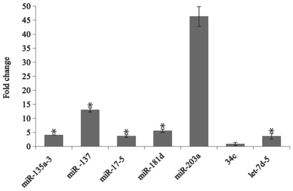
miRNAs are differentially expressed in
ependymomas
We performed individual assays for group of miRNAs
and the results revealed that miR-135a-3p, miR-137, miR-17-5p,
miR-181d and let-7d-5p are differentially expressed in ependymomas
when compared to control group (P<0.05). All of those miRNAs
were overexpressed (Fig. 1, Table III). The miR-203 was found to be
overexpressed with the highest difference as compared to control
group (fold change 46.3), but the result was not statistically
significant (P=0.08). Our results also indicate that the miR-34c
belonging to the tumor suppressor family miR-34 is (15,16)
slightly downregulated in ependymoma samples although the
difference was not statistically significant (RQ 0.9, P=0.75).
| Table III.Table presenting the up- vs.
downregulation of miRNAs expression in normal vs. disease samples
and grade III vs. grade II cohort. |
Table III.
Table presenting the up- vs.
downregulation of miRNAs expression in normal vs. disease samples
and grade III vs. grade II cohort.
|
| N vs. Ep | G3 vs. G2 |
|---|
|
|
|
|
|---|
| Assay | Fold change | P-value | Fold change | P-value |
|---|
| miR-135a-3p | 4.0 | 0.01 | 1.0 | 0.15 |
| miR-137 | 13.0 | 0.01 | 0.8 | 0.10 |
| miR-17-5p | 3.7 | 0.01 | 0.8 | 0.50 |
| miR-181d | 5.6 | 0.001 | 0.6 | 0.08 |
| miR-203a | 46.3 | 0.08 | 6.6 | 0.01 |
| 34c |
0.9 | 0.75 | 1.0 | 0.40 |
| let-7d-5p |
3.6 | 0.01 | 1.0 | 0.06 |
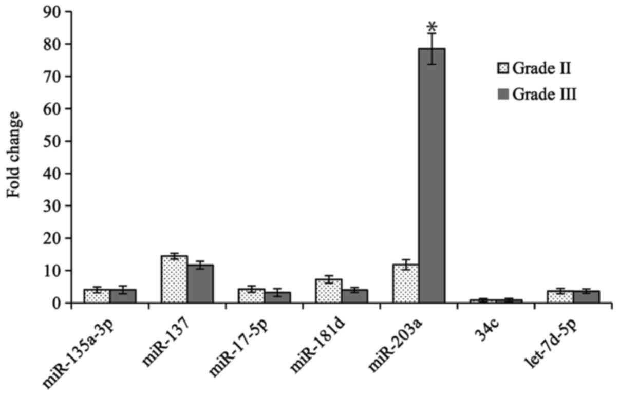
MiR-203a is overexpressed in grade III
tumors
Statistical analysis was performed in order to find
differences in expression of miRNAs between grade II and grade III
ependymomas. We found miR-203a to have higher expression in grade
III ependymomas (Fig. 2, Table III) suggesting its possible role in
distinguishing more aggressive tumors. Others miRNAs that are
differentially expressed in tumor samples when compared to normal
controls did not show statistically significant differences between
grade II and grade III tumors.
Discussion
Ependymomas exhibit wide range of biological
behavior extending from low-grade lesions to highly malignant
tumors. Prognostic stratification based solely on morphology or
immunohistochemistry remains elusive and brings only poor results
in regards to patient outcome.
In this study we aim at studying expression levels
of various miRNAs using individual qPCR assays in order to identify
new prognostic and/or diagnostic markers of ependymomas. We have
chosen miRNAs based on results of our preliminary experiment (data
not shown) where more than 20 miRNAs were involved. Out of this
preliminary experiment we have chosen seven miRNAs which were
showing differential expression between normal and ependymoma
samples or at least trend towards significance for this study.
We have identified miR-135a-3p, miR-137, miR-17-5p,
miR-181d and let-7d-5p to be differentially expressed in ependymoma
samples. To our knowledge this is the first report of
overexpression of miR-137 in pediatric intracranial ependymoma. The
let-7d has been previously described to act both as a
tumor-suppressor and oncogene (17,18). In
ependymomas it was previously shown (12) that let-7d correlates with overall
survival. However, we have not found significant difference in
let-7d expression between grade II and grade III samples. This
result underlines the fact that miRNA are pluripotent molecules and
their role in tumorigenesis in given tumor type needs to be
elucidated by more studies especially in tumors of genetically
heterogeneous nature.
Tumor grading in ependymoma reveals high
interindividual variability (19).
This fact calls for new markers which would make distinguishing
between low-grade and high-grade lesion more convenient. We have
demonstrated increased expression of miR-203a in grade III tumors
compared to grade II tumors. In recent years there has been
considerable interest in miR-203. This miRNA located on chromosome
14q32.33 was shown to have aberrant expression in many types of
cancers. However, the role of miR-203 in various cancer types is
not straightforward. It was previously reported that overexpression
of miR-203a in glioblastoma (20)
increased apoptosis of cancer cells thus acting as tumor-suppressor
gene. Similarly, it was shown that miR-203 upregulation inhibits
epithelial-mesenchymal transition in glioblastoma cells (21). miR-203a also suppress hepatocellular
carcinoma tumorigenesis by targeting HOXD3 via EGFR-related cell
signaling pathways (22). On the
other hand, some studies clearly indicated significant correlation
between the elevated miR-203 expression and poor overall survival
in colorectal adenocarcinoma (23,24) or
pancreatic carcinoma (25). miR-203a
was also described as a predictor of a poor prognosis in renal cell
carcinoma as its silencing inhibits cell proliferation, migration
and induces apoptosis (26). Despite
relatively common incidence of ependymomas and increased evidence
of miRNAs role in various types of cancer, not much attention has
been given to role of miRNAs in ependymoma (27). It was shown that miR-17-5p, miR19a-3p
and miR-106b-5p differentiate between grade II and III ependymomas
(12). Margolin-Miller et al
demonstrated that high expression of miR-124-3p in ependymomas
significantly correlated with the lower progression-free survival
(28).
Our observations of miR-203 expression in
ependymomas are in contrast with previous findings (13) which shown that lower expression of
miR-203 in ependymoma patients correlated to a trend to develop
recurrences. Some evidences suggest that miR-203 might not have
sufficient power to predict overall or event-free survival of
patient with different types of cancer (29). It was shown that methodology employed
in assuming miR-203 expression as well as the ethnicity of a
patients might influence the correlation of miR-203 expression and
clinical outcome of the patient (24). This could be one of the explanations
of discrepancies and needs to be further elucidated.
It was previously shown that PCR amplification
results from fresh-frozen material and FFPE samples are similar
(30). Routinely archived FFPE
samples in pathological departments are therefore suitable for
retrospective analysis of miRNAs expression. We have demonstrated
that FFPE samples of ependymoma archived for more than 25 years
still provide high yields of RNA of excellent purity.
In conclusion, we have shown several miRNAs to be
differentially expressed in ependymoma and outlined the role of
miR-203. These findings may add to a growing body of literature on
our understanding of miRNA expression and their role in ependymoma
biology.
Acknowledgements
Not applicable.
Funding
This study was supported by The Charles University
Grant Agency, grant no. GAUK190315.
Availability of data and materials
The datasets used and analyzed during this study are
available from the corresponding author on reasonable request.
Authors' contributions
All authors read and approved the final manuscript.
ŠC contributed to evaluation of tumor grade, for RNA extraction,
quantification of miRNAs and writing of the manuscript, MB
contributed to RNA extraction, quantification of miRNAs, TE
contributed to the project design and writing of manuscript, JZ
contributed to evaluation of tumor grade, the project design and
writing of manuscript. All authors read and approved the final
manuscript.
Ethics approval and consent to
participate
This study was approved by Ethical committee of
Second Faculty of Medicine, Charles University and University
hospital Motol, Prague, Czech Republic. Due to the retrospective
nature of this study the right to informed consent was waived.
Patient consent for publication
Not applicable.
Competing interests
The authors declare that they have no competing
interests.
References
|
1
|
Gupta A and Dwivedi T: A simplified
overview of World Health Organization classification update of
central nervous system tumors 2016. J Neurosci Rural Pract.
8:629–641. 2017. View Article : Google Scholar : PubMed/NCBI
|
|
2
|
PDQ Pediatric Treatment Editorial Board:
Childhood ependymoma treatment (PDQ®): Health
professional version. 2002.
|
|
3
|
Zamecnik J, Snuderl M, Eckschlager T,
Chanova M, Hladikova M, Tichy M and Kodet R: Pediatric intracranial
ependymomas: Prognostic relevance of histological,
immunohistochemical, and flow cytometric factors. Mod Pathol.
16:980–991. 2003. View Article : Google Scholar : PubMed/NCBI
|
|
4
|
Hübner JM, Kool M, Pfister SM and Pajtler
KW: Epidemiology, molecular classification and WHO grading of
ependymoma. J Neurosurg Sci. 62:46–50. 2018.PubMed/NCBI
|
|
5
|
Pajtler KW, Witt H, Sill M, Jones DT,
Hovestadt V, Kratochwil F, Wani K, Tatevossian R, Punchihewa C,
Johann P, et al: Molecular classification of ependymal tumors
across all CNS compartments, histopathological grades, and age
groups. Cancer Cell. 27:728–743. 2015. View Article : Google Scholar : PubMed/NCBI
|
|
6
|
Pajtler KW, Mack SC, Ramaswamy V, Smith
CA, Witt H, Smith A, Hansford JR, von Hoff K, Wright KD, Hwang E,
et al: The current consensus on the clinical management of
intracranial ependymoma and its distinct molecular variants. Acta
Neuropathol. 133:5–12. 2017. View Article : Google Scholar : PubMed/NCBI
|
|
7
|
Tihan T, Zhou T, Holmes E, Burger PC,
Ozuysal S and Rushing EJ: The prognostic value of histological
grading of posterior fossa ependymomas in children: A Children's
Oncology Group study and a review of prognostic factors. Mod
Pathol. 21:165–177. 2008. View Article : Google Scholar : PubMed/NCBI
|
|
8
|
Hayes J, Peruzzi PP and Lawler S:
MicroRNAs in cancer: Biomarkers, functions and therapy. Trends Mol
Med. 20:460–469. 2014. View Article : Google Scholar : PubMed/NCBI
|
|
9
|
Bentwich I, Avniel A, Karov Y, Aharonov R,
Gilad S, Barad O, Barzilai A, Einat P, Einav U, Meiri E, et al:
Identification of hundreds of conserved and nonconserved human
microRNAs. Nat Genet. 37:766–770. 2005. View Article : Google Scholar : PubMed/NCBI
|
|
10
|
Vannini I, Fanini F and Fabbri M: Emerging
roles of microRNAs in cancer. Curr Opin Genet Dev. 48:128–133.
2018. View Article : Google Scholar : PubMed/NCBI
|
|
11
|
Filipowicz W, Bhattacharyya SN and
Sonenberg N: Mechanisms of post-transcriptional regulation by
microRNAs: Are the answers in sight? Nat Rev Genet. 9:102–114.
2008. View
Article : Google Scholar : PubMed/NCBI
|
|
12
|
Zakrzewska M, Fendler W, Zakrzewski K,
Sikorska B, Grajkowska W, Dembowska-Bagińska B, Filipek I,
Stefańczyk Ł and Liberski PP: Altered MicroRNA expression is
associated with tumor grade, molecular background and outcome in
childhood infratentorial ependymoma. PLoS One. 11:e01584642016.
View Article : Google Scholar : PubMed/NCBI
|
|
13
|
Costa FF, Bischof JM, Vanin EF, Lulla RR,
Wang M, Sredni ST, Rajaram V, Mde Bonaldo F, Wang D, Goldman S, et
al: Identification of microRNAs as potential prognostic markers in
ependymoma. PLoS One. 6:e251142011. View Article : Google Scholar : PubMed/NCBI
|
|
14
|
Chen C, Ridzon DA, Broomer AJ, Zhou Z, Lee
DH, Nguyen JT, Barbisin M, Xu NL, Mahuvakar VR, Andersen MR, et al:
Real-time quantification of microRNAs by stem-loop RT-PCR. Nucleic
Acids Res. 33:e1792005. View Article : Google Scholar : PubMed/NCBI
|
|
15
|
Wiggins JF, Ruffino L, Kelnar K, Omotola
M, Patrawala L, Brown D and Bader AG: Development of a lung cancer
therapeutic based on the tumor suppressor microRNA-34. Cancer Res.
70:5923–5930. 2010. View Article : Google Scholar : PubMed/NCBI
|
|
16
|
Misso G, Di Martino MT, De Rosa G, Farooqi
AA, Lombardi A, Campani V, Zarone MR, Gullà A, Tagliaferri P,
Tassone P and Caraglia M: Mir-34: A new weapon against cancer? Mol
Ther Nucleic Acids. 3:e1942014. View Article : Google Scholar : PubMed/NCBI
|
|
17
|
Boyerinas B, Park SM, Hau A, Murmann AE
and Peter ME: The role of let-7 in cell differentiation and cancer.
Endocr Relat Cancer. 17:F19–F36. 2010. View Article : Google Scholar : PubMed/NCBI
|
|
18
|
Kolenda T, Przybyła W, Teresiak A,
Mackiewicz A and Lamperska KM: The mystery of let-7d-a small RNA
with great power. Contemp Oncol (Pozn). 18:293–301. 2014.PubMed/NCBI
|
|
19
|
Leeper H, Felicella MM and Walbert T:
Recent advances in the classification and treatment of ependymomas.
Curr Treat Options Oncol. 18:552017. View Article : Google Scholar : PubMed/NCBI
|
|
20
|
Yang CH, Wang Y, Sims M, Cai C, He P,
Häcker H, Yue J, Cheng J, Boop FA and Pfeffer LM: MicroRNA203a
suppresses glioma tumorigenesis through an ATM-dependent interferon
response pathway. Oncotarget. 8:112980–112991. 2017. View Article : Google Scholar : PubMed/NCBI
|
|
21
|
Liao H, Bai Y, Qiu S, Zheng L, Huang L,
Liu T, Wang X, Liu Y, Xu N, Yan X and Guo H: MiR-203 downregulation
is responsible for chemoresistance in human glioblastoma by
promoting epithelial-mesenchymal transition via SNAI2. Oncotarget.
6:8914–8928. 2015. View Article : Google Scholar : PubMed/NCBI
|
|
22
|
Wang L, Sun H, Wang X, Hou N, Zhao L, Tong
D, He K, Yang Y, Song T, Yang J and Huang C: EGR1 mediates miR-203a
suppress the hepatocellular carcinoma cells progression by
targeting HOXD3 through EGFR signaling pathway. Oncotarget.
7:45302–45316. 2016.PubMed/NCBI
|
|
23
|
Schetter AJ, Leung SY, Sohn JJ, Zanetti
KA, Bowman ED, Yanaihara N, Yuen ST, Chan TL, Kwong DL, Au GK, et
al: MicroRNA expression profiles associated with prognosis and
therapeutic outcome in colon adenocarcinoma. JAMA. 299:425–436.
2008. View Article : Google Scholar : PubMed/NCBI
|
|
24
|
Bovell LC, Shanmugam C, Putcha BD,
Katkoori VR, Zhang B, Bae S, Singh KP, Grizzle WE and Manne U: The
prognostic value of microRNAs varies with patient race/ethnicity
and stage of colorectal cancer. Clin Cancer Res. 19:3955–3965.
2013. View Article : Google Scholar : PubMed/NCBI
|
|
25
|
Greither T, Grochola LF, Udelnow A,
Lautenschläger C, Würl P and Taubert H: Elevated expression of
microRNAs 155, 203, 210 and 222 in pancreatic tumors is associated
with poorer survival. Int J Cancer. 126:73–80. 2010. View Article : Google Scholar : PubMed/NCBI
|
|
26
|
Hu G, Lai P, Liu M, Xu L, Guo Z, Liu H, Li
W, Wang G, Yao X, Zheng J and Xu Y: miR-203a regulates
proliferation, migration, and apoptosis by targeting glycogen
synthase kinase-3β in human renal cell carcinoma. Tumour Biol.
35:11443–11453. 2014. View Article : Google Scholar : PubMed/NCBI
|
|
27
|
Birks DK, Barton VN, Donson AM, Handler
MH, Vibhakar R and Foreman NK: Survey of MicroRNA expression in
pediatric brain tumors. Pediatr Blood Cancer. 56:211–216. 2011.
View Article : Google Scholar : PubMed/NCBI
|
|
28
|
Margolin-Miller Y, Yanichkin N, Shichrur
K, Toledano H, Ohali A, Tzaridis T, Michowitz S, Fichman-Horn S,
Feinmesser M, Pfister SM, et al: Prognostic relevance of miR-124-3p
and its target TP53INP1 in pediatric ependymoma. Genes Chromosomes
Cancer. 56:639–650. 2017. View Article : Google Scholar : PubMed/NCBI
|
|
29
|
Liang Y, Yang W, Zhu Y and Yuan Y:
Prognostic role of microRNA-203 in various carcinomas: Evidence
from a meta-analysis involving 13 studies. Springerplus.
5:15382016. View Article : Google Scholar : PubMed/NCBI
|
|
30
|
Siebolts U, Varnholt H, Drebber U, Dienes
HP, Wickenhauser C and Odenthal M: Tissues from routine pathology
archives are suitable for microRNA analyses by quantitative PCR. J
Clin Pathol. 62:84–88. 2009. View Article : Google Scholar : PubMed/NCBI
|