Introduction
Colorectal cancer (CRC) is one of the most common
cancers types worldwide (1,2). Within the last ten years in particular,
the prognosis for patients with metastatic CRC has markedly
improved due to patients undergoing advanced surgical resection of
localized metastases and advanced systemic chemotherapy (3,4).
Leucovorin (FOL) and fluorouracil (5-FU) plus oxaliplatin (l-OHP;
FOLFOX), or FOL and 5-FU plus irinotecan (SN-38; FOLFIRI),
administered in combination with molecularly-targeted drugs, are
used as first-line chemotherapy regimens in the treatment of
patients with advanced CRC worldwide (5,6). In recent
years, various studies have revealed that the median survival time
(MST) of patients with advanced CRC undergoing chemotherapy is
>30 months when patients are simultaneously administered a
combination of numerous cytotoxic agents and molecularly-targeted
therapies (7–10).
In previous years, the use of primary tumor location
in CRC as a predictive factor for response to therapy has attracted
attention. Numerous studies have demonstrated the predictive impact
of primary tumor location in CRC (11–13). In
embryonic development, the right colon (the cecum, the ascending
colon and the transverse colon) and the left colon (the descending
colon and the sigmoid colon) originate from the midgut and the
hindgut, respectively (14). Improved
clinical outcomes for patients with left-sided colon cancer (CC)
compared with patients with right-sided CC have been previously
reported (11–14). Improved outcomes for patients with
left-sided CC are dependent upon molecular tumor biology,
particularly when molecularly-targeted agent regimens for
palliative chemotherapies are used (15–22).
Therefore, in aforementioned studies, the administered chemotherapy
regimens always included molecularly-targeted agents. To the best
of our knowledge, the impact of primary tumor location as a
predictive factor in cytotoxic anticancer agent alone
(administration of FOLFOX/FOLFIRI in the absence of
molecularly-targeted agents) remains to be determined.
The aim of the present study was to clarify the
impact of primary tumor location as a predictive factor for the
efficacy of cytotoxic anticancer agents when administered in the
absence of molecular target agents using collagen gel
droplet-embedded drug sensitivity test (CD-DST).
Materials and methods
Patients
Between March 2008 and April 2017, tumor specimens
were obtained from 133 patients with CRC. Lymph node metastasis
with and without distant metastasis was reported in these patients.
All patients included in the present study had not received
preoperative chemotherapy or chemoradiotherapy prior to enrollment.
Written, informed consent for the determination of individual
chemosensitivity was obtained from all patients. Approval for the
present study was granted by the Tobu Chiiki Hospital Institutional
Review Board (Tokyo, Japan; grant no. 02.03.29. #1).
Methods
The concept of the CD-DST method, it is in
vitro assay, is to reproduce the minimum in vivo tumor
environment and predict the effect of anticancer drugs on the
original primary tumor in vivo. This method was approved by
the Japanese Ministry of Health, Labor and Welfare as Practical
Diagnostic Assay in 2008 as an assay reimbursed with public medical
insurance after assessing the validity, safety and reliability of
the assay. CD-DST was performed using a Human Cancer Primary
Culture System kit; Primastarä (Kurabo Industries, Ltd., Osaka,
Japan). All tumor tissues were excised from primary surgical
specimens and subjected to CD-DST. CD-DST analysis was performed to
investigate the drug sensitivity of isolated tumor cells cultured
in a three-dimensional manner in a small collagen gel droplet, and
to determine the sensitivity of the tumors to 5-FU, which was
performed in accordance with a protocol previously published by
Kobayashi et al (23,24). Each specimen was washed five times
with 50 ml saline, which was followed by with an additional five
washes with 50 ml antibiotic fluid containing 1.0 mg/ml
piperacillin and 0.5 mg/ml kanamycin. The transport centrifuge tube
was filled with 30 ml of the culture medium containing 1.0 mg/ml
piperacillin, 0.5 mg/ml kanamycin and 2.5 µg/ml amphotericin B.
Tissue samples (1 g) were incubated for 2 h at 37°C with a cocktail
containing 1.0% dispersion enzyme EZ™ (Kurabo Industries, Ltd.).
Dispersed cell suspensions were inoculated in pre-culture media in
collagen-coated flasks (CG-flusk™; Kurabo Industries, Ltd.)
overnight. Surviving tumor cells were subsequently recovered via
treatment with 0.05% collagenase and then embedded in 30 µl
collagen gel droplets.
Embedded cells were then incubated for 24 h at 37°C
in culture media containing either 5-FU (6.0 µg/ml) and l-OHP (3.0
µg/ml; FOLFOX regimen), or 5-FU (6.0 µg/ml) and SN-38 (0.2 µg/ml;
FOLFIRI regimen). Following the removal of the anticancer
agent-containing media, cells were additionally cultured for 7 days
in serum-free culture media (PCM-2™; Kurabo Industries, Ltd.) to
prevent the growth of fibroblasts. Surviving cells were stained
with neutral red solution and counted using the imaging
colorimetric quantification method (Primage™; Kurabo Industries,
Ltd.). The viable cell number ratio between the drug-treated group
and the control group, which received no drug treatment, was
calculated. A growth rate of <0.8 was considered to indicate a
successful culture.
Collecting cancer cells
A viable region was taken by a skilled surgeon while
avoiding the necrotic area from the excised tumor tissue. It was
confirmed by a pathologist that the excised tumor was definitely a
cancer tissue. Then, the viable cancer cells were collected through
the means of digestion by enzyme and pre-culture method. Cell lines
from collected cancer cells were not established.
Validity of the assay
For the control group, when the required minimum
number of cells (measured value of the Image Analysis System; 0.1
or more) was present and the relative proliferation ratio in the
assay period was 0.8 or more, it was regarded as being performed
correctly.
Treatment
Cetuximab or panitumumab was administered prior to
cytotoxic chemotherapies: 400 mg/m2 of cetuximab was
infused intravenously over 2 h on day 1, then 250 mg/m2
over 1 h every week, and 6 mg/kg of panitumumab was infused
intravenously over 1 h every 2 weeks. Bevacizumab was administered
prior to cytotoxic chemotherapies: 5 mg/kg of bevacizumab was
infused over 90 min every 2 weeks. Assuming no adverse reactions,
subsequent infusions were administered over a half-hour to 1 h
every other week.
Both histograms and associations between growth
inhibition rate (IR) values for each condition were investigated.
Cancers proximal or distal of the splenic flexure were designated
as right-side or left-side CRC, respectively. The association
between the side of the tumor and IR values was determined. The
prognosis for patients with right CC vs. patients with left CC was
also investigated using patients undergoing palliative chemotherapy
who additionally received molecularly-targeted treatment.
Statistical analysis
Histograms were analyzed using the
D'Agostino-Pearson omnibus normality test. Data are presented as
the median, mean, standard deviation (SD) and standard error (SE)
of the mean of IR values. The association between each condition
was determined via linear regression analysis. The associations
between IR values of patients with right-side tumors and left-side
tumors were investigated using the Student's t-test. MST values
were calculated using the Kaplan Meier method. Overall survival
curve groups were compared using the log rank test. Data are
presented as the mean ± standard deviation and were analyzed using
GraphPad Prism software (version 5.04; GraphPad Software, Inc., La
Jolla, CA, USA). P<0.05 was considered to indicate a
statistically significant difference.
Results
Patients
Patient characteristics are presented in Table I. A total of 42 of the 133 patients
received palliative chemotherapy following surgery, the
characteristics of whom are presented in Table II. Of the 42 patients, 25 CC patients
[right CC (n=7) and left CC (n=18)] received palliative
chemotherapy with molecularly-targeted agents. The number of
patients with right CC who received bevacizumab (Genentech, Inc.,
South San Francisco, CA, USA) and both bevacizumab and cetuximab
(Merck KGaA, Darmstadt, Germany) were five and two, respectively.
Of the patients with left CC, nine patients received bevacizumab,
two patients received cetuximab, one patient received panitumumab
(Amgen, Inc., Thousand Oaks, CA, USA), five patients received both
bevacizumab and cetuximab and one patient received both bevacizumab
and panitumumab.
 | Table I.Patient characteristics. |
Table I.
Patient characteristics.
| Primary tumor
location | Cecum | Ascending
colon | Transverse
colon | Descending
colon | Sigmoid colon | Rectum | Total |
|---|
| Age, years, mean
(range) | 63.1 | 66.9 | 69.0 | 68.1 | 66.5 | 66.0 | 65.5 |
|
| (42–79) | (46–81) | (36–84) | (55–82) | (47–82) | (37–79) | (36–84) |
| Sex,
male/female | 2/5 | 15/8 | 2/5 | 5/5 | 32/8 | 28/18 | 84/49 |
| Histological
type |
|
Papillary adenocarcinoma |
|
|
|
| 1 | 1 | 2 |
| Well
differentiated adenocarcinoma | 3 | 5 | 2 | 2 | 6 | 8 | 26 |
|
Moderately differentiated
adenocarcinoma | 4 | 14 | 3 | 6 | 28 | 33 | 88 |
| Poorly
differentiated adenocarcinoma |
|
| 1 |
| 2 | 2 | 5 |
|
Mucinous adenocarcinoma |
| 4 | 1 | 2 | 3 | 1 | 11 |
|
Squamous cell carcinoma |
|
|
|
|
| 1 | 1 |
| Total | 7 | 23 | 7 | 10 | 40 | 46 | 133 |
| Table II.Palliative chemotherapy patient
characteristics. |
Table II.
Palliative chemotherapy patient
characteristics.
| Primary tumor
location | Cecum | Ascending
colon | Transverse
colon | Descending
colon | Sigmoid colon | Rectum | Total |
|---|
| Age, years, mean
(range) | 54 | 63.5 | 74.5 | 68.4 | 64.6 | 64.7 | 65.1 |
|
|
| (52–78) | (70–79) | (55–80) | (51–80) | (51–78) | (51–80) |
| Sex,
male/female | 0/1 | 5/1 | 0/2 | 2/3 | 10/3 | 12/3 | 29/13 |
| Histological
type |
|
Papillary adenocarcinoma |
|
|
|
| 1 | 1 | 2 |
| Well
differentiated adenocarcinoma |
| 1 |
| 1 | 1 | 1 | 4 |
|
Moderately differentiated
adenocarcinoma | 1 | 5 | 1 | 3 | 9 | 11 | 30 |
| Poorly
differentiated adenocarcinoma |
|
| 1 |
| 1 |
| 2 |
|
Mucinous adenocarcinoma |
|
|
| 1 | 1 | 1 | 3 |
|
Squamous cell carcinoma |
|
|
|
|
| 1 | 1 |
| Total | 1 | 6 | 2 | 5 | 13 | 15 | 42 |
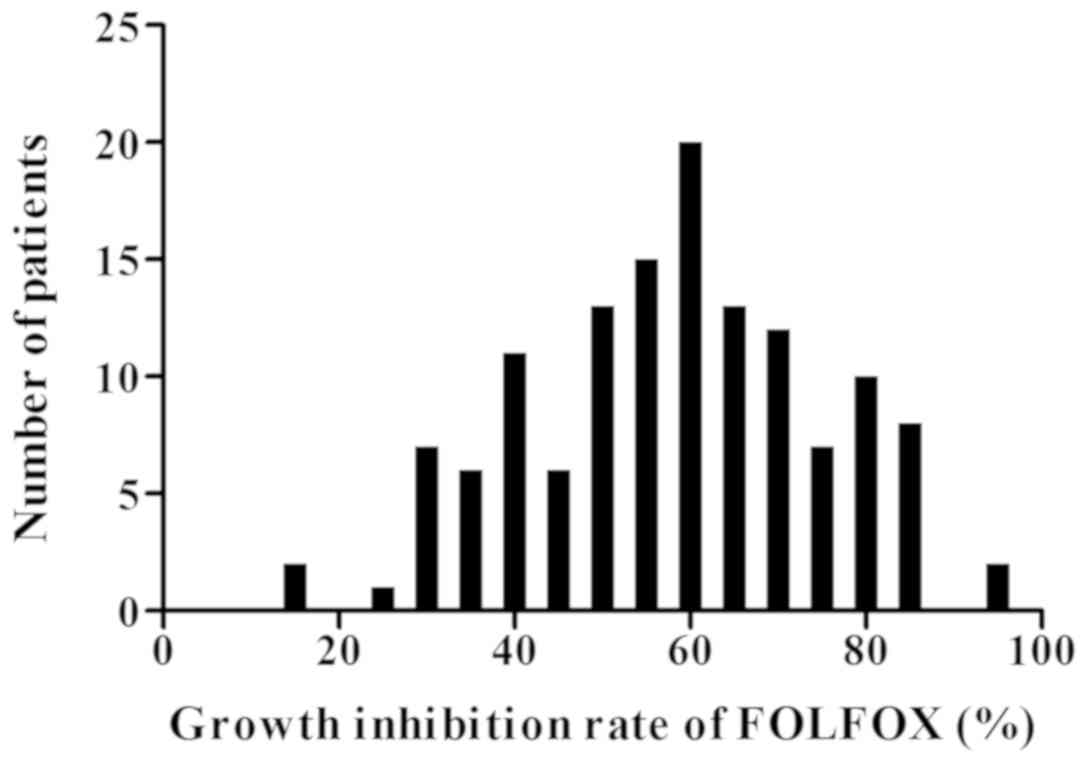
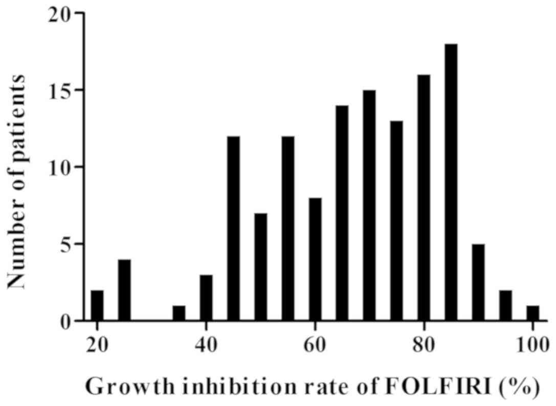
Histograms of the individual growth IR
values
Histograms of the individual growth IRs (%) under
the conditions of the FOLFOX and FOLFIRI regimens are presented in
Figs. 1 and 2, respectively. The median, mean, SD and SE
of the mean in the FOLFOX regimen were 58.7, 58.5, 16.7 and 1.45,
respectively. The median, mean, and SD and SE of the mean in the
FOLFIRI regimen were 69.1, 66.2, 17.1 and 1.48, respectively. The
histograms passed the normality test (α=0.05; FOLFOX regimen,
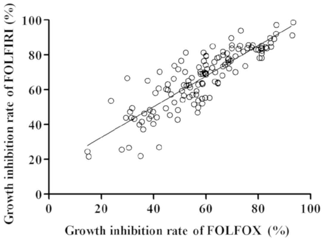
P=0.68; FOLFIRI regimen, P=0.06). There was a strong correlation
between the IRs (%) of the FOLFOX and FOLFIRI regimens (Fig. 3; y=0.88×+14.98,
R2=0.74).
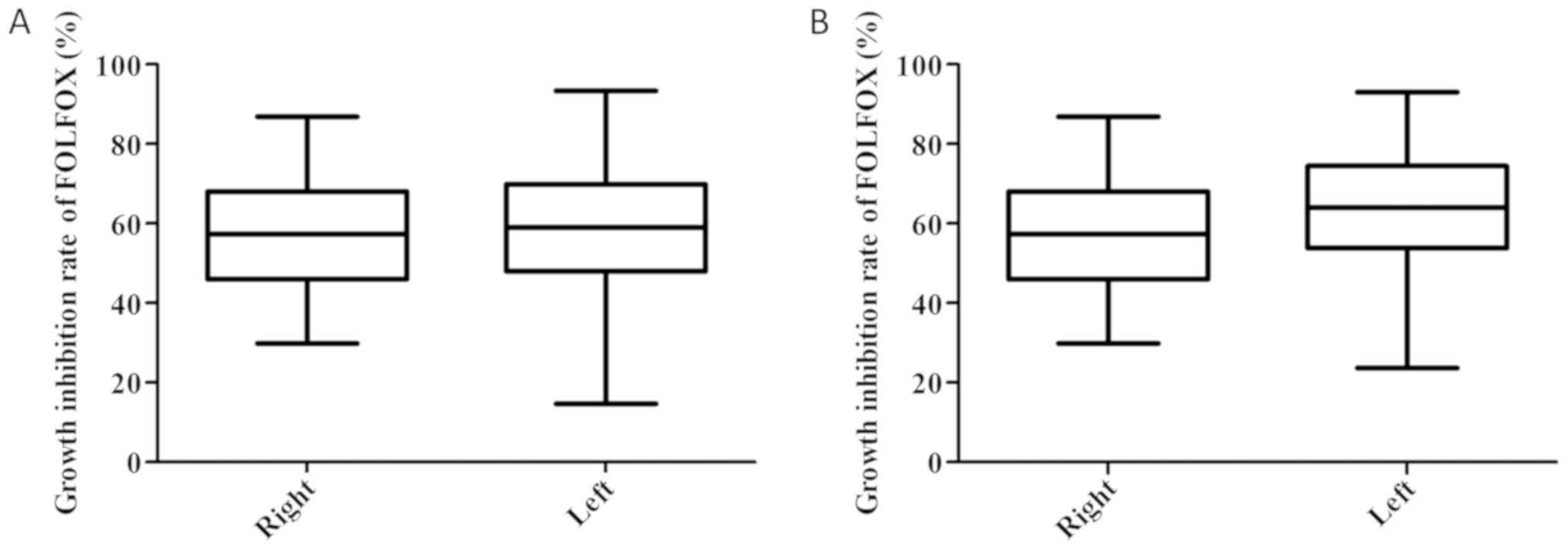
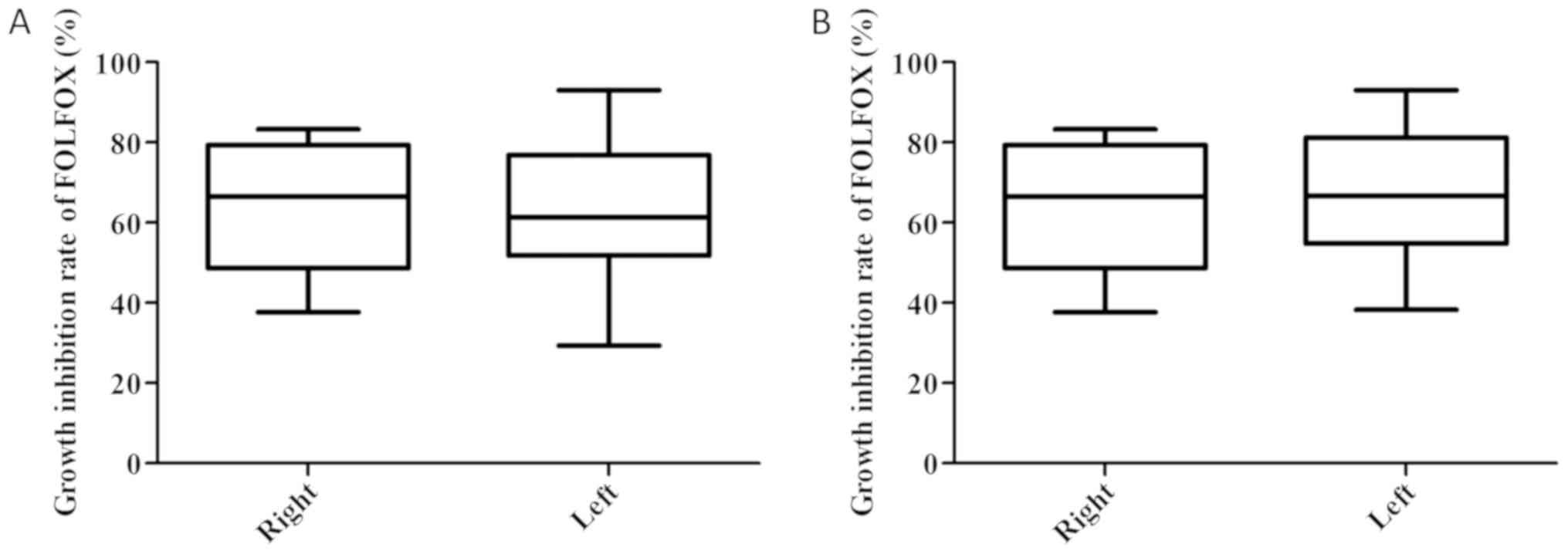
IR values of all patients in the
FOLFOX regimen
The IRs (%) of right- and left-sided tumors
including the rectum, were 57.4±2.5 and 58.5±1.8, respectively
(P=0.72; Fig. 4A). The IRs (%) of
right- and left-sided tumors excluding the rectum (n=87) were
57.4±2.5 and 61.6±2.4, respectively (P=0.23; Fig. 4B).
IR values of all patients in the
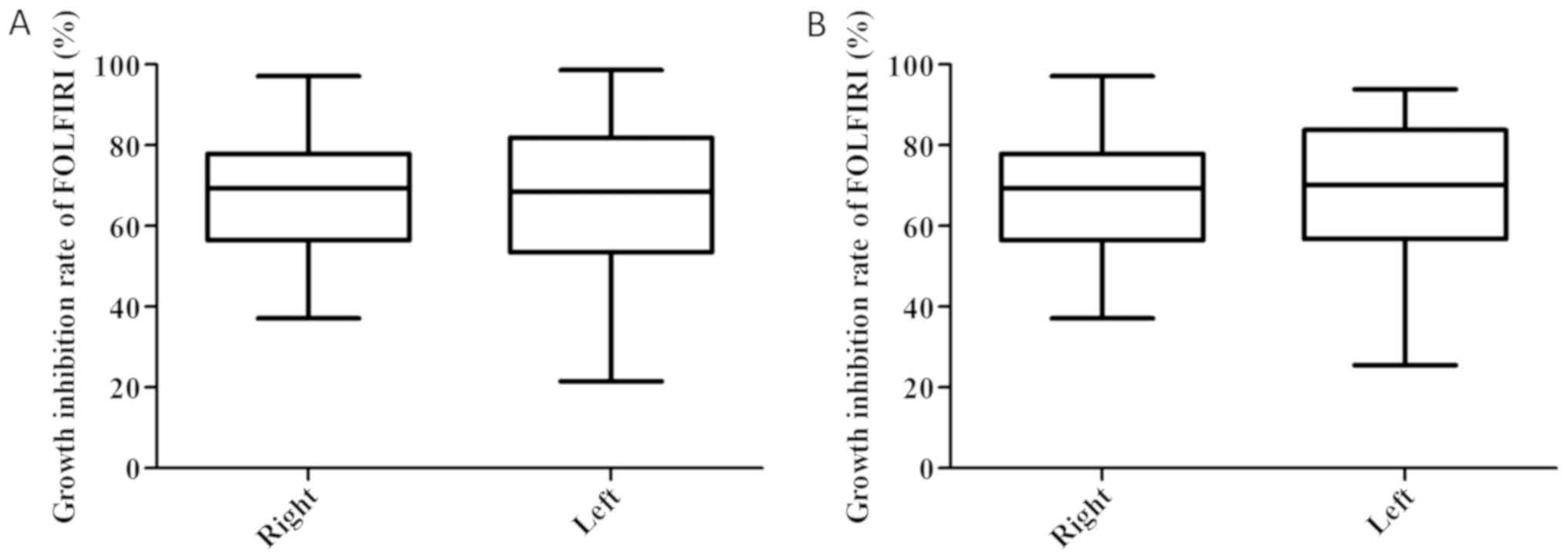
FOLFIRI regimen
The IRs (%) of right- and left-sided tumors
including the rectum, were 67.0±2.3 and 65.8±1.9, respectively
(P=0.69; Fig. 5A). The IRs (%) of
right- and left-sided tumors excluding the rectum (n=87) were
67.0±2.3 and 69.1±2.4, respectively (P=0.53; Fig. 5B).
IR values of 42 patients receiving
palliative chemotherapy in the FOLFOX regimen
The IRs (%) of right- and left-sided tumors
including the rectum, were 63.8±5.4 and 62.9±2.8, respectively
(P=0.88; Fig. 6A). The IRs (%) of
right- and left-sided tumors excluding the rectum (n=27) were
63.8±5.4 and 67.0±3.8, respectively (P=0.64; Fig. 6B).
IR values of 42 patients receiving
palliative chemotherapy in the FOLFIRI regimen
The IRs (%) of right- and left-sided tumors
including the rectum, were 74.0±2.6 and 69.2±2.8, respectively
(P=0.22; Fig. 7A). The IRs (%) of
right- and left-sided tumors excluding the rectum (n=27) were
74.0±2.6 and 72.7±3.5, respectively (P=0.76; Fig. 7B).
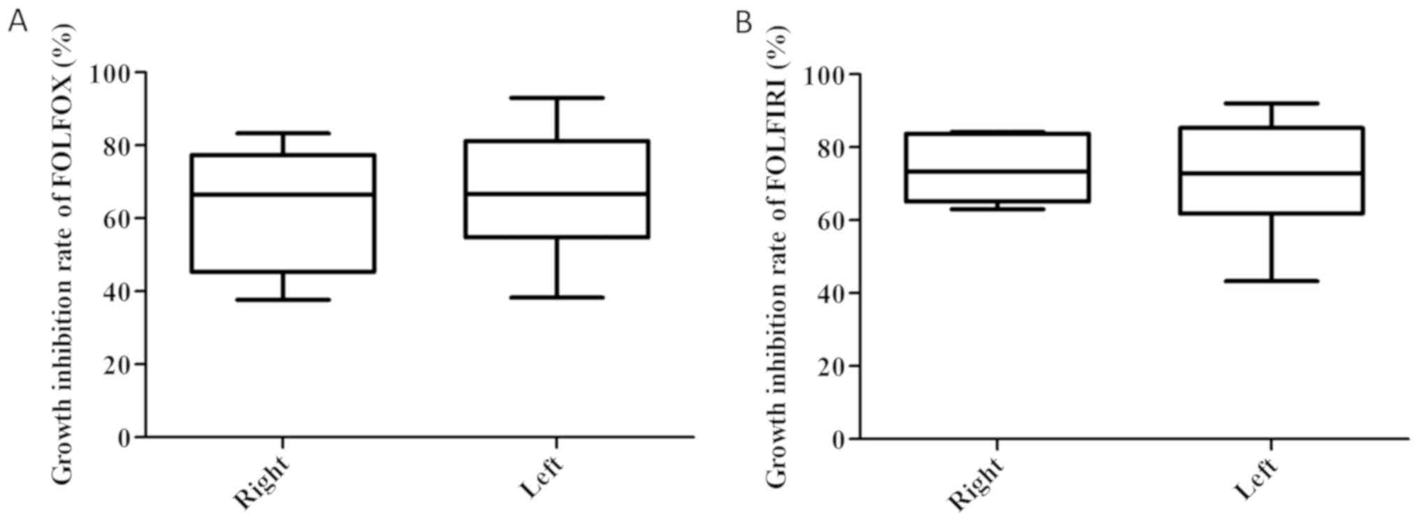
IR values of 25 patients with CC
receiving palliative chemotherapy and treated with
molecularly-targeted agents in the FOLFOX regimen
The IRs (%) of right- and left-sided tumors in the
FOLFOX regimen were 61.9±6.5 and 67.0±3.8, respectively (P=0.52;
Fig. 8A).
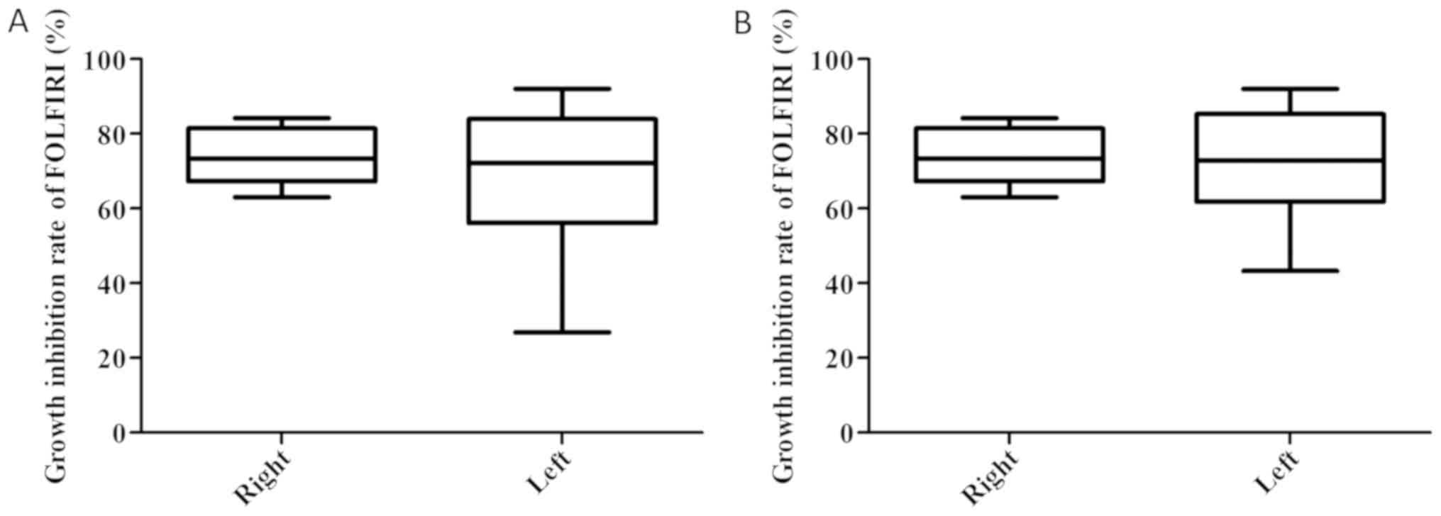
IR values of 25 patients with CC
receiving palliative chemotherapy and treated with
molecularly-targeted agents in the FOLFIRI regimen
The IRs (%) of right- and left-sided tumors in the
FOLFIRI regimen were 74.1±3.2 and 72.7±3.5, respectively (P=0.77;
Fig. 8B).
Among all patients, there was no significant
difference in the IRs (%) of the FOLFOX and FOLFIRI regimens using
CD-DST between right- and left-sided tumors, including or excluding
the rectum.
Prognosis of 25 patients receiving
palliative chemotherapy and treated with molecularly-targeted
agents
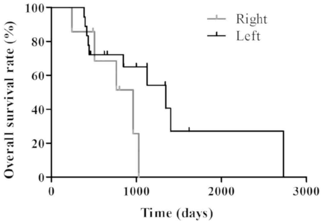
The median follow-up time period of patients
suffering from CC who had been administered palliative chemotherapy
and treatment with molecularly-targeted agents (n=25) was 800 days.
Furthermore, the MSTs in patients with right CC and patients with
left CC were 960 and 1,348 days, respectively (Fig. 9). However, there were no significant
differences (P=0.11).
Discussion
The CD-DST method is an examination for evaluating
the effect of a cross productive anticancer drug on tumors under a
culturing condition that minimally reproduces tumor
microenvironments in vivo. This reproduction of this
microenvironment is to carry out three-dimensional primary culture
on a coexistence of cancer cells and fibroblasts derived from tumor
tissue with a serum-free medium containing various cell growth
factors in the collagen gel which is an extracellular matrix (ECM).
In order to accurately evaluate chemosensitivity against cancer
cells, it is evaluated by ‘Image Analysis System’, which utilizes
the difference in growth morphology between cancer cells and
fibroblasts (25). In the CD-DST
method, the primary cancer cells were cultured in serum-free
medium. In addition, the serum-free medium does not contain cell
growth factors necessary for proliferation of fibroblasts and
vascular endothelial cells that may be contaminated. Unlike on
collagen gel (2D-culture), fibroblasts have the property of
suppressing their proliferation in collagen gel (3D-culture). In
the collagen gel, the epithelium-derived cancer cell and the
mesenchymal normal cell clearly differ in their proliferation form.
The former takes a spherical or thick dendritic form; the latter
universally takes a bipolar shape. The fact that spherical colonies
are cancer cells has been confirmed by the immunostaining method at
the time of development of this assay method (25). Macrophages and lymphocytes, which are
typical inflammatory cells, are not adhered on collagen gel coat
flasks during preliminary culture within 72 h, so they are always
removed by medium change. The cancer cells are thereby purified. As
described above, in the CD-DST method, the chemosensitivity of each
anticancer agent is evaluated in the primary cell-culture condition
of cancer cells which have been collected and purified from fresh
tumor tissue.
As mentioned earlier, this assay is intended to
‘predict the direct effect of anticancer drugs on tumor tissue’, so
it is not possible to evaluate the adverse reactions caused by
chemotherapy. Many side effects caused by administration of
anticancer drugs are caused by disorders such as bone marrow,
peripheral nerve, gastrointestinal tract, not tumor part.
Therefore, the influence of anticancer agents on normal cells on
the premise of side effect prediction deviates from our objective.
Regarding the influence on normal cells in the tumor tissue, in the
CD-DST method, fibroblasts derived from tumor tissue are mixed in
the collagen gel. The proliferation of these cells is suppressed by
the culture environment and a serum-free medium, but it does not
lead to cell death and its physiological activity is maintained.
Under this coexisting condition, we can evaluate the effect of
anticancer drugs on cancer. Usually, when chemotherapy is
performed, patients are monitored for the occurrence and degree of
adverse reactions, symptomatic treatment is administered at any
time, and symptoms are also managed (26). As we already reported (27–29), the
prognosis of the high-sensitive group is good, so the prediction of
adverse reactions is not so important, but it seems to be most
important to extract the group that can be expected to be
effective.
FOLFOX or FOLFIRI in combination with
molecularly-targeted drugs represent first-line chemotherapy
regimens used for the treatment of advanced CRC globally (3,4). It has
been reported that the clinical response rates to FOLFOX and
FOLFIRI are equivalent (~50%) (30–33). In
the present study, there were a number of small differences between
the IRs (%) of the FOLFOX and FOLFIRI regimens in numerous
patients. However, there was a strong overall correlation between
the IRs (%) of the two regimen exhibited by the majority of
patients (R2=0.74). Therefore, this result might show
that the efficacies of FOLFOX and FOLFIRI are approximately
equivalent in individual. This result also supports the findings of
previous studies (30–33).
Improved clinical outcomes for patients with
left-sided CC compared with patients with right-sided CC have been
reported worldwide (11–13). It has been previously demonstrated
that improved outcomes for patients with left-sided CC is dependent
upon molecular tumor biology, particularly when
molecularly-targeted agent regimens are used (15–22). The
molecular differences between the right colon and the left colon
are notable. Molecularly, right-sided CC and left-sided CC are two
different diseases (14). Right-sided
CC is associated with defective mismatch repair (MMR) genes, as
well as mutations in K-Ras, B-Raf and microRNA-31, whereas
left-sided CC is associated with p53, chromosomal instability and
expression of N-Ras, microRNA-146a, microRNA-147b and microRNA-1288
(34–36). In clinical practice,
molecularly-targeted agents corresponding to identified molecular
tumor biology are improving prognostic prediction (15–22).
Therefore, primary tumor location may be considered to represent a
predictive factor in molecularly-targeted agents.
Molecularly-targeted agents should be administered according to
molecular tumor biology. However, to the best of our knowledge,
there is only one study that has previously investigated whether
primary tumor location alone is a predictive factor for the correct
use of cytotoxic anticancer agents (15). Boisen et al reported that
patients with left-sided CC treated with capecitabin and
oxaliplatin (CAPEOX) with bevacizumab exhibited improved clinical
outcomes compared with patients suffering from right-sided CC.
However, no correlation between primary tumor location and outcome
was observed in patients treated with CAPEOX without bevacizumab
(15). In recently performed
chemotherapy trials, molecularly-targeted agent regimens were
revealed to be necessary, particularly in patients receiving
palliative chemotherapy (5,6). Therefore, at present, it is difficult to
investigate primary tumor location as a predictive factor in
patients treated with cytotoxic anticancer agents alone via
prospective randomized studies, particularly in patients undergoing
palliative chemotherapy. Previously, we have reported the
effectiveness of CD-DST for the individualization of first-line
treatment in CRC. Therefore, the present study aimed to determine
the importance of primary tumor location as a predictive factor in
cytotoxic anticancer agent treatment using CD-DST (27–29).
In the present study, there were no significant
differences in the IRs (%) of the FOLFOX and FOLFIRI regimens using
CD-DST between right- and left-sided tumors, including or excluding
the rectum. These results were consistent with Boisen's study
(15). Cytotoxic anticancer agents
inhibit the cellular division of cancer cells as well as normal
cells (26). 5-FU is an
S-phase-specific drug and is active only during specific stages of
the cell cycle. The target enzyme of 5-FU is thymidylate synthase,
which is the main enzyme associated with DNA synthesis. Therefore,
5-FU inhibits DNA synthesis, which results in RNA dysfunction
(27,37,38).
Irinotecan is also an S-phase-specific drug that is active only
during specific stages of the cell cycle. Irinotecan is metabolized
by carboxylesterases to its active metabolite,
7-ethyl-10-hydroxycamptothecin (SN-38). SN-38 functions as a
classic topoisomerase I inhibitor by stabilizing the topoisomerase
I/DNA cleavable complex, which subsequently blocks DNA replication
and results in DNA strand breakages (39). Furthermore, oxaliplatin is a
cell-cycle non-specific antineoplastic agent (40). Oxaliplatin is classified as a
third-generation platinum compound, following cisplatin and
carboplatin. Similar to other platinum compounds, oxaliplatin,
cisplatin and carboplatin form crosslinks with DNA strands within
cancer cells and inhibit DNA replication and transcription
(41). It has been well established
that cancer cells attenuate this effect by removing cisplatin from
DNA via an MMR mechanism (42).
However, it has been previously revealed that oxaliplatin, which is
larger in structure than cisplatin, is not removed by MMR and it is
difficult to acquire a tolerance to (43–45). It
can be suggested that molecular tumor biology may not significantly
affect the efficacy of cytotoxic anticancer agents during the cell
division stage of cancer cells. Therefore, in patients receiving
adjuvant chemotherapy in the absence of treatment with
molecularly-targeted agents, improved outcomes for patients
suffering from left-sided CC compared with right-sided CC are not
observed (46–48). However, of the 25 patients that
received palliative chemotherapy as well as treatment with
molecularly-targeted agents in the present study, the MSTs in
patients suffering from right-sided CC and left-sided CC were 960
and 1,348 days, respectively. This result supports the hypothesis
that when administered molecularly-targeted agent regimens,
patients suffering from left-sided CC exhibit improved clinical
outcomes compared with patients suffering from right-sided CC,
depending upon molecular tumor biology, as Boisen et al
reported (15). In the CD-DST, there
were no significant differences in the IRs (%) of cytotoxic
anticancer agents without molecularly-targeted agents between
right- and left-sided colon cancer tumors. Conversely, clinical
outcomes of left-sided colorectal cancer patients treated with
cytotoxic agents and molecularly-targeted agents were better than
those of right-sided colon cancer patients. Thus, impact on the
prognosis may be due to the effect of molecular target agents.
Therefore, primary tumor location may represent a predictive factor
for the efficacy of molecularly-targeted agents, rather than a
prognostic factor, in patients with CRC.
However, there were several limitations in the
present study. Firstly, the sample size was small. However, the
proportion of rectal cancers in left-sided colorectal cancers was
not small, which might have influenced statistical analysis.
Therefore, we had to evaluate the prognosis of only colon cancer
patients in palliative chemotherapy with molecularly-targeted
agents. A larger sample size would have improved the quality of the
data. Secondly, observation periods were short for overall survival
rates. The small sample size and the short observation periods may
have affected the validity of statistical analyses. Thirdly, we did
not evaluate the effects of cytotoxic anticancer agents on normal
cell division. Investigation of these effects would have improved
the quality of the data.
In conclusion, primary tumor location was revealed
to not represent a predictive factor in cytotoxic anticancer agent
regimens for patients with CRC. However, improved clinical outcomes
for patients with left-sided CC compared with right-sided CC were
demonstrated when patients were administered molecularly-targeted
agent regimens. Therefore, the results of the present study
suggested that molecularly-targeted agents rather than cytotoxic
anticancer agents may result in improved clinical outcomes for
patients with CC suffering from left-sided tumors.
Acknowledgements
Not applicable.
Funding
No funding was received.
Availability of data and materials
The datasets used and/or analyzed during the current
study are available from the corresponding author on reasonable
request.
Authors' contributions
TO and IN contributed to the conception and design
of the present study. KNi, TW, MK, AN, KNa, NS, TS, KK, YA and TO
acquired the patient data. TW, MK, AN, KNa, NS, TS, KK, YA and TO
performed the preparation of viable regions from excised tumor
tissues. TO analyzed and interpreted the data. KNi and IN were
major contributors in writing the manuscript. TO wrote the
manuscript. All authors read and approved the final manuscript.
Ethics approval and consent to
participate
Approval for the present study was obtained from the
Tobu Chiiki Hospital Institutional Review Board (approval no.
02.03.29. #1; Tokyo, Japan). Written informed consent for this
study was obtained from all patients.
Patient consent for publication
The patients provided written informed consent for
the publication of any associated data.
Competing interests
The authors declare that they have no competing
interests.
References
|
1
|
Ferlay J, Soerjomataram I, Dikshit R, Eser
S, Mathers C, Rebelo M, Parkin DM, Forman D and Bray F: Cancer
incidence and mortality worldwide: Sources, methods and major
patterns in GLOBOCAN 2012. Int J Cancer. 136:E359–E386. 2015.
View Article : Google Scholar : PubMed/NCBI
|
|
2
|
Siegel RL, Miller KD, Fedewa SA, Ahnen DJ,
Meester RG, Barzi A and Jemal A: Colorectal cancer statistics,
2017. CA Cancer J Clin. 67:177–193. 2017. View Article : Google Scholar : PubMed/NCBI
|
|
3
|
Adam R, De Gramont A, Figueras J, Guthrie
A, Kokudo N, Kunstlinger F, Loyer E, Poston G, Rougier P,
Rubbia-Brandt L, et al: The oncosurgery approach to managing liver
metastases from colorectal cancer: A multidisciplinary
international consensus. Oncologist. 17:1225–1239. 2012. View Article : Google Scholar : PubMed/NCBI
|
|
4
|
Jones RP, Stättner S, Sutton P, Dunne DF,
McWhirter D, Fenwick SW, Malik HZ and Poston GJ: Controversies in
the oncosurgical management of liver limited stage IV colorectal
cancer. Surg Oncol. 23:53–60. 2014. View Article : Google Scholar : PubMed/NCBI
|
|
5
|
NCCN: NCCN Clinical Practice Guidelines in
Oncology. Colon Cancer. Version 2. 2017, http://www.nccn.orgFebruary 24–2018
|
|
6
|
Yoshino T, Arnold D, Taniguchi H,
Pentheroudakis G, Yamazaki K, Xu RH, Kim TW, Ismail F, Tan IB, Yeh
KH, et al: Pan-Asian adapted ESMO consensus guidelines for the
management of patients with metastatic colorectal cancer: A
JSMO-ESMO initiative endorsed by CSCO KACO, MOS, SSO and TOS. Ann
Oncol. 29:44–70. 2018. View Article : Google Scholar : PubMed/NCBI
|
|
7
|
Fakih MG: Metastatic colorectal cancer:
Current state and future directions. J Clin Oncol. 33:1809–1824.
2015. View Article : Google Scholar : PubMed/NCBI
|
|
8
|
Venook A, Niedzwiecki D, Lenz H, Mahoney
M, Innocenti F, O'Neil B, Shaw J, Polite B, Hochster H, Goldberg R,
et al: CALGB/SWOG 80405: Analysis of patients undergoing surgery as
part of treatment strategy. Ann Oncol. 25 Suppl 4:S1–S41. 2014.
View Article : Google Scholar
|
|
9
|
Recondo G Jr, Díaz-Cantón E, de la Vega M,
Greco M, Recondo G Sr and Valsecchi ME: Advances and new
perspectives in the treatment of metastatic colon cancer. World J
Gastrointest Oncol. 6:211–224. 2014. View Article : Google Scholar : PubMed/NCBI
|
|
10
|
Yamada Y, Denda T, Gamoh M, Iwanaga I,
Yuki S, Shimodaira H, Nakamura M, Yamaguchi T, Ohori H, Kobayashi
K, et al: S-1 and irinotecan plus bevacizumab versus mFOLFOX6 or
CapeOX plus bevacizumab as first-line treatment in patients with
metastatic colorectal cancer (TRICOLORE): A randomized, open-label,
phase III, noninferiority trial. Ann Oncol. 29:624–631. 2018.
View Article : Google Scholar : PubMed/NCBI
|
|
11
|
Wolmark N, Wieand HS, Rockette HE, Fisher
B, Glass A, Lawrence W, Lerner H, Cruz AB, Volk H, Shibata H, et
al: The prognostic significance of tumor location and bowel
obstruction in Dukes B and C colorectal cancer. Findings from the
NSABP clinical trials. Ann Surg. 198:743–752. 1983. View Article : Google Scholar : PubMed/NCBI
|
|
12
|
Loupakis F, Yang D, Yau L, Feng S,
Cremolini C, Zhang W, Maus MK, Antoniotti C, Langer C, Scherer SJ,
et al: Primary tumor location as a prognostic factor in metastatic
colorectal cancer. J Natl Cancer Inst. 107(pii):
dju4272015.PubMed/NCBI
|
|
13
|
Petrelli F, Tomasello G, Borgonovo K,
Ghidini M, Turati L, Dallera P, Passalacqua R, Sgroi G and Barni S:
Prognostic survival associated with left-sided vs right-sided colon
cancer: A systematic review and meta-analysis. JAMA Oncol. Oct
27–2016.(Epub ahead of print).
|
|
14
|
Boeckx N, Janssens K, Van Camp G,
Rasschaert M, Papadimitriou K, Peeters M and Op de Beeck K: The
predictive value of primary tumor location in patients with
metastatic colorectal cancer: A systematic review. Crit Rev Oncol
Hematol. 121:1–10. 2018. View Article : Google Scholar : PubMed/NCBI
|
|
15
|
Boisen MK, Johansen JS, Dehlendorff C,
Larsen JS, Osterlind K, Hansen J, Nielsen SE, Pfeiffer P, Tarpgaard
LS, Holländer NH, et al: Primary tumor location and bevacizumab
effectiveness in patients with metastatic colorectal cancer. Ann
Oncol. 24:2554–2559. 2013. View Article : Google Scholar : PubMed/NCBI
|
|
16
|
Brulé SY, Jonker DJ, Karapetis CS,
O'Callaghan CJ, Moore MJ, Wong R, Tebbutt NC, Underhill C, Yip D,
Zalcberg JR, et al: Location of colon cancer (right-sided versus
left-sided) as a prognostic factor and a predictor of benefit from
cetuximab in NCIC CO.17. Eur J Cancer. 51:1405–1414. 2015.
View Article : Google Scholar : PubMed/NCBI
|
|
17
|
Shen H, Yang J, Huang Q, Jiang MJ, Tan YN,
Fu JF, Zhu LZ, Fang XF and Yuan Y: Different treatment strategies
and molecular features between right-sided and left-sided colon
cancers. World J Gastroenterol. 21:6470–6478. 2015. View Article : Google Scholar : PubMed/NCBI
|
|
18
|
Holch JW, Ricard I, Stintzing S, Modest DP
and Heinemann V: The relevance of primary tumour location in
patients with metastatic colorectal cancer: A meta-analysis of
first-line clinical trials. Eur J Cancer. 70:87–98. 2017.
View Article : Google Scholar : PubMed/NCBI
|
|
19
|
Tejpar S, Stintzing S, Ciardiello F,
Tabernero J, Van Cutsem E, Beier F, Esser R, Lenz HJ and Heinemann
V: Prognostic and predictive relevance of primary tumor location in
patients with RAS wild-type metastatic colorectal cancer:
Retrospective analyses of the CRYSTAL and FIRE-3 trials. JAMA
Oncol. Oct 10–2016.(Epub ahead of print).
|
|
20
|
Aljehani MA, Morgan JW, Guthrie LA, Jabo
B, Ramadan M, Bahjri K, Lum SS, Selleck M, Reeves ME, Garberoglio C
and Senthil M: Association of primary tumor site with mortality in
patients receiving bevacizumab and cetuximab for metastatic
colorectal cancer. JAMA Surg. 153:60–67. 2018. View Article : Google Scholar : PubMed/NCBI
|
|
21
|
Ishihara S, Murono K, Sasaki K, Yasuda K,
Otani K, Nishikawa T, Tanaka T, Kiyomatsu T, Kawai K, Hata K, et
al: Impact of primary tumor location on postoperative recurrence
and subsequent prognosis in nonmetastatic colon cancers: A
multicenter retrospective study using a propensity score analysis.
Ann Surg. 267:917–921. 2018. View Article : Google Scholar : PubMed/NCBI
|
|
22
|
Yamashita S, Brudvik KW, Kopetz SE, Maru
D, Clarke CN, Passot G, Conrad C, Chun YS, Aloia TA and Vauthey JN:
Embryonic origin of primary colon cancer predicts pathologic
response and survival in patients undergoing resection for colon
cancer liver metastases. Ann Surg. 267:514–520. 2018. View Article : Google Scholar : PubMed/NCBI
|
|
23
|
Kobayashi H, Tanisaka K, Doi O, Kodama K,
Higashiyama M, Nakagawa H, Miyake M, Taki T, Hara S, Yasutomi M, et
al: An in vitro chemosensitivity test for solid tumors using
collagen gel droplet embedded cultures. Int J Oncol. 11:449–455.
1997.PubMed/NCBI
|
|
24
|
Kobayashi H, Higashiyama M, Minamigawa K,
Tanisaka K, Takano T, Yokouchi H, Kodama K and Hata T: Examination
of in vitro chemosensitivity test using collagen droplet culture
method with colorimetric endpoint quantification. Jpn J Cancer Res.
92:203–210. 2001. View Article : Google Scholar : PubMed/NCBI
|
|
25
|
Kobayashi H: Development of a new in vitro
chemosensitivity test using collagen gel droplet embedded culture
and image analysis for clinical usefulness. Recent Results Cancer
Res. 161:48–61. 2003. View Article : Google Scholar : PubMed/NCBI
|
|
26
|
Corrie PG: Cytotoxic chemotherapy:
Clinical aspects. Medicine. 36:24–28. 2008. View Article : Google Scholar
|
|
27
|
Ochiai T, Nishimura K, Watanabe T,
Kitajima M, Hashiguchi T, Nakatani A, Marusasa T, Muraki A, Nagaoka
I and Futagawa S: Leucovorin and fluorouracil plus oxaliplatin or
leucovorin and fluorouracil plus irinotecan as individualized
first-line therapy based on a drug sensitivity test. Exp Ther Med.
1:325–329. 2010. View Article : Google Scholar : PubMed/NCBI
|
|
28
|
Ochiai T, Nishimura K, Watanabe T,
Kitajima M, Nakatani A, Inou T, Washio M, Sakuyama N, Sato T,
Kishine K, et al: Individualized chemotherapy for colorectal cancer
based on the collagen gel droplet-embedded drug sensitivity test.
Oncol Lett. 4:621–624. 2012. View Article : Google Scholar : PubMed/NCBI
|
|
29
|
Ochiai T, Nishimura K, Watanabe T,
Kitajima M, Nakatani A, Nagayasu K, Naito S, Sato T, Kishine K, Abe
Y, et al: Impact of the individualization of the first-line
chemotherapy for advanced colorectal cancer based on collagen gel
droplet-embedded drug sensitivity test. Oncol Lett. 14:6045–6052.
2017.PubMed/NCBI
|
|
30
|
Tournigand C, André T, Achille E, Lledo G,
Flesh M, Mery-Mignard D, Quinaux E, Couteau C, Buyse M, Ganem G, et
al: FOLFIRI followed by FOLFOX6 or the reverse sequence in advanced
colorectal cancer: A randomized GERCOR study. J Clin Oncol.
22:229–237. 2004. View Article : Google Scholar : PubMed/NCBI
|
|
31
|
de Gramont A, Figer A, Seymour M, Homerin
M, Hmissi A, Cassidy J, Boni C, Cortes-Funes H, Cervantes A, Freyer
G, et al: Leucovorin and fluorouracil with or without oxaliplatin
as first-line treatment in advanced colorectal cancer. J Clin
Oncol. 18:2938–2947. 2000. View Article : Google Scholar : PubMed/NCBI
|
|
32
|
Goldberg RM, Sargent DJ, Morton RF, Fuchs
CS, Ramanathan RK, Williamson SK, Findlay BP, Pitot HC and Alberts
SR: A randomized controlled trial of fluorouracil plus leucovorin,
irinotecan, and oxaliplatin combinations in patients with
previously untreated metastatic colorectal cancer. J Clin Oncol.
22:23–30. 2004. View Article : Google Scholar : PubMed/NCBI
|
|
33
|
Douillard JY, Cunningham D, Roth AD,
Navarro M, James RD, Karasek P, Jandik P, Iveson T, Carmichael J,
Alakl M, et al: Irinotecan combined with fluorouracil compared with
fluorouracil alone as first-line treatment for metastatic
colorectal cancer: A multicentre randomized trial. Lancet.
355:1041–1047. 2000. View Article : Google Scholar : PubMed/NCBI
|
|
34
|
Lee MS, McGuffey EJ, Morris JS, Manyam G,
Baladandayuthapani V, Wei W, Morris VK, Overman MJ, Maru DM, Jiang
ZQ, et al: Association of CpG island methylator phenotype and
EREG/AREG methylation and expression in colorectal cancer. Br J
Cancer. 114:1352–1361. 2016. View Article : Google Scholar : PubMed/NCBI
|
|
35
|
Kim SE, Paik HY, Yoon H, Lee JE, Kim N and
Sung MK: Sex- and gender-specific disparities in colorectal cancer
risk. World J Gastroenterol. 21:5167–5175. 2015. View Article : Google Scholar : PubMed/NCBI
|
|
36
|
Sinicrope FA, Mahoney MR, Smyrk TC,
Thibodeau SN, Warren RS, Bertagnolli MM, Nelson GD, Goldberg RM,
Sargent DJ and Alberts SR: Prognostic impact of deficient DNA
mismatch repair in patients with stage III colon cancer from a
randomized trial of FOLFOX-based adjuvant chemotherapy. J Clin
Oncol. 31:3664–3672. 2013. View Article : Google Scholar : PubMed/NCBI
|
|
37
|
Ochiai T, Nishimura K, Noguchi H, Kitajima
M, Watanabe T, Nagaoka I and Futagawa S: Prognotic impact of
orotate phosphoribosyl transferase among 5-fluorouracil metabolic
enzymes in resectable colorectal cancers treated by oral
5-fluorouracil-based adjuvant chemotherapy. Int J Cancer.
118:3084–3088. 2006. View Article : Google Scholar : PubMed/NCBI
|
|
38
|
Ochiai T, Umeki M, Miyake H, Iida T,
Okumura M, Ohno K, Sakamoto M, Miyoshi N, Takahashi M, Tsumura H,
et al: Impact of 5-fluorouracil metabolizing enzymes on
chemotherapy in patients with resectable colorectal cancer. Oncol
Rep. 32:887–892. 2014. View Article : Google Scholar : PubMed/NCBI
|
|
39
|
Minderman H, Conroy JM, O'Loughlin KL,
McQuaid D, Quinn P, Li S, Pendyala L, Nowak NJ and Baer MR: In
vitro and in vivo irinotecan-induced changes in expression profiles
of cell cycle and apoptosis-associated genes in acute myeloid
leukemia cells. Mol Cancer Ther. 4:885–900. 2005. View Article : Google Scholar : PubMed/NCBI
|
|
40
|
Lévi F, Metzger G, Massari C and Milano G:
Oxaliplatin: Pharmacokinetics and chronopharmacological aspects.
Clin Pharmacokinet. 38:1–21. 2000. View Article : Google Scholar : PubMed/NCBI
|
|
41
|
Fink D, Zheng H, Nebel S, Norris PS, Aebi
S, Lin TP, Nehmé A, Christen RD, Haas M, MacLeod CL and Howell SB:
In vitro and in vivo resistance to cisplatin in cells that have
lost DNA mismatch repair. Cancer Res. 57:1841–1845. 1997.PubMed/NCBI
|
|
42
|
Fink D, Nebel S, Aebi S, Zheng H, Cenm B,
Nehmé A, Christen RD and Howell SB: The role of DNA mismatch repair
in platinum drug resistance. Cancer Res. 56:4881–4886.
1996.PubMed/NCBI
|
|
43
|
Chaney SG, Campbell SL, Bassett E and Wu
Y: Recognition and processing of cisplatin- and oxaliplatin-DNA
adducts. Crit Rev Oncol Hematol. 53:3–11. 2005. View Article : Google Scholar : PubMed/NCBI
|
|
44
|
Kiyonari S, Iimori M, Matsuoka K, Watanabe
S, Morikawa-Ichinose T, Miura D, Niimi S, Saeki H, Tokunaga E, Oki
E, et al: The 1,2-diaminocyclohexane carrier ligand in oxaliplatin
induces p53-dependent transcriptional repression of factors
involved in thymidylate biosynthesis. Mol Cancer Ther.
14:2332–2342. 2015. View Article : Google Scholar : PubMed/NCBI
|
|
45
|
Ramachandran S, Temple B, Alexandrova AN,
Chaney SG and Dokholyan NV: Recognition of platinum-DNA adducts by
HMGB1a. Biochemistry. 51:7608–7617. 2012. View Article : Google Scholar : PubMed/NCBI
|
|
46
|
Yoshida M, Ishiguro M, Ikejiri K,
Mochizuki I, Nakamoto Y, Kinugasa Y, Takagane A, Endo T, Shinozaki
H, Takii Y, et al: S-1 as adjuvant chemotherapy for stage III colon
cancer: A randomized phase III study (ACTS-CC trial). Ann Oncol.
25:1743–1749. 2014. View Article : Google Scholar : PubMed/NCBI
|
|
47
|
André T, de Gramont A, Vernerey D,
Chibaudel B, Bonnetain F, Tijeras-Raballand A, Scriva A, Hickish T,
Tabernero J, Van Laethem JL, et al: Adjuvant fluorouracil,
leucovorin, and oxaliplatin in stage II to III colon cancer:
Updated 10-year survival and outcomes according to BRAF mutation
and mismatch repair status of the MOSAIC study. J Clin Oncol.
33:4176–4187. 2015. View Article : Google Scholar : PubMed/NCBI
|
|
48
|
Hamaguchi T, Shimada Y, Mizusawa J,
Kinugasa Y, Kanemitsu Y, Ohue M, Fujii S, Takiguchi N, Yatsuoka T,
Takii Y, et al: Capecitabine versus S-1 as adjuvant chemotherapy
for patients with stage III colorectal cancer (JCOG0910): An
open-label, non-inferiority, randomised, phase 3, multicentre
trial. Lancet Gastroenterol Hepatol. 3:47–56. 2018. View Article : Google Scholar : PubMed/NCBI
|