Introduction
Photodynamic therapy (PDT) is a minimally invasive
treatment with fewer side effects (1). PDT is known to be effective on many
types of cancers including esophageal and gastric cancer (2). Early gastric cancer with a nominal risk
of metastatic malignancy can be cured by local treatment such as
endoscopic resections, including endoscopic submucosal dissection
(ESD) (3) or PDT (2). Compared to endoscopic resections, PDT
controls technical quality more easily. However, PDT using the
first-generation photosensitizer, porfimer sodium, and excimer dye
laser has several problems such as a high occurrence of skin
phototoxicity, a long sun shade period and the necessity of an
expensive and large laser system for excitation (2). In contrast, the second-generation
photosensitizer, talaporfin sodium (TS) is rapidly cleared from the
skin, requiring shorter sun shade period. Furthermore, the
antitumor effect can be deeper through the submucosal layer even to
the muscularis propria because the excited wavelength of diode
laser is longer (664 nm) than excimer dye laser (630 nm). Recently,
Yano et al successfully performed a phase II study to
evaluate the efficacy and safety of TS-PDT using a diode laser for
local failure after chemoradiotherapy (CRT) or radiotherapy (RT)
alone, against esophageal cancer (4). In 2015, TS-PDT was approved by Ministry
of Health, Labour and Welfare (MHLW) in Japan for this indication
setting. In addition, it has been already adapted for brain
glioblastoma and lung cancer; however, TS-PDT has not been approved
for gastric cancer by MHLW despite its medical needs.
Matsumoto et al established an experimental
system to evaluate antitumor effect of TS-PDT for biliary tract
cancer cells in vitro, and reported that TS-PDT induced
rather high tumor necrosis and apoptosis; it also inhibited
cellular proliferation efficiently (5). However, TS-PDT effect has not been
evaluated in gastric cancer cells in vitro.
In this study, we used the above established in
vitro system to evaluate the antitumor effect of TS-PDT on
gastric cancer cells, MKN45 and MKN74. As there were differences of
the antitumor effect between these two cell lines, we assessed the
underlying mechanisms especially in the viewpoint of low-density
lipoprotein (LDL) receptor mediated-uptake of TS. Since porphyrins
have high affinity to the LDL receptor (6), TS could be bound by the LDL receptor as
well.
Furthermore, we used GW3965 and simvastatin to
evaluate the effect of LDL receptor expression. GW3965 is
agonist/activator of Liver X Receptor (LXR) which inhibits the LDL
receptor pathway through transcriptional induction of inducible
degrader of the LDL receptor (7,8).
Simvastatin is an HMG-CoA (hydroxymethylglutaryl-Coenzyme A)
reductase inhibitor, which is a therapeutic agent for
hypercholesterolemia by virtue of enhancing the expression of LDL
receptor and absorbing blood cholesterol (9).
Materials and methods
Human gastric cancer cell lines and
cultures
MKN45-Luc and MKN74/CMV-Luc cells were obtained from
JCRB cell bank. Cells were grown in RPIM-1640 medium supplemented
with 10% fetal bovine serum and 1% L-glutamine solution without
antibiotics. The cells were cultured in a humidified incubator with
5% CO2 at 37°C.
Reagents
TS, GW3965 (10054) and simvastatin (196–17801) were
purchased from Meiji Seika Pharma Co., Ltd. (Tokyo, Japan), Cayman
Chemical Co. (Ann Arbor, Michigan, USA), and Fujifilm Wako Pure
Chemical Co., Ltd. (Osaka, Japan), respectively. Rabbit monoclonal
anti-LDL-receptor antibody (ab52818; Abcam PLC, Tokyo, Japan),
rabbit monoclonal anti-β-actin (D6A8) antibody (8457; CST Japan
Co., Ltd., Tokyo, Japan) and horseradish peroxidase
(HRP)-conjugated goat anti-Rabbit IgG H&L (ab97051; Abcam PLC)
were purchased for western blotting analysis.
Microscopic imaging
Cells were visualized under a fluorescent microscope
(BZ-X710; Keyence Co., Osaka, Japan) with the filters included BZ-X
filter GFP and for TS (OP-87763 and OP-87767; Keyence Co.). The
latter has excitation filter (405BP20) and fluorescence filter
(RPE630LP). The software BZ-analyzer (Keyence Co.) was used for
merging, reducing noise and enhancing the signal intensity.
PDT protocol and proliferation
assay
Cells were treated with GW3965 and simvastatin
reagent for 22 h as this is the earliest time at which the effect
can be observed and cultured for 4 h with TS in serum-free medium,
660 nm light (LEDR-660DL; Optocode Co., Ltd., Tokyo, Japan) was
irradiated at 2.53 J/cm2 (5) and cell viability was measured by MTS
(3-(4,5-dimethylthiazol-2-yl)-5-(3-carboxymethoxy-phenyl)-2-(4-sulfophenyl)-2H-tetrazolium)
assay. We usually evaluate the effect of TS-PDT 24 h after LED
irradiation, but for simvastatin, the effect was clearly observed
48 h after LED irradiation. MTS Assay was performed as below; 20 µl
proliferation assay solution (G3580, CellTiter 96®
AQueous One Solution Cell Proliferation Assay; Promega Co., Tokyo,
Japan) added to 100 µl culture medium, and after an hour,
absorbance of 490 nm was measured by microplate reader (Vientonano;
DS Pharma Biochemical Co., Ltd., Osaka, Japan). Finally, we
calculated the viability against control cell.
Fluorescent staining of intracellular
organelle
Cells were treated by lysosome staining reagent
(C10507, CellLight™ Lysosome-GFP, BacMam 2.0; Thermo Fisher
Scientific, Inc.). This reagent is a fusion constructed with
lysosomal associated membrane protein 1 and emGFP, providing
specific targeting to cellular lysosomes, and is packaged in the
insect virus baculovirus. We added this reagent to cells, incubated
the cells overnight, and then observed GFP-tagged lysosomes in the
cells using a fluorescent microscopy and a standard GFP filter set.
We observed that TS had a porphyrin structure showing fluorescence,
and emitted red light at 630 nm when excitation light irradiation
was at 405 nm.
Western blotting analysis
Cultured cells were directly lysed for 15 min on ice
with RIPA Lysis and Extraction Buffer (89900; Thermo Fisher
Scientific Inc., Tokyo, Japan) containing with cOmplete™ ULTRA
Tablets, Mini, EASYpack Protease Inhibitor Cocktail and PhoSTOP
(05892970001 and 4906845001; Roche Diagnostics Co., Ltd., Tokyo,
Japan). After centrifugation at 21,500 × g for 15 min, protein
concentrations were measured using Pierce 660 nm Protein Assay
Reagent (1861426; Thermo Fisher Scientific, Inc.), and protein was
denatured by boiling for 5 min. Equal weights of protein (40 µg)
protein was loaded onto sodium dodecyl sulfate-polyacrylamide gels
for electrophoresis and then transferred onto nitrocellulose
membranes. After blocking with 5% milk in TBST (150 mmol/l NaCl and
50 mmol/l Tris-HCl containing 0.05% Tween-20), the membranes were
incubated with anti-LDL receptor antibody (dilution, 1:1,000) and
anti-β-Actin antibody (dilution, 1:1,000) at 4°C overnight. After
washing with TBST 3 times (5 min each), the membranes were
incubated with their corresponding HRP-conjugated secondary
antibodies (dilution, 1:5,000) at room temperature for 1 h. After
washing with TBST 3 times (5 min each), bound antibodies were
visualized using Clarity Western ECL Substrate (1705061; Bio-Rad
Laboratories, Inc., Tokyo, Japan) and image analyzer (LAS-3000
mini; Fujifilm Co. Ltd., Tokyo, Japan).
RNA extraction
Total RNA from cultured cells was extracted using
miRNeasy Mini Kit (217004; Qiagen Co., Ltd., Tokyo, Japan). The RNA
was quantified using a NanoDrop 1000 spectrophotometer (Thermo
Fisher Scientific, Inc.). Extracted RNA samples were stored at
−80°C until used.
Reverse transcription-quantitative
polymerase chain reaction (RT-qPCR)
cDNAs were prepared from total RNA using
High-Capacity cDNA Reverse Transcription Kit (4374966; Thermo
Fisher Scientific, Inc.). The RT reactions were performed in
aliquots containing 2,000 ng of total RNA, 1X RT buffer, 4 mM dNTP
mix, 1X RT random primer, 50 units multiscribe reverse
transcriptase, 20 units RNase inhibitor, and nuclease-free water
added up to 20 µl at 25°C for 10 min, followed by 37°C for 120 min
and 85°C for 5 min. Primer sequences for quantitative PCR are
below, LDL receptor forward, CCCGACCCCTACCCACTT and reverse,
AATAACACAAATGCCAAATGTACACA; and β-actin forward,
GCATCCTCACCCTGAAGTA and reverse, TGTGGTGCCAGATTTTCTCC. qPCR
reaction was performed in 20 µl aliquots containing 1 µl RT
products with 4 µl LightCycler® FastStart DNA MasterPLUS
SYBR-Green I (03515869001; Roche Diagnostics Co., Ltd.), 0.5 µM
each primer and 14.6 µl nuclease-free water and run on the
Real-Time PCR Lightcycler 1.5 Complete System (Roche Diagnostics
Co., Ltd.). Thermal cycling was initiated with a first denaturation
step at 95°C for 10 min, followed by 45 cycles of 95°C for 10 sec,
60°C, 10 sec and 72°C, 10 sec. The cycle passing threshold
(Ct) was recorded for mRNA by LightCycler Software
version 3.5.28 (Roche Diagnostics Co., Ltd.) and β-actin was used
as the endogenous control for data normalization. Relative
expression was calculated using the formula
2−ΔΔCt=2−(ΔCt, reagent treatment−ΔCt,
control) (10).
Statistical analysis
The differences between groups were analyzed using
the paired, two-tailed, Student's t-test. The differences among
three groups were analyzed using one-way analysis of variance
followed by Tukey's post hoc test. Data were expressed as means ±
standard error. Differences were considered statistically
significant at P<0.05.
Results
Differences of PDT efficacy between
MKN-45 and MKN-74 cells
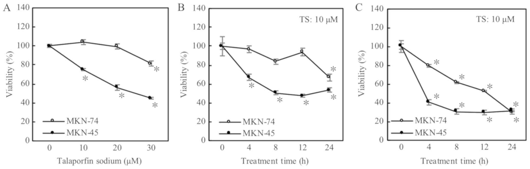
As shown in Fig. 1A,
the effect of TS-PDT at 2.53 J/cm2 on MKN74 cells was
less than on MKN45; cellular viabilities were not decreased within
20 µM of TS concentration. Using higher concentration (30 µM) of
TS, MKN74 cells showed enhancement of the effect (Fig. 1A). In the longest treatment time of
24 h at 10 µM of TS, cellular viability was decreased significantly
(Fig. 1B). In addition, when
irradiation power of LED raised up to 5.06 J/cm2 at 10
µM of TS, TS-PDT induced the decrease in cellular viabilities even
on MKN74 cells (Fig. 1C). In
summary, MKN74 cells seemed rather resistant to TS-PDT compared to
MKN45 cells.
LDL receptor is associated with uptake
of TS
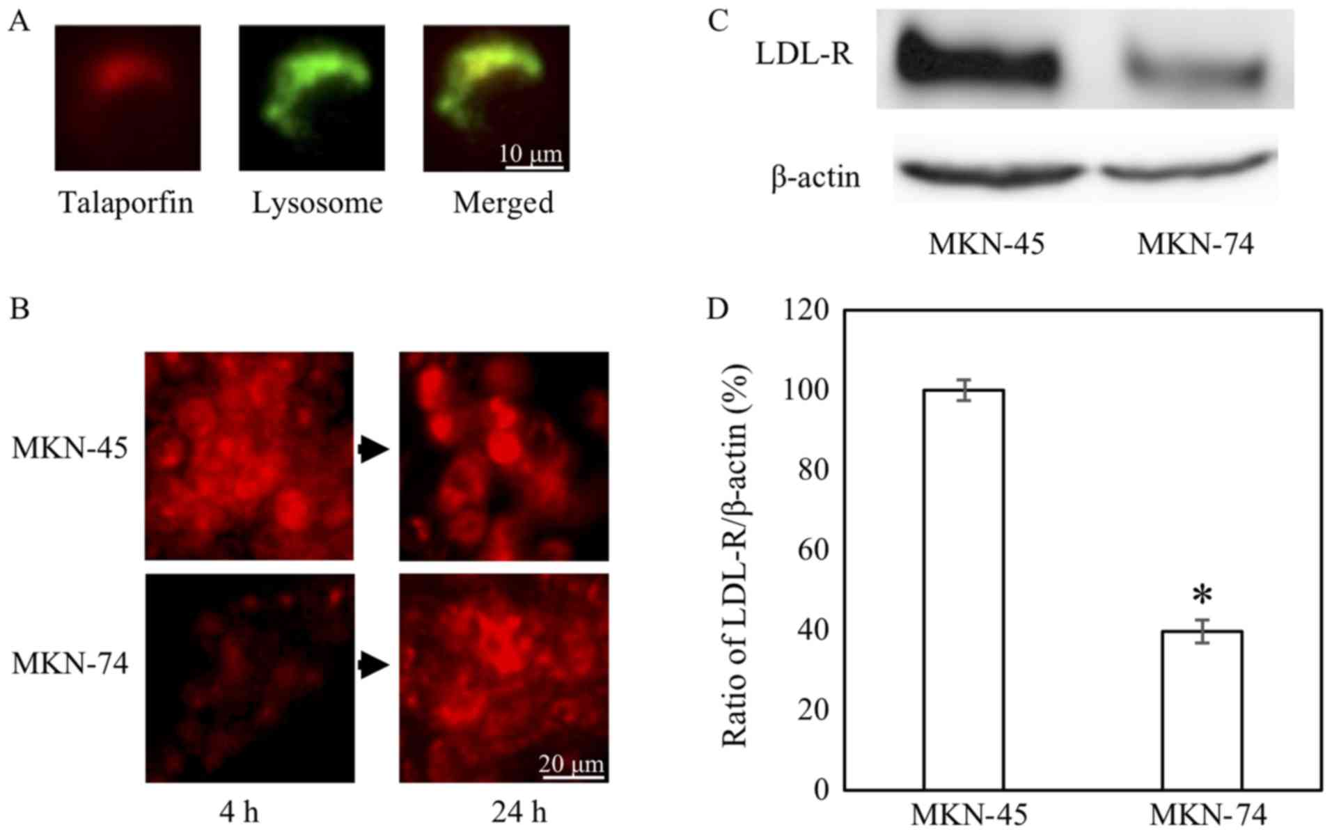
As shown with longer treatment time with TS, the
difference in the effects between MKN45 and MKN74 cells could be
due to the ability of cellular uptake of TS. Considering the
correspondence of location and their mergence in color, similar to
LDL intracellular movement (11), TS
was carried into the lysosome (Fig.
2A). In fact, uptake of TS in MKN74 cells at 4 h was lower than
MKN45 cells at this particular time-point (Fig. 2B). The uptake tended to be increased
at the time-point of 24 h. In addition, we confirmed that the
expression levels of LDL receptor protein and mRNA in MKN74 cells
were lower than MKN45 cells, respectively (Fig. 2C and D).
Decreased LDL receptor by GW3965
induces PDT-resistant
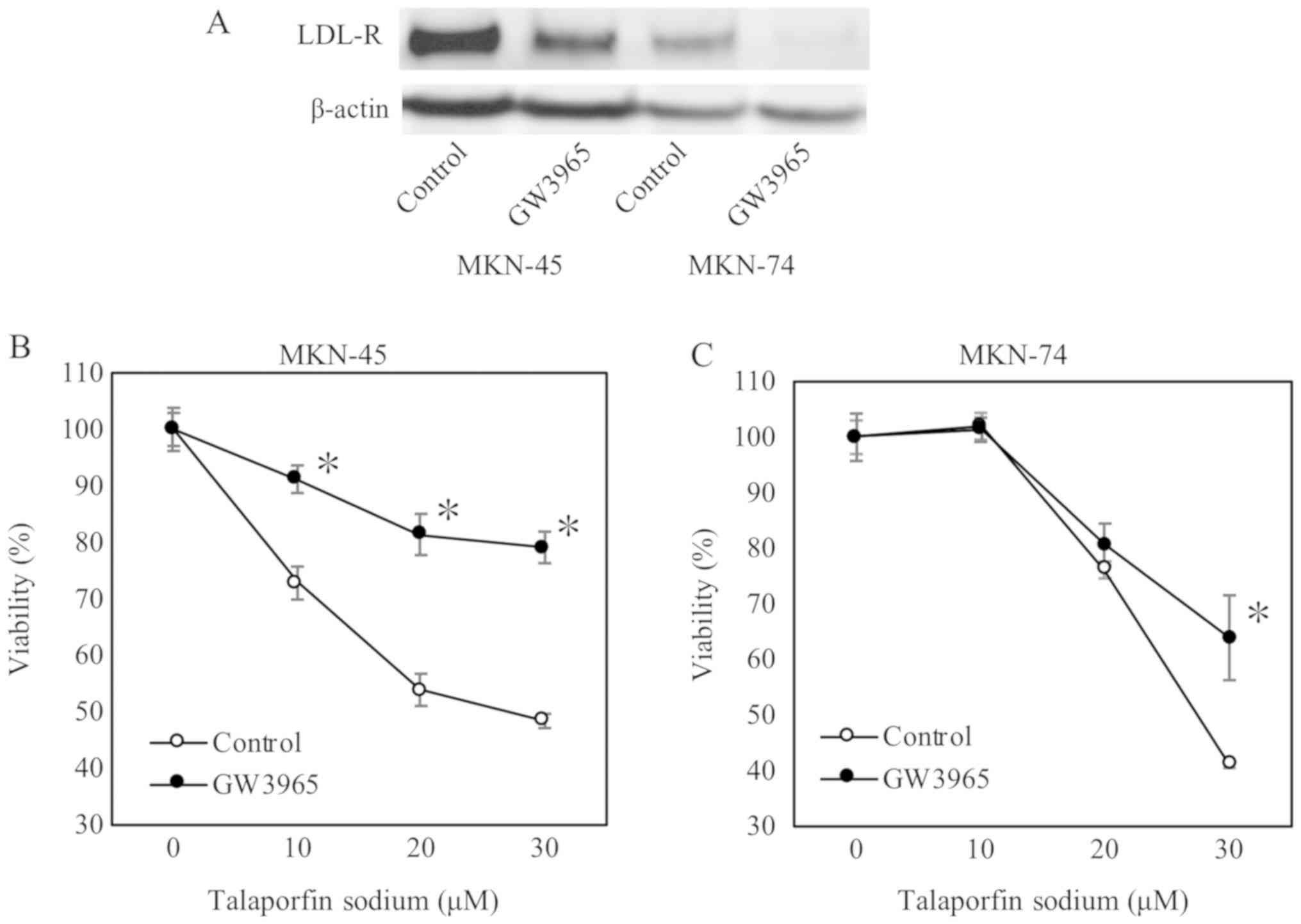
Furthermore, we used GW3965 to confirm whether LDL
receptor could be related to uptake of TS. As expected, LDL
receptor expression was reduced by GW3965 treatment as shown in
Fig. 3A. Subsequently, GW3965
treatment significantly increased cellular viabilities on MKN45
cells (from 10 to 30 µM concentration of TS) and MKN74 cells at 30
µM TS (Fig. 3B and C).
Increased LDL receptor by simvastatin
enhances efficacy of PDT
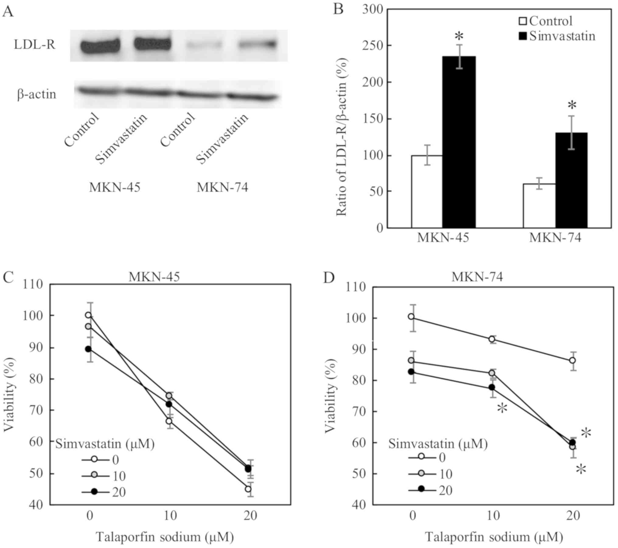
When these cells were treated with simvastatin at 20
µM, both protein and mRNA expression levels of LDL receptor were
substantially increased (Fig. 4A and
B, respectively). Simvastatin significantly decreased cellular
viabilities, and enhanced the PDT effects, on MKN74 cells (Fig. 4D). On the other hand, simvastatin did
not affect cellular viabilities on MKN45 cells as shown in Fig. 4C.
Discussion
In this paper, we elucidated that LDL receptor
expression is involved in the effect with TS-PDT, and simvastatin
enhances the therapeutic effect on TS-PDT resistant cell.
PDT has been shown to be a safe and effective
treatment for early gastric cancer, not only for the intramucosal
type, but also for the submucosal invasion. PDT using the
excimer-dye laser and Photofrin® (porfimer sodium, the
first-generation photosensitizer) can be an endoscopic treatment
option for gastric cancer particularly in a rapidly aging society
like Japan (2). In clinical
practice, PDT is administered at 40 mg/m2 of TS per
person and 100 J/cm2 of laser light. This concentration
is similar to that in in vitro conditions. However, the LED
power is different. This is because we need to eradicate cancer
cells in clinical practice completely, but need fewer effective
conditions for evaluating reagents in vitro. In addition,
cells are cultured in a monolayer fashion in vitro.
Therefore, we think that we need less LED power in in vitro
system compared to that in clinical practice.
As PDT using TS on gastric cancer is yet to be
approved by MHLW in Japan, and currently is not available in
clinical settings, therefore we investigated the effects of TS-PDT
on gastric cancer cell. In the present study, MKN74 cells were
shown to be resistant to TS-PDT. There were substantial differences
in expression levels of LDL receptor between MKN74 cells and
TS-PDT-sensitive MKN45 cells. TS was localized in the lysosome
possibly after being taken by LDL receptor. Downregulation of LDL
receptor by GW3965 significantly decreased the effect of TS-PDT in
gastric cancer cells. Furthermore, LDL receptor expression was
decreased by siRNA knockdown (Fig.
S1). The viability was increased from 52.0±6.25 to 56.8±5.11%
for MKN45 cells (n=6). We were able to observe an up-trend of
viability although not statistically significant while LDL receptor
expression was decreased by siRNA. The effect of GW3965 might have
been caused by other molecules and receptor contributing to the
resistance. On the other hand, simvastatin-mediated LDL receptor
upregulation enhanced the effect of PDT in MKN74 cells but not in
MKN45 cells, in which LDL receptor could be sufficiently expressed.
Therefore, the difference of LDL receptor expression between these
two gastric cell lines could affect TS-PDT efficacy.
In our experiments, simvastatin did not have such a
dramatic effect on TS-PDT low-sensitive MKN74 cells. Firstly, in
regard of the uptake of TS, there could be expressing several other
receptors including albumin receptor (12) and heme-carrier protein-1 (13) in addition to LDL receptor (6). Although LDL receptor could be used for
uptake of TS in the gastric cancers, upregulation by simvastatin
gave just the partial effect of TS-PDT. Secondly, recent research
documented that simvastatin itself has an antitumor effect
(14), therefore, we did not use
higher concentration of simvastatin. In contrast, simvastatin at
moderately higher concentrations decreased the viability of MKN74
cells in the TS-PDT in vitro model (Fig. 4D). However, by adding simvastatin to
TS-PDT in an in vitro system, we could observe that TS-PDT
was effective even on resistant cells. The viability (60%) was
close to the equivalent with TS-PDT sensitive cell (50%). This
datum shows the possibility of combining TS-PDT and simvastatin in
clinical practice. Under the criteria of approval by Japanese
Universal Health Insurance Coverage System, the efficacy of PDT
with Photofrin® for gastric cancer with superficial
early gastric cancer was good, with 42 patients (73.7%) out of 57
patients (70 lesions) showed a complete response (Fulfills both 1
and 2). i) No residual tumor at the original lesion examined
endoscopically. ii) Biopsy specimen shows no carcinoma cells)
(2). In the resistant residual or
remnant cases despite PDT treatments, simvastatin or similar
derivatives might show enhanced/additive effects of PDT, paving the
way for novel combination therapy.
On the other hand, remaining cancer stem cells could
cause a relapse after anticancer drugs and radiation therapy
(15). In fact, TS-PDT was rather
effective for local failure after CRT or RT alone against
superficially localized esophageal cancer (16). TS-PDT might be rather effective for
aggressive gastric cancer with cancer stem cell appearance,
considering the origin of these cell lines; MKN45 cell line was
derived from undifferentiated-type gastric cancer, whereas MKN74
cell line was established from differentiated-type gastric cancer.
ESD is currently in widespread use for early gastric cancer
(17). However, when tumors invade
the submucosa or have potential risks of metastatic malignancy,
additional therapies including surgery would be necessary following
ESD (18). With a rapidly aging
population in Japan, additional surgical intervention is still
debatable in particular for elderly patients with high risk
comorbidities, suggesting an urgent need for a safer and more
efficient therapy in gastric cancer. Therefore, further studies are
necessary to evaluate underlying mechanisms of PDT with TS.
In conclusion, LDL receptor expression is involved
in the efficacy of TS-PDT. Therefore, simvastatin has the
possibility to enhance the effects of TS-PDT as a novel combination
therapy.
Supplementary Material
Supporting Data
Acknowledgements
Not applicable.
Funding
No funding was received.
Availability of data and materials
All data generated or analyzed during this study are
included in this published article.
Authors' contributions
TK and TSu acquired data, analyzed and interpreted
the data, and drafted the manuscript. TT also analyzed and
interpreted the data. YM, HKi, TSa, TH and HKu assisted in
acquiring the data. YI made contributions to the conception and
design of the study. TM established MKN45-Luc and MKN74/CMV-Luc
cells. HI made substantial contributions to the conception and
design of the study and drafted the manuscript.
Ethics approval and consent to
participate
Not applicable.
Patient consent for publication
Not applicable.
Competing interests
The authors declare that they have no competing
interests.
References
|
1
|
Shafirstein G, Bellnier D, Oakley E,
Hamilton S, Potasek M, Beeson K and Parilov E: Interstitial
photodynamic therapy-a focused review. Cancers (Basel). 9(pii):
E122017. View Article : Google Scholar : PubMed/NCBI
|
|
2
|
Oinuma T, Nakamura T and Nishiwaki Y:
Report on the National survey of photodynamic therapy (PDT) for
gastric cancer in Japan (a secondary publication). Laser Ther.
25:87–98. 2016. View Article : Google Scholar : PubMed/NCBI
|
|
3
|
Isomoto H, Shikuwa S, Yamaguchi N, Fukuda
E, Ikeda K, Nishiyama H, Ohnita K, Mizuta Y, Shiozawa J and Kohno
S: Endoscopic submucosal dissection for early gastric cancer: A
large-scale feasibility study. Gut. 58:331–336. 2009. View Article : Google Scholar : PubMed/NCBI
|
|
4
|
Yano T, Kasai H, Horimatsu T, Yoshimura K,
Teramukai S, Morita S, Tada H, Yamamoto Y, Kataoka H, Kakushima N,
et al: A multicenter phase II study of salvage photodynamic therapy
using talaporfin sodium (ME2906) and a diode laser (PNL6405EPG) for
local failure after chemoradiotherapy or radiotherapy for
esophageal cancer. Oncotarget. 8:22135–22144. 2017. View Article : Google Scholar : PubMed/NCBI
|
|
5
|
Matsumoto J, Suzuki K, Yasuda M, Yamaguchi
Y, Hishikawa Y, Imamura N and Nanashima A: Photodynamic therapy of
human biliary cancer cell line using combination of phosphorus
porphyrins and light emitting diode. Bioorg Med Chem. 25:6536–6541.
2017. View Article : Google Scholar : PubMed/NCBI
|
|
6
|
Shibata Y, Matsumura A, Yoshida F,
Yamamoto T, Nakai K, Nose T, Sakata I and Nakajima S: Competitive
uptake of porphyrin and LDL via the LDL receptor in glioma cell
lines: Flow cytometric analysis. Cancer Lett. 166:79–87. 2001.
View Article : Google Scholar : PubMed/NCBI
|
|
7
|
Gabbi C, Warner M and Gustafsson JÅ:
Action mechanisms of liver X receptors. Biochem Biophys Res Commun.
446:647–650. 2014. View Article : Google Scholar : PubMed/NCBI
|
|
8
|
Zelcer N, Hong C, Boyadjian R and Tontonoz
P: LXR regulates cholesterol uptake through idol-dependent
ubiquitination of the LDL receptor. Science. 325:100–104. 2009.
View Article : Google Scholar : PubMed/NCBI
|
|
9
|
Bard JM, Luc G, Douste-Blazy P, Drouin P,
Ziegler O, Jacotot B, Dachet C, De Gennes JL and Fruchart JC:
Effect of simvastatin on plasma lipids, apolipoproteins and
lipoprotein particles in patients with primary
hypercholesterolaemia. Eur J Clin Pharmacol. 37:545–550. 1989.
View Article : Google Scholar : PubMed/NCBI
|
|
10
|
Livak KJ and Schmittgen TD: Analysis of
relative gene expression data using real-time quantitative PCR and
the 2(-Delta Delta C(T)) method. Methods. 25:402–408. 2001.
View Article : Google Scholar : PubMed/NCBI
|
|
11
|
Underwood KW, Jacobs NL, Howley A and
Liscum L: Evidence for a cholesterol transport pathway from
lysosomes to endoplasmic reticulum that is independent of the
plasma membrane. J Biol Chem. 273:4266–4274. 1998. View Article : Google Scholar : PubMed/NCBI
|
|
12
|
Ogawa E, Motohashi S, Ito A and Arai T:
Effects of albumin binding on photocytotoxicity of extracellular
photosensitization reaction using talaporfin sodium to rat
myocardial cells. Photodiagnosis Photodyn Ther. 12:252–257. 2015.
View Article : Google Scholar : PubMed/NCBI
|
|
13
|
Cho MR, Han JH, Lee HJ, Park YK and Kang
MH: Mitochondrial reactive oxygen species accelerate the expression
of heme carrier protein 1 and enhance photodynamic cancer therapy
effect. J Clin Biochem Nutr. 56:49–56. 2015. View Article : Google Scholar : PubMed/NCBI
|
|
14
|
Pavan LM, Rêgo DF, Elias ST, De Luca Canto
G and Guerra EN: In vitro antitumor effects of statins on head and
neck squamous cell carcinoma: A systematic review. PLoS One.
10:e01304762015. View Article : Google Scholar : PubMed/NCBI
|
|
15
|
Dragu DL, Necula LG, Bleotu C, Diaconu CC
and Chivu-Economescu M: Therapies targeting cancer stem cells:
Current trends and future challenges. World J Stem Cells.
7:1185–1201. 2015.PubMed/NCBI
|
|
16
|
Nagai K, Muto M, Ezoe Y, Yamamoto T, Niimi
M, Yoshimura K, Yoda Y, Yano T, Higashino K, Iishi H, et al: A
phase I study of salvage photodynamic therapy (PDT) using
talaporfin sodium and a diode laser for local failure of esophageal
carcinoma (EC) after chemoradiotheapy (CRT). J Clin Oncol. 1:1–7.
2012.
|
|
17
|
Iizuka T, Kikuchi D, Hoteya S, Kajiyama Y
and Kaise M: Efficacy and safety of endoscopic submucosal
dissection for superficial cancer of the cervical esophagus. Endosc
Int Open. 5:E736–E741. 2017. View Article : Google Scholar : PubMed/NCBI
|
|
18
|
Ikeda A, Hoshi N, Yoshizaki T, Fujishima
Y, Ishida T, Morita Y, Ejima Y, Toyonaga T, Kakechi Y, Yokosaki H
and Azuma T: Endoscopic submucosal dissection (ESD) with additional
therapy for superficial esophageal cancer with submucosal invasion.
Intern Med. 54:2803–2813. 2015. View Article : Google Scholar : PubMed/NCBI
|