Introduction
Endometrial carcinoma is the most common gynecologic
malignancy worldwide; moreover, in Japan, there has been a
continual annual increase in its incidence, which is associated
with lifestyle changes (1).
Diagnosis at an early stage leads to high survival rates, whereas
patients diagnosed with advanced-stage or recurrent disease have a
poor prognosis (2). Thus,
considerable research attention has focused on identifying specific
markers for early-stage endometrial carcinoma (3).
Ki-67 is a widely used proliferation marker and
prognostic factor in cancer (4),
since Ki-67 antibodies recognize a nuclear protein that is
expressed only in proliferating cells (5). Moreover, the Ki-67-positive population
is detected mainly among proliferating cells in diverse cell types
during the active phases of the cell cycle (G1, S, G2, and M
phases), and is absent among cells in the resting/quiescent phase
(G0 phase) (5,6). Ki-67 expression is strongly associated
with cell proliferation and tumor growth and is thus widely used in
routine pathological investigations as a proliferation marker;
moreover, Ki-67 is well characterized at the molecular level and is
extensively used as a prognostic and predictive marker for cancer
diagnosis and treatment (7). Ki-67
has also been recognized as a potential prognostic biomarker in
endometrial carcinoma (8–10), and is increasingly used in
presurgical studies of endometrial cancer as a primary outcome
measure; however, unlike its use in breast cancer, there are no
guidelines for standardizing its measurement and the clinical
relevance of Ki-67 as a biomarker in endometrial cancer remains
undetermined (11).
Our previous study on estrogen receptor
(ER)-transfected endometrial cancer cells suggested that the
stimulatory effect of estrogen on cell proliferation is exerted
through the increased expression of cyclin D1 and cyclin A
(12). We have also investigated
various cyclins as prognostic indicators using clinical specimens
of endometrial cancer, which revealed an association of cyclin A
expression with progression to malignancy and a correlation with
the proliferative activity and prognostic features, including
histological grade (13).
Moreover, we found that cyclin A and p53 are both
expressed in cells obtained from patients with endometrial
carcinoma at more advanced clinical stages, using liquid-based
cytology (14). In other studies,
the expression of cyclins D1 and E was significantly correlated
with the histological grade of endometrial cancer, but not with
other clinicopathological parameters (15,16).
Among cyclins, cyclin A is most strongly associated with DNA
replication, as the complex of cyclin A with CDK2 induces the G1/S
transition (17). Accordingly, we
selected cyclin A as a proliferation marker candidate.
Notably, several other studies have also addressed
the clinical significance of elevated cyclin A expression in
endometrial carcinoma (18). For
example, cyclin A has been identified as an independent prognostic
factor in endometrial endometrioid adenocarcinoma, and its
expression correlates with the cancer grade and, to a lesser
degree, with the International Federation of Gynecology and
Obstetrics (FIGO) stage (19).
Additionally, a multivariate analysis showed that high expression
of cyclin A is linked to poor prognosis for patients at advanced
stages of endometrial cancer, indicating the suitability of cyclin
A expression as a prognostic factor for endometrial cancer
(20).
Cyclin A overexpression has been reported in several
other types of cancer, demonstrating prognostic value (i.e., a poor
prognosis), such as in the prediction of survival or early relapse,
and it has also been correlated with carcinogenesis (21–24).
Furthermore, high expression of cyclin A in endometrial carcinoma
has been widely linked to tumor carcinogenesis, progression, and
prognosis prediction. However, a consensus has not yet been reached
regarding the clinical significance of these findings.
As described above, cyclin A regulates the cell
cycle and is overexpressed in cancer cells. Moreover, it has been
reported as a prognostic/predictive factor in endometrial tissues
(18–20). However, only a few studies have
directly addressed the relationship between cyclin A and cell
proliferation (25), and cyclin A
has not yet been established as a proliferation marker.
Accordingly, the aim of the present study was to
clarify the value of cyclin A as a marker of the cell proliferation
ability in endometrioid carcinoma. For this purpose, we used two
distinct types of differentiated endometrial cancer cell lines,
Ishikawa and HEC-50B cells, derived from low-grade and high-grade
endometrial carcinomas, respectively. We compared the expression of
cyclin A with that of Ki-67 in both cell lines to clarify the
usefulness of cyclin A expression as a marker of cell proliferation
in endometrial cancer. Specifically, we used flow cytometry and
immunocytochemical staining to compare cyclin A and Ki-67
expression in the two endometrial cancer cell lines and we
investigated the efficacy of cyclin A as a proliferation
marker.
Materials and methods
Cell culture and cell number
count
The human endometrial cancer cell lines, Ishikawa
and HEC-50B, were kindly provided by Dr Kuramoto (Kitasato
University, Japan) and Dr Nishida (Tsukuba University, Japan).
Ishikawa cells were established from a patient with low-grade
endometrial carcinoma (grade 1), and these cells express ER and
progesterone receptor (26,27). HEC-50B cells were established from
the ascitic fluid of a patient with recurrent high-grade
endometrioid carcinoma (grade 3) (28).
The cells were cultured in RPMI-1640 medium (Thermo
Fisher Scientific, Inc., Waltham, MA, USA) supplemented with 10%
fetal bovine serum and 1% penicillin-streptomycin (Thermo Fisher
Scientific, Inc.) at 37°C in a humidified incubator containing 5%
CO2. At 1, 2, 4, 6, 8 and 10 days after seeding the
cultured cells, they were harvested with 0.05% trypsin-0.02%
ethylenediaminetetraacetic acid, and the cells from three dishes
were counted using a Burker-Turk counter plate under an inverted
phase-contrast microscope. The cell numbers were used for
generating growth curves and calculating the doubling time.
Flow cytometric analysis of cyclin A
and Ki-67
HEC-50B and Ishikawa cells were seeded in 60-mm
culture dishes at a density of 5×10⁵ cells/dish,
collected at various time points, and fixed using ice-cold 100%
methanol. Subsequently, the expression of cyclin A was analyzed
using direct-immunofluorescence flow cytometry, which was performed
through double-staining with a combination of fluorescein
isothiocyanate (FITC)-conjugated anti-human cyclin A2 mouse
antibody (clone 11B2G3) and 7-amino-actinomycin D (7-AAD); both
reagents were from Beckman Coulter (Brea, CA, USA). Ki-67 analysis
was performed through single staining with an anti-mouse Ki-67
monoclonal antibody (clone PP-67, 1:100) and anti-mouse IgG/FITC
(1:200); both antibodies were from Abcam (Tokyo, Japan). Cell
suspensions were incubated with anti-cyclin A2 and 7-AAD for 20 min
at room temperature (20-25°C) and washed; suspensions were then
incubated with anti-Ki-67 for 20 min at room temperature and
washed. Lastly, the cells were incubated with anti-mouse IgG/FITC
for 20 min at room temperature and then washed. The percentage of
cells in each cell-cycle phase was determined through flow
cytometry performed on a Cytomics FC 500 system (Beckman Coulter).
In each experiment, an isotype-matched irrelevant mouse antibody
was used as a negative control.
Immunocytochemical staining of cyclin
A and Ki-67
Harvested cells were resuspended in
phosphate-buffered saline and cytocentrifuged at 1,500 rpm for 5
min using an Auto Smear (CF-120; Sakura, Tokyo, Japan). The cells
on the glass slides were immersed in 95% ethanol, and
immunocytochemical staining was performed using an
EnVision™ detection system (DakoCytomation A/S,
Glostrup, Denmark) according to the manufacturer's recommendations.
Endogenous peroxidases in the specimens were blocked with
Peroxidase-Blocking Solution (DakoCytomation A/S) for 5 min at room
temperature, and after incubation with Protein Block Serum-Free
reagent (DakoCytomation A/S), the following primary antibodies were
applied: anti-mouse Ki-67 monoclonal antibody (clone PP-67, 1:200;
Abcam) and anti-mouse cyclin A2 monoclonal antibody (clone 6E6,
1:15; Abcam). Following incubation for 90 min at room temperature
and washing, the cells on the slides were incubated with
EnVision™/HRP Rabbit/Mouse secondary antibodies (DakoCytomation
A/S) for 30 min at room temperature, and then with the chromogen,
3,3′-diaminobenzidine. Nuclei were counterstained using Mayer's
hematoxylin (Wako Pure Chemical Industries, Ltd., Osaka, Japan).
The specimens were examined and photographed at ×200 magnification
using a digital microscope camera (Olympus AX80 DP21; Olympus,
Tokyo, Japan) interfaced with a computer. Cyclin A and Ki-67
expression was evaluated using the nuclear labeling index (%),
recorded as the percentage of positively stained nuclei in 100
cells in the hot spot.
Double immunofluorescence staining of
cyclin A and Ki-67
Specimens were prepared according to the methodology
given in the immunocytochemical staining section above. The primary
antibodies (anti-mouse cyclin A monoclonal antibody (clone 6E6,
1:15; Abcam) and anti-rabbit Ki-67 polyclonal antibody (1:100;
Abcam) were applied, incubated for 90 min at room temperature, and
washed. Then, the cells on the slides were incubated with
FITC-labeled anti-mouse IgG and tetramethylrhodamine
(TRITC)-labeled anti-rabbit IgG (1:40; Abcam) for 30 min at room
temperature. Cell nuclei were counterstained with
4′,6-diamidino-2-phenylindole (DAPI). Immediately after treatment,
the cells were observed under a fluorescence microscope (Bz-x700;
Keyence Corporation, Tokyo, Japan).
Statistical analysis
All statistical tests were conducted using BellCurve
for Excel ver. 2.15 software (Social Survey Research Information,
Tokyo, Japan). For multiple comparison of more than three
experimental groups ANOVA analysis with the Tukey post hoc test
were performed (Figs. 1 and 2), while t-tests were used for comparisons
between two groups (Fig. 3).
P<0.05 was considered to indicate statistical significance. Data
are presented as mean percentages of positive cells ± standard
deviation.
Results
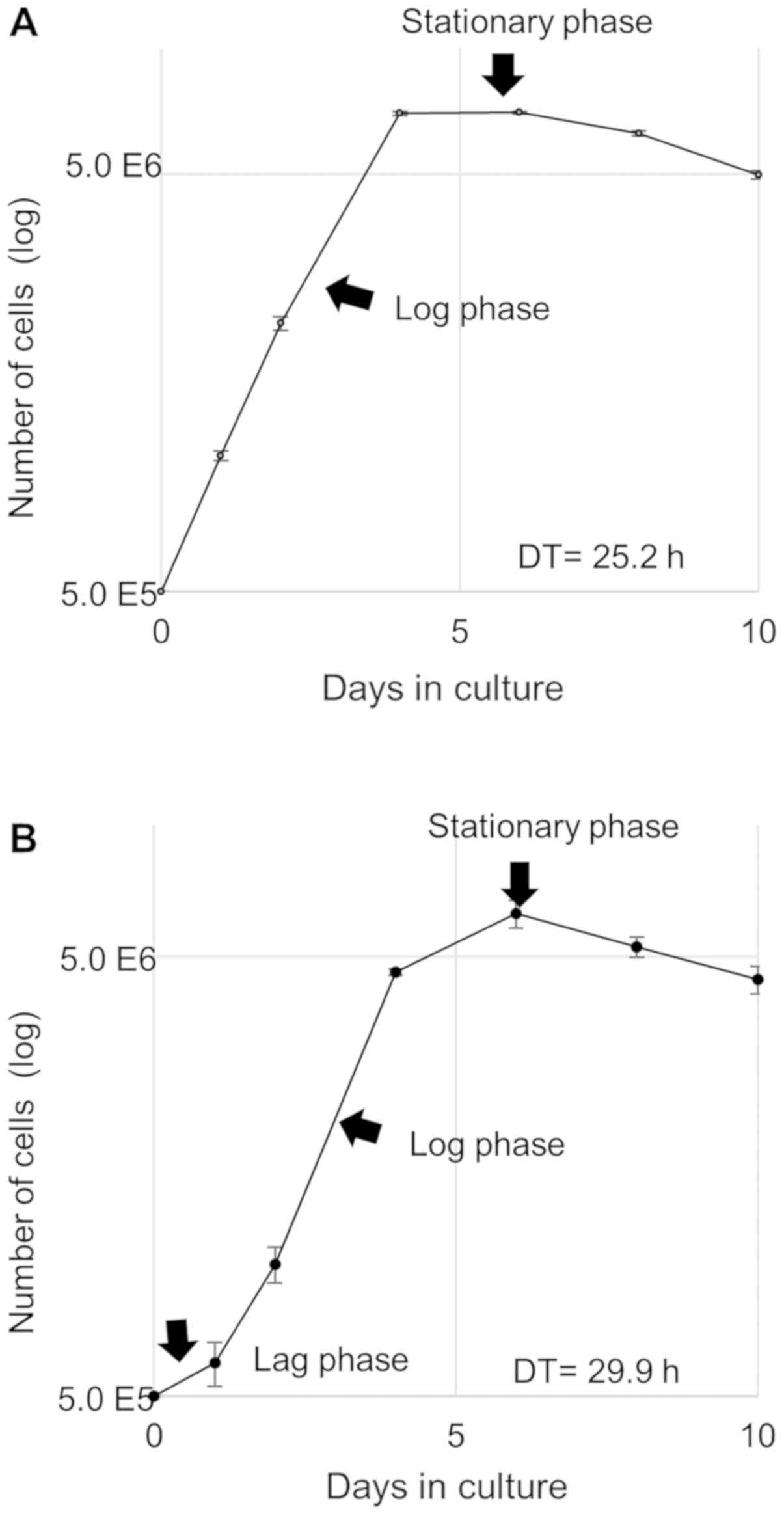
Cell growth curves
To validate the difference in proliferative ability
between the two cell lines, we first constructed cell growth curves
for HEC-50B (Fig. 4A) and Ishikawa
cells (Fig. 4B). The log-phase
growth of HEC-50B cells lasted for approximately four days (from
day 0 to day 4) and was followed by a stationary phase. In
contrast, the lag phase of the Ishikawa cells lasted for one day,
and the log phase lasted for three days (from day 1 to day 4); the
cell number then continued to increase until day 6, after which it
decreased. Calculation of the doubling time from the log phase in
the growth curve revealed that the doubling time was shorter for
HEC-50B cells (25.2 h) than for Ishikawa cells (29.9 h), thereby
confirming the greater proliferative ability of the high-grade cell
line.
Flow cytometric analysis of the cell
cycle
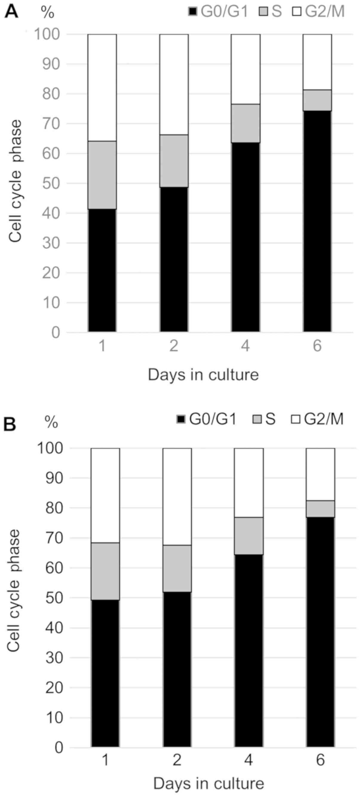
For cell cycle analysis, the cellular DNA content
was monitored by performing flow cytometry on cells stained with
7-AAD (Fig. 5). The cellular DNA
content frequency histograms revealed the cell distribution in the
three major phases of the cell cycle (G0/G1, S and G2/M). Both cell
lines exhibited a reduction in the percentage of cells in the S and
G2/M phases and an increase in that in the G0/G1 phase as the
culture time increased.
Expression of cyclin A and Ki-67
evaluated using flow cytometry
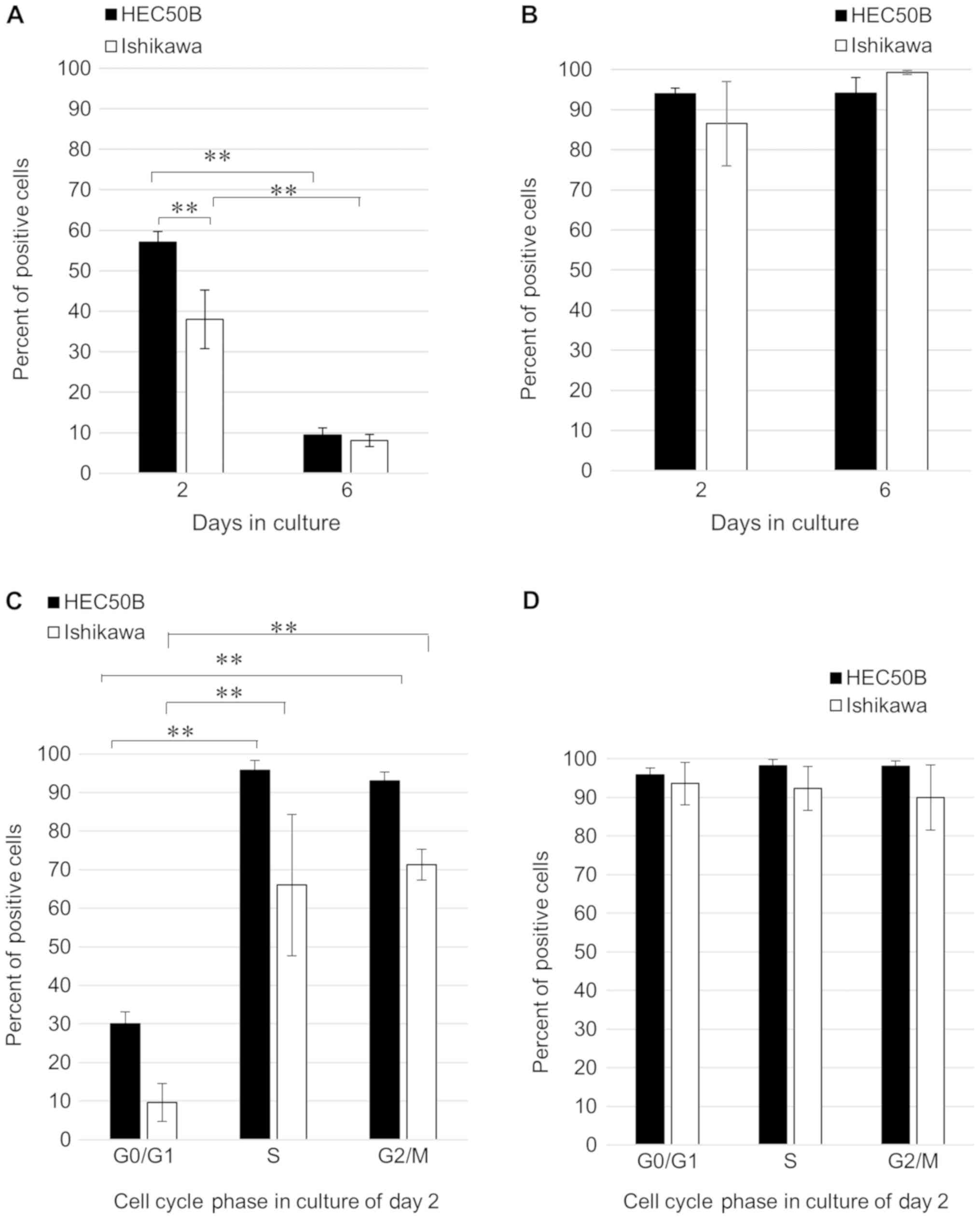
The flow cytometry results confirmed the expression
of cyclin A and Ki-67 in both HEC-50B and Ishikawa cells. For this
analysis, the cells were collected on the second (logarithmic
growth phase) and sixth day (stationary phase) of culture. Cyclin A
expression was significantly higher in HEC-50B cells than in
Ishikawa cells on day 2 (P=0.0017) and was consistently higher on
day 2 than on day 6 in both cell lines (P<0.001; Fig. 1A). Conversely, no remarkable
differences in the expression of Ki-67 were observed between Hec50B
and Ishikawa cells on day 2 and day 6 (Fig. 1B). Examination of cyclin A and Ki-67
expression during the cell cycle on day 2 revealed that, in both
HEC-50B and Ishikawa cells, cyclin A expression was significantly
higher in S and G2/M phases than in G0/G1 phase (P<0.001;
Fig. 1C). In contrast, Ki-67 was
expressed by roughly 90% of both the HEC-50B and Ishikawa cells and
showed no notable difference in expression in the different phases
of the cell cycle (Fig. 1D).
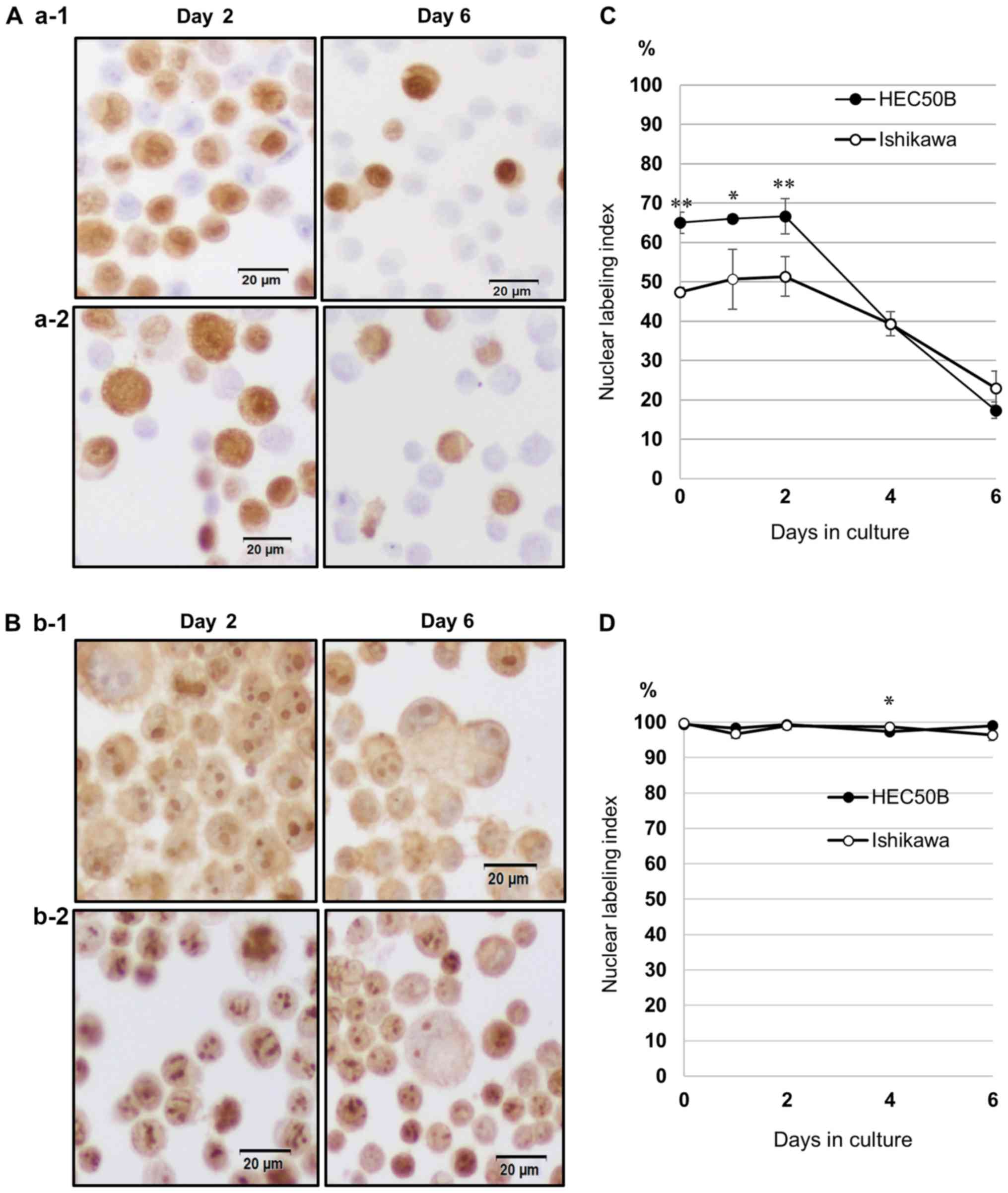
Expression of cyclin A and Ki-67
evaluated using immunocytochemical staining
Immunocytochemical staining confirmed the expression
of cyclin A (Fig. 3A) and Ki-67
(Fig. 3B) in HEC-50B and Ishikawa
cells. Both proteins were expressed mainly in the nucleus, although
they were also partially detected in the cytoplasm in the two cell
lines. Determination of the nuclear labeling index of cyclin A
(Fig. 3C) revealed that cyclin A
expression was higher in HEC-50B cells than in Ishikawa cells from
day 0 until day 2 and showed no significant difference after day 4.
Conversely, the nuclear labeling index for Ki-67 was nearly 100%,
and Ki-67 expression showed no notable difference between HEC-50B
and Ishikawa cells (Fig. 3D).
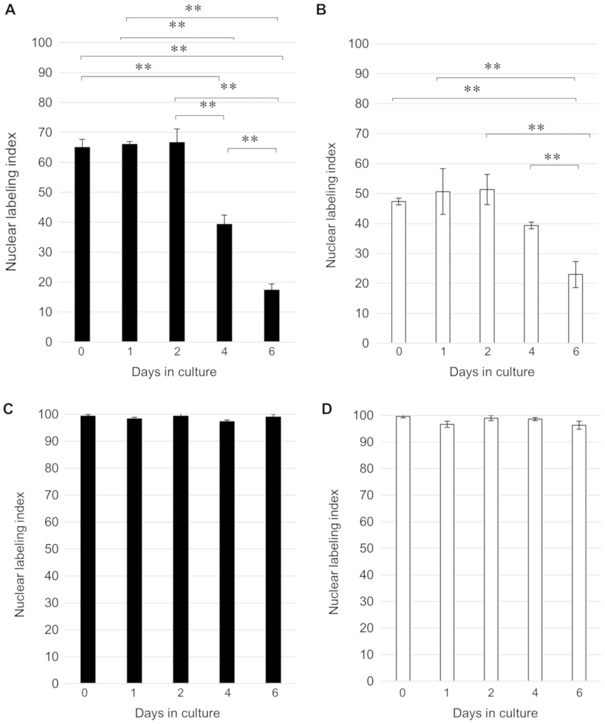
Comparison of the time courses of cyclin A and Ki-67 expression in
HEC-50B and Ishikawa cells (Fig. 2)
indicated that cyclin A expression was significantly higher on days
0–4 than on day 6 in both cell lines (P<0.001). Furthermore, we
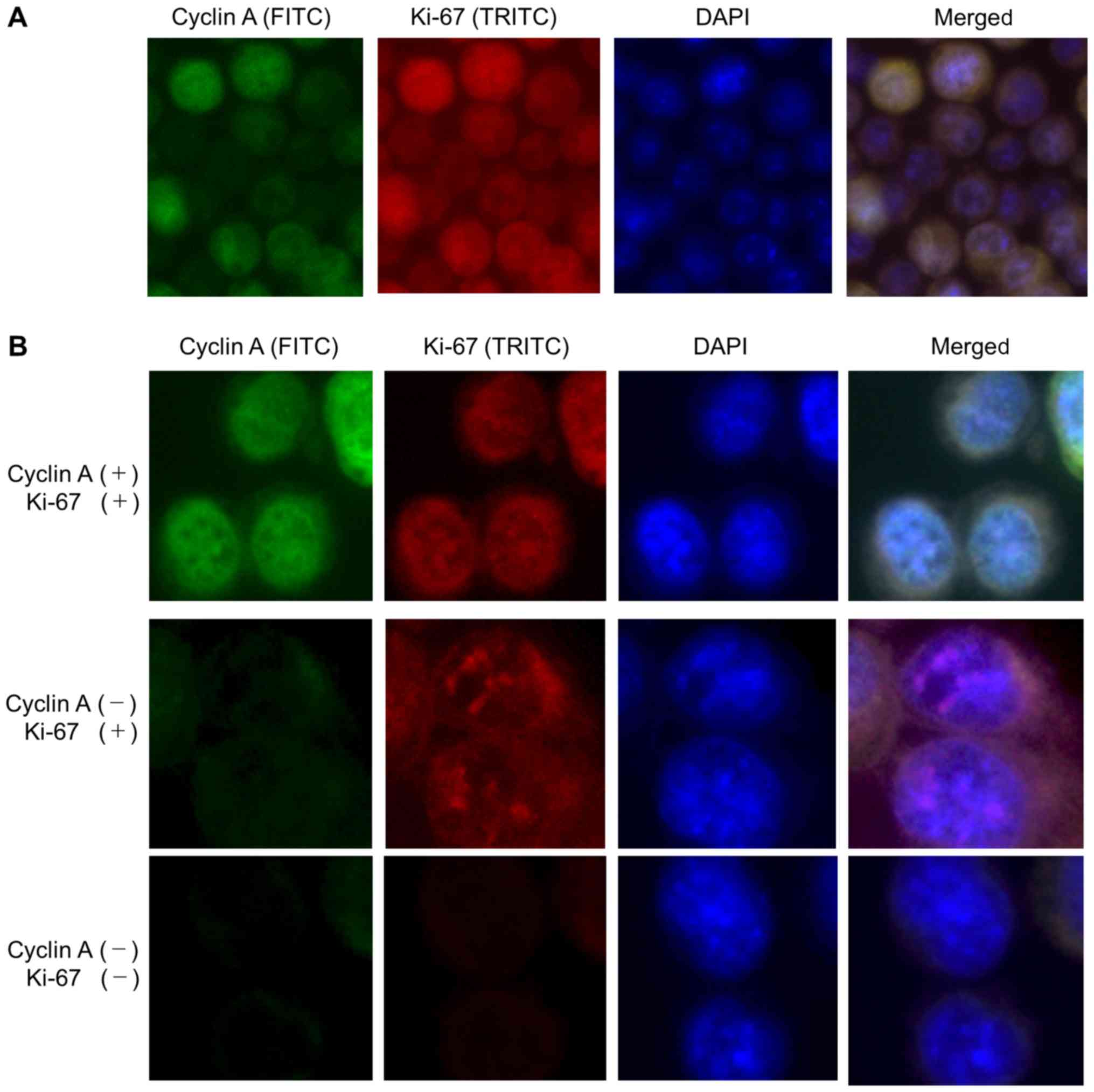
performed double immunofluorescence staining of cyclin A and Ki-67
to show different expression patterns between these two proteins
(Fig. 6). Both cyclin A and Ki-67
were expressed in the nuclei of endometrial carcinoma cells; where
nuclear cyclin A expression was observed, the expression of Ki-67
was also found.
Discussion
In this study, we used flow cytometry to analyze the
cell cycle in two endometrial cancer cell lines featuring distinct
types of differentiation. Indeed, compared to that of Ishikawa
cells, HEC-50B cells had a shorter doubling time and higher
proliferation rate, which is consistent with the fact that HEC-50B
and Ishikawa cells were derived from grade 3 and grade 1 tumors,
respectively.
Flow cytometric analysis of the cell cycle showed
that both cell lines exhibited a reduction in S and G2/M phases and
had a longer G0/G1 phase with prolongation of culture. These
results indicated that cell proliferation ceased during culture,
and that both cell lines shifted from the log phase to the
stationary phase. The S-phase fraction of cells is considered to be
the strongest predictor of survival, provided that the flow
cytometric methods have been carefully standardized (29). Thus, our results confirmed that high
cyclin A expression in the S-phase was correlated with the high
proliferation ability of endometrial cancer cell lines.
Furthermore, both flow cytometric analysis and
immunocytochemical staining revealed that cyclin A expression was
markedly higher in HEC-50B cells than in Ishikawa cells on day 2 of
culture. Cyclin A expression was also significantly higher in both
cell lines in S and G2/M phases than in G0/G1 phase. This confirms
previous studies showing that cyclin A is specifically expressed
after the S phase in endometrial carcinoma cells (18,21).
In contrast, Ki-67 was expressed by roughly 90% of
both HEC-50B and Ishikawa cells and showed no notable difference in
expression in distinct phases of the cell cycle, which is also
consistent with previous reports indicating that Ki-67 expression
can be observed in all phases of the cell cycle, except G0
(5,6).
Therefore, cyclin A expression is markedly elevated
in cells exhibiting increased proliferative ability and it was
consistently higher on day 2 than on day 6 of culture, whereas
there was no such pattern for Ki-67 expression. In fact, Ki-67
expression was higher in Ishikawa cells than in HEC-50B cells on
day 6. This is because Ishikawa cells proliferate more slowly than
HEC-50B cells, and thus continued to grow for longer periods. The
immunocytochemical staining confirmed the flow cytometry results,
showing cyclin A expression that was higher on days 0–2 than on day
6 in both cell lines, while Ki-67 expression did not differ
markedly over time. This is because the cells enter the stationary
phase and cell growth declines after four days of culture. Thus,
although Ki-67 is widely recognized as a useful cell proliferation
marker and prognostic factor in endometrial carcinoma (8–10), our
results showed no association of Ki-67 expression with changes in
the cell cycle or established variations in differentiation between
the cell lines. These differences confirm that cyclin A expression
is more relevant than Ki-67 expression in endometrial carcinoma
cells for detecting cells with increased proliferative ability.
Furthermore, in double immunofluorescence staining, many
Ki-67-expressing cells were observed and a subset of these was
cyclin A positive. There is a strong possibility that cyclin A has
proliferative activity.
Several studies have addressed the usefulness of
cyclin A expression in endometrial carcinoma tissues as a
prognostic factor using immunohistochemical staining (19,20). We
showed that cyclin A expression markedly increases depending on the
cellular state, and that cyclin A is a more accurate indicator of
elevated proliferation ability in endometrial cancer cell lines
than Ki-67. To the best of our knowledge, this is the first study
focused on cyclin A expression as a proliferation marker in this
context. Thus, cyclin A expression could serve as a biomarker for
the proliferation of endometrial cancer cells to provide more
accurate prognostic predictions.
Acknowledgements
Not applicable.
Funding
This research was partially supported by a Hirosaki
University Institutional Research Grant for Young Investigators,
Research Funding Granted by Hirosaki University President and SCOPE
of the Japan Ministry of Internal Affairs and Communications.
Availability of data and materials
The datasets used and/or analyzed during the current
study are available from the corresponding author on reasonable
request.
Authors' contributions
KH designed the study. HYa, KK, KT, HiY and HaY
performed the experiments and analyzed the data. KH wrote the
manuscript. JW contributed to analysis of the data. All authors
read and approved the final manuscript.
Ethics approval and consent to
participate
Not applicable.
Patient consent for publication
Not applicable.
Competing interests
The authors declare that they have no competing
interests.
References
|
1
|
Yamagami W, Nagase S, Takahashi F, Ino K,
Hachisuga T, Aoki D and Katabuchi H: Clinical statistics of
gynecologic cancers in Japan. J Gynecol Oncol. 28:e322017.
View Article : Google Scholar : PubMed/NCBI
|
|
2
|
Tran AQ and Gehrig P: Recent advances in
endometrial cancer. F1000Res. 6:812017. View Article : Google Scholar : PubMed/NCBI
|
|
3
|
Żyła MM, Wilczyński JR, Kostrzewa M,
Księżakowska-Łakoma K, Nowak M, Stachowiak G, Szyłło K and
Stetkiewicz T: The significance of markers in the diagnosis of
endometrial cancer. Prz Menopauzalny. 15:176–185. 2016.PubMed/NCBI
|
|
4
|
Salvesen HB, Iversen OE and Akslen LA:
Prognostic significance of angiogenesis and Ki-67, p53, and p21
expression: A population-based endometrial carcinoma study. J Clin
Oncol. 17:1382–1390. 1999. View Article : Google Scholar : PubMed/NCBI
|
|
5
|
Gerdes J, Lemke H, Baisch H, Wacker HH,
Schwab U and Stein H: Cell cycle analysis of a cell
proliferation-associated human nuclear antigen defined by the
monoclonal antibody Ki-67. J Immunol. 133:1710–1715.
1984.PubMed/NCBI
|
|
6
|
Schwarting R, Gerdes J, Niehus J, Jaeschke
L and Stein H: Determination of the growth fraction in cell
suspensions by flow cytometry using the monoclonal antibody Ki-67.
J Immunol Methods. 90:65–70. 1986. View Article : Google Scholar : PubMed/NCBI
|
|
7
|
Li LT, Jiang G, Chen Q and Zheng JN: Ki67
is a promising molecular target in the diagnosis of cancer
(review). Mol Med Rep. 11:1566–1572. 2015. View Article : Google Scholar : PubMed/NCBI
|
|
8
|
Kosmas K, Stamoulas M, Marouga A,
Kavantzas N, Patsouris E and Athanassiadou P: Expression of Ki-67
as proliferation biomarker in imprint smears of endometrial
carcinoma. Diagn Cytopathol. 41:212–217. 2013. View Article : Google Scholar : PubMed/NCBI
|
|
9
|
Masjeed NMA, Khandeparkar SGS, Joshi AR,
Kulkarni MM and Pandya N: Immunohistochemical study of ER, PR, Ki67
and p53 in endometrial hyperplasias and endometrial carcinomas. J
Clin Diagn Res. 11:EC31–EC34. 2017.PubMed/NCBI
|
|
10
|
Yang B, Shan B, Xue X, Wang H, Shan W,
Ning C, Zhou Q, Chen X and Luo X: Predicting lymph node metastasis
in endometrial cancer using serum CA125 combined with
immunohistochemical markers PR and Ki67, and a comparison with
other prediction models. PLoS One. 11:e01551452016. View Article : Google Scholar : PubMed/NCBI
|
|
11
|
Kitson S, Sivalingam VN, Bolton J, McVey
R, Nickkho-Amiry M, Powell ME, Leary A, Nijman HW, Nout RA, Bosse
T, et al: Ki-67 in endometrial cancer: Scoring optimization and
prognostic relevance for window studies. Mod Pathol. 30:459–468.
2017. View Article : Google Scholar : PubMed/NCBI
|
|
12
|
Watanabe J, Kamata Y, Seo N, Okayasu I and
Kuramoto H: Stimulatory effect of estrogen on the growth of
endometrial cancer cells is regulated by cell-cycle regulators. J
Steroid Biochem Mol Biol. 107:163–171. 2007. View Article : Google Scholar : PubMed/NCBI
|
|
13
|
Kyushima N, Watanabe J, Hata H, Jobo T,
Kameya T and Kuramoto H: Expression of cyclin A in endometrial
adenocarcinoma and its correlation with proliferative activity and
clinicopathological variables. J Cancer Res Clin Oncol.
128:307–312. 2002. View Article : Google Scholar : PubMed/NCBI
|
|
14
|
Watanabe J, Nishimura Y, Tsunoda S,
Kawaguchi M, Okayasu I and Kuramoto H: Liquid-based preparation for
endometrial cytology-usefulness for predicting the prognosis of
endometrial carcinoma preoperatively. Cancer. 117:254–263.
2009.PubMed/NCBI
|
|
15
|
Kato N, Watanabe J, Jobo T, Nishimura Y,
Fujisawa T, Kamata Y and Kuramoto H: Immunohistochemical expression
of cyclin E in endometrial adenocarcinoma (endometrioid type) and
its clinicopathological significance. J Cancer Res Clin Oncol.
129:222–226. 2003.PubMed/NCBI
|
|
16
|
Nishimura Y, Watanabe J, Jobo T, Kato N,
Fujisawa T, Kamata Y and Kuramoto H: Cyclin D1 expression in
endometrioid-type endometrial adenocarcinoma is correlated with
histological grade and proliferative activity, but not with
prognosis. Anticancer Res. 24:2185–2191. 2004.PubMed/NCBI
|
|
17
|
Sobczak-Thepot J, Harper F, Florentin Y,
Zindy F, Brechot C and Puvion E: Localization of cyclin A at the
sites of cellular DNA replication. Exp Cell Res. 206:43–48. 1993.
View Article : Google Scholar : PubMed/NCBI
|
|
18
|
Desdouets C, Sobczak-Thépot J, Murphy M
and Bréchot C: Cyclin A: Function and expression during cell
proliferation. Prog Cell Cycle Res. 1:115–123. 1995. View Article : Google Scholar : PubMed/NCBI
|
|
19
|
Santala S, Talvensaari-Mattila A, Soini Y,
Honkavuori-Toivola M and Santala M: High expression of cyclin A is
associated with poor prognosis in endometrial endometrioid
adenocarcinoma. Tumour Biol. 35:5395–5399. 2014. View Article : Google Scholar : PubMed/NCBI
|
|
20
|
Shih HC, Shiozawa T, Kato K, Imai T,
Miyamoto T, Uchikawa J, Nikaido T and Konishi I:
Immunohistochemical expression of cyclins, cyclin-dependent
kinases, tumor-suppressor gene products, Ki-67, and sex steroid
receptors in endometrial carcinoma: Positive staining for cyclin A
as a poor prognostic indicator. Hum Pathol. 34:471–478. 2003.
View Article : Google Scholar : PubMed/NCBI
|
|
21
|
Yam CH, Fung TK and Poon RY: Cyclin A in
cell cycle control and cancer. Cell Mol Life Sci. 59:1317–1326.
2002. View Article : Google Scholar : PubMed/NCBI
|
|
22
|
Masaki T, Shiratori Y, Rengifo W, Igarashi
K, Yamagata M, Kurokohchi K, Uchida N, Miyauchi Y, Yoshiji H,
Watanabe S, et al: Cyclins and cyclin-dependent kinases:
Comparative study of hepatocellular carcinoma versus cirrhosis.
Hepatology. 37:534–543. 2003. View Article : Google Scholar : PubMed/NCBI
|
|
23
|
Husdal A, Bukholm G and Bukholm IR: The
prognostic value and overexpression of cyclin A is correlated with
gene amplification of both cyclin A and cyclin E in breast cancer
patient. Cell Oncol. 28:107–116. 2006.PubMed/NCBI
|
|
24
|
Mrena J, Wiksten JP, Kokkola A, Nordling
S, Haglund C and Ristimäki A: Prognostic significance of cyclin A
in gastric cancer. Int J Cancer. 119:1897–1901. 2006. View Article : Google Scholar : PubMed/NCBI
|
|
25
|
Paterlini P, Suberville AM, Zindy F, Melle
J, Sonnier M, Marie JP, Dreyfus F and Bréchot C: Cyclin A
expression in human hematological malignancies: A new marker of
cell proliferation. Cancer Res. 53:235–238. 1993.PubMed/NCBI
|
|
26
|
Nishida M, Kasahara K, Kaneko M, Iwasaki H
and Hayashi K: Establishment of a new human endometrial
adenocarcinoma cell line, Ishikawa cells, containing estrogen and
progesterone receptors. Nihon Sanka Fujinka Gakkai Zasshi.
37:1103–1111. 1985.(In Japanese). PubMed/NCBI
|
|
27
|
Nishida M, Kasahara K, Oki A, Satoh T,
Arai Y and Kubo T: Establishment of eighteen clones of Ishikawa
cells. Hum Cell. 9:109–116. 1996.PubMed/NCBI
|
|
28
|
Kuramoto H, Hamamo M, Nishida M, Toguchi
A, Jobo T, Suzuki M and Osanai K: Establishment of a cell line of
human endometrial carcinoma originated from ascitic fluid. Acta
Obstet Gynaecol Jpn. 28:1405–1406. 1976.
|
|
29
|
Nordström B, Strang P, Bergström R,
Nilsson S and Tribukait B: A comparison of proliferation markers
and their prognostic value for women with endometrial carcinoma.
Ki-67, proliferating cell nuclear antigen, and flow cytometric
S-phase fraction. Cancer. 78:1942–1951. 1996. View Article : Google Scholar : PubMed/NCBI
|