Introduction
The breast cancer type 1 susceptibility protein
(BRCA1) gene is a tumor suppressor gene that is ~100 kb in length
(1). BRCA1 contains 24 exons that
encode a large multi-domain protein, which consists of 1,863 amino
acids and is 220 kDa (1). The BRCA1
protein is predominantly present in the nucleus and is
phosphorylated by various kinases, including the DNA damage sensor
proteins ataxia-telangiectasia mutated (ATM), ataxia
telangiectasia, Rad3-related protein and checkpoint kinase 2
(2). As a tumor suppressor, BRCA1
serves an important role in the response to hazardous DNA damage,
including DNA double strand breaks, which are repaired by
error-free homologous recombination (2). In addition, BRCA1 interacts with a
number of proteins involved in chromatin remodeling,
transcriptional regulation and the cell cycle to maintain genome
integrity (3). In total, >500
different BRCA1 mutations have been identified throughout the
coding region and untranslated region (4). A mutation or alteration in BRCA1
results in DNA replication errors and mutations, which induce tumor
growth (2). Germline mutations in
BRCA1 and breast cancer type 2 susceptibility protein are
responsible for hereditary breast-ovarian cancer syndromes (HBOCs).
Patients with a HBOC are at an increased risk of breast, ovarian
and fallopian tube cancer, and, to a lesser extent, other cancer
types, including pancreatic, stomach, laryngeal and prostate cancer
(5). In addition, decreased
expression or loss of BRCA1 has been reported in sporadic breast
cancer and ovarian cancer (6,7). The
decrease or loss of BRCA1 expression can be explained by a mutation
of the BRCA1 gene, BRCA1 promoter hypermethylation, or
overexpression of microRNAs that target BRCA1 mRNA (8–12).
Previously, clinical studies reported an association
between a low BRCA1 expression level or BRCA1 mutation and the
incidence and prognosis of gastric cancer (13,14).
Patients with a high BRCA1 expression level demonstrate a longer
overall survival time (14–16). By contrast, patients with a
BRCA1-negative status are more likely to have a high tumor grade
according to The American Joint Committee on Cancer, a high
Tumor-Node-Metastasis (TNM) stage, or a poorly differentiated tumor
(14–17). In addition, patients with a BRCA1
single nucleotide polymorphism (SNP) were identified to possess a
predisposition for gastric cancer. In the BRCA1 coding sequence, a
rs799917 T>C SNP increases the risk of gastric cancer and this
SNP is associated with shorter overall survival and
progression-free survival times (18,19).
Platinum agents, including cisplatin and
oxaliplatin, are popular anticancer drugs in clinical practice
(20). Cisplatin exerts cytotoxic
effects by forming DNA adducts and inducing DNA lesions (20,21). The
predominant mechanism that repairs DNA adducts is the nucleotide
excision repair pathway; however, the mismatch repair pathway can
also serve a role (20). Each repair
pathway typically arrests the cell cycle and resolves the DNA
lesion; however, if the damage is excessive the cell will transduce
signals to initiate apoptosis (22).
In addition, cisplatin has the ability to deplete methionine and
cysteine-containing peptides, including glutathione, which depletes
antioxidant molecules and induces oxidative stress (23). Reactive oxygen species and nitric
oxide induce cytotoxicity via mitochondrial outer membrane
permeabilization, which promotes apoptosis via the intrinsic
pathway (24). The mechanism of
action of oxaliplatin is similar to that of cisplatin; however, it
produces fewer adducts and demonstrates a higher cytotoxicity
(25). For the treatment of gastric
cancer, platinum agents can be used as a monotherapy or in the
following combinations: Cisplatin and 5-fluorouracil (5-FU);
epirubicin, cisplatin and 5-FU; epirubicin, cisplatin and
capecitabine; mitomycin, cisplatin and 5-FU; docetaxel, cisplatin
and 5-FU; and 5-FU, leucovorin and oxaliplatin (26). A number of studies have revealed that
BRCA1-negative gastric cancer is associated with a poor prognosis
and is more sensitive to platinum-based adjuvant chemotherapy
compared with BRCA1-positive gastric cancer (15,16).
These findings indicate that patients with BRCA1-negative gastric
cancer have a longer overall survival time and improved prognosis,
which suggests an important association between BRCA1 expression
and platinum-based chemotherapy.
In summary, clinical studies have revealed an
association between BRCA1 expression and gastric cancer; however,
to the best of our knowledge, a comprehensive in vitro study
has not been performed to support this clinical observation.
Therefore, the present study investigated whether BRCA1 expression
is correlated with chemosensitivity to platinum agents, including
cisplatin and oxaliplatin, in a number of gastric cancer cell
lines. The current study revealed that the BRCA1 expression level
is variable in different types of gastric cancer and is positively
correlated with the treatment response to platinum-based
chemotherapy. This suggests that BRCA1 may serve as a therapeutic
marker to predict the effectiveness of platinum-based chemotherapy
in gastric cancer.
Materials and methods
Cell culture
The human suspension gastric cancer cell lines SNU1,
SNU5, SNU16 and SNU620 were purchased from the Korean Cell Line
Bank (Seoul, Korea) and cultured in RPMI-1640 (Welgene, Inc.,
Gyeongsan, South Korea) supplemented with 20% fetal bovine serum
(Welgene, Inc.), 1 mM sodium pyruvate (Welgene, Inc.), minimal
essential medium non-essential amino acids (Gibco; Thermo Fisher
Scientific, Inc., Waltham, MA, USA), 10 mM HEPES (Gibco; Thermo
Fisher Scientific, Inc.), 100 U/ml penicillin and 100 µg/m
streptomycin (Gibco; Thermo Fisher Scientific, Inc.). The human
adherent gastric cancer cell lines SNU216, SNU484, SNU601, AGS and
NCI-N87, and the human mixed type gastric cancer cell line KATO III
(all from the Korean Cell Line Bank) were cultured in RPMI-1640
medium supplemented with 10% fetal bovine serum, 100 U/ml
penicillin and 100 µg/ml streptomycin. The human gastric adherent
gastric cancer cell line Hs746T was obtained from the Korean Cell
Line Bank. The human normal gastric cell line HFE-145 was kindly
provided by Professor Won Sang Park (Catholic University, Seoul,
Korea) with permission from Professor Hassan Ashktorab (Howard
University, Washington, DC, USA) who had originally established the
cell line. Hs746T and HFE-145 cells were cultured in Dulbecco's
modified Eagle's medium (Welgene, Inc.) supplemented with 10% fetal
bovine serum, 100 U/ml penicillin and 100 µg/ml streptomycin. All
cells were cultured at 37°C and 5% CO2.
Cell viability assay
All gastric cancer cell lines and HFE-145 cells were
seeded at a density of 5×10³ cells/75 µl per well in a
96-well plate. Following incubation overnight, the cells were
treated with 0.000, 0.025, 0.076, 0.228, 0.685, 2.060, 6.170,
18.500, 55.500 and 167.000 µM cisplatin (Selleck Chemicals,
Houston, TX, USA) or 0.000, 0.019, 0.058, 0.173, 0.518, 1.550,
4.660, 14.000, 42.000 and 126 µM oxaliplatin (Selleck Chemicals).
Cisplatin and oxaliplatin powders were obtained, and 167 µM
cisplatin and 126 µM oxaliplatin stock solutions were prepared in
dimethyl sulfoxide (Sigma-Aldrich; Merck KGaA) and subsequently
three-fold diluted in culture medium as aforementioned. Following
48 h of treatment at 37°C, 20 µl MTT (5 mg/ml; Sigma-Aldrich; Merck
KGaA) was added. Cells were incubated for 6 h and then 150 µl
acidic isopropanol (0.04 N HCl final concentration) was added to
dissolve the formazan crystals. To quantify the viable cells, the
optical density was measured at 540 nm using an EMax microplate
reader (Molecular Devices, LLC, Sunnyvale, CA, USA).
Western blotting
All gastric cancer cell lines and HFE-145 cells were
harvested and resuspended in lysis buffer containing 0.0625 M
Tris-HCl (pH 6.8), 20% glycerol, 2% SDS and 5% b-mercaptoethanol in
distilled water. The protein concentration was measured using a
Pierce™ BCA Protein assay kit (Thermo Fisher Scientific,
Inc.), according to the manufacturer's protocol. Total protein (50
µg) was then loaded onto an 8% SDS-PAGE gel and transferred to an
Immune-Blot® polyvinylidene difluoride membrane (Bio-Rad
Laboratories, Inc., Hercules, CA, USA). The membrane was blocked
for 1 h at room temperature with TBS containing 0.1% Tween-20
(TBS-Tween; Amresco, LLC, Solon, OH, USA) and 5% skim milk powder
(Bioworld Technology, Inc., St. Louis Park, MN, USA). Following
blocking, the membrane was washed and incubated with an anti-BRCA1
mouse monoclonal antibody (catalog no. OP92; 1:1,000; EMD
Millipore, Billerica, MA, USA) in TBS-Tween containing 5% bovine
serum albumin at 4°C overnight. Following washing, the membranes
were incubated with horseradish peroxidase (HRP)-conjugated horse
anti-mouse IgG antibody (catalog no. 7076S; 1:3,000; Cell Signaling
Technology, Inc., Danvers, MA, USA) for 1 h at room temperature.
For the detection of β-tubulin, an anti-β-tubulin rabbit monoclonal
antibody (catalog no. 2128S; 1:2,000; Cell Signaling Technology,
Inc.) was used at 4°C overnight, followed by incubation with goat
anti-rabbit IgG-HRP-conjugated antibody (catalog no. 1706515;
1:3,000; Bio-Rad Laboratories, Inc.) for 1 h at room temperature.
West-Q Pico enhanced chemiluminescent solution (GenDEPOT, Barker,
TX, USA) was used to visualize the protein bands on the membrane. A
ChemiDoc XRS densitometer (Bio-Rad Laboratories, Inc.) and Quantity
One software (version 4.6.3; Bio-Rad Laboratories, Inc.) were used
to detect and quantify the protein bands.
Statistical analysis
All data are presented as the mean ± standard
deviation. Each experiment was performed a minimum of three times
and representative data were obtained. Pearson's correlation
coefficients were calculated using Microsoft Excel 2013 (Microsoft
Corporation, Redmond, WA, USA). Statistical significance was
assessed by one-way analysis of variance followed by Dunnett's
multiple comparison test. Statistical analysis was performed using
GraphPad Prism 5 (GraphPad Software Inc., La Jolla, CA, USA) or R
3.5.2 (The R Foundation for Statistical Computing, Vienna,
Austria). P<0.05 was considered to indicate a statistically
significant difference.
Results
Expression of BRCA1 at the protein
level
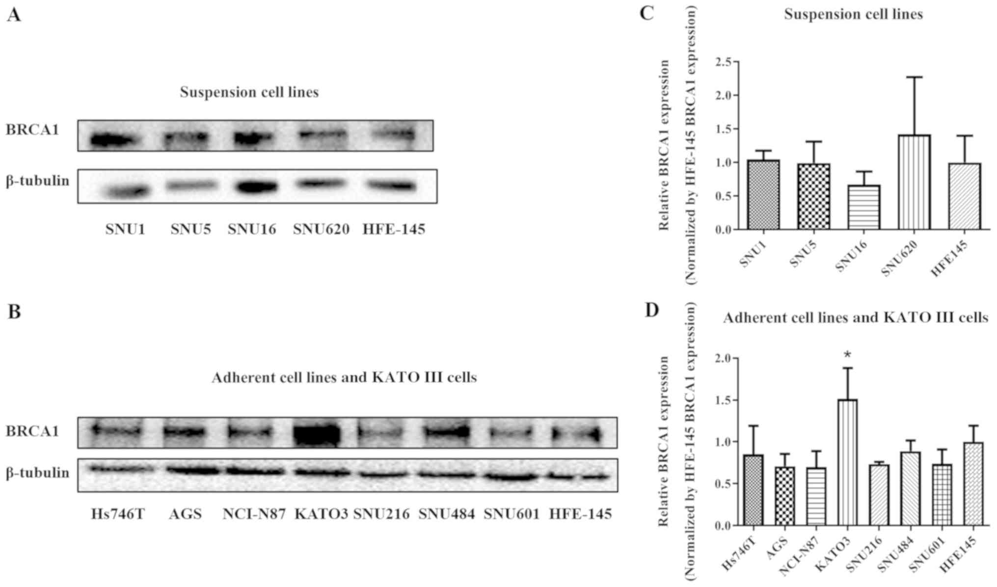
To measure BRCA1 protein expression levels in
various gastric cancer cell lines western blot analysis was
performed. The gastric cancer cell lines were categorized into
suspension cell lines and adherent cell lines, and protein
expression in these two categories was analyzed separately. KATO
III cells are mixed type gastric cancer cells, growing as mixed
adherent and suspension cultures. The BRCA1 expression in the mixed
type KATO III cells was also analyzed. As presented in Fig. 1, the BRCA1 protein expression level
varies depending on the cell type. No significant differences were
identified in the BRCA1 expression in the suspension or adherent
gastric cancer cell lines compared with the HFE-145 control cell
line. However, BRCA1 protein expression was significantly higher in
the mixed type gastric cancer KATO III cell line compared with that
in the HFE-145 cell line (P<0.05).
Effects of platinum agents against
gastric cancer cell lines
To investigate the anticancer effects of platinum
agents on gastric cancer cells, the IC50 values of
cisplatin or oxaliplatin in each cell line were measured using an
MTT assay. As presented in Tables I
and II, the IC50 values
of cisplatin or oxaliplatin depend on the type of gastric cancer
cell. Among the suspension cell lines, the IC50 values
for both platinum agents were higher in the SNU1 and SNU620 cells
compared with the normal HFE-145 gastric cell line. The
IC50 value of cisplatin was lower in SNU5 and SNU16
cells compared with HFE-145 cells; however, the IC50
value of oxaliplatin was higher in the SNU5 and SNU16 cells
compared with HFE-145 cells. Among the adherent cell lines, the
IC50 values for both platinum agents were lower in AGS,
SNU216, SNU484, SNU601 and NCI-N87 cells compared with the normal
HFE-145 cells. The IC50 value of cisplatin was lower in
Hs746T cells compared with HFE-145 cells; however, the
IC50 value of oxaliplatin was higher in Hs746T cells
compared with HFE-145 cells. The mixed type cell line KATO III was
identified to exhibit a higher resistance to both platinum agents
compared with HFE-145 cells.
 | Table I.IC50 values of cisplatin
and oxaliplatin in suspended cells and control HFE-145 cells. |
Table I.
IC50 values of cisplatin
and oxaliplatin in suspended cells and control HFE-145 cells.
| Cell line | IC50 of
cisplatin, µMa |
P-valueb | IC50 of
oxaliplatin, µMa |
P-valueb |
|---|
| SNU1 | 19.78±1.20 | 0.01770 | 23.13±4.52 | 0.01140 |
| SNU5 | 11.28±2.07 | 0.05880 | 49.2±3.44 |
1.8×10⁻⁵ |
| SNU16 | 9.66±0.47 | 0.00160 | 29.62±0.27 | 0.00024 |
| SNU620 | 25.15±1.28 | 0.00001 | 32.11±2.50 | 0.00001 |
| HFE-145 | 15.68±1.57 | – | 14.64±2.00 | – |
| Table II.IC50 values of cisplatin
and oxaliplatin in adherent cells, mixed type KATO III cells, and
control HFE-145 cells. |
Table II.
IC50 values of cisplatin
and oxaliplatin in adherent cells, mixed type KATO III cells, and
control HFE-145 cells.
| Cell line | IC50 of
cisplatin, µMa |
P-valueb | IC50 of
oxaliplatin, µMa |
P-valueb |
|---|
| Hs746T | 10.37±2.61 | 0.0329 | 15.75±0.79 | 0.90159 |
| AGS | 14.60±2.20 | 0.9794 | 9.87±0.81 | 0.00741 |
| SNU216 | 14.77±1.38 | 0.9918 | 13.43±2.02 | 0.85964 |
| SNU484 | 9.06±0.75 | 0.0071 | 13.45±0.85 | 0.87080 |
| SNU601 | 14.44±1.62 | 0.9249 | 9.11±1.72 | 0.00250 |
| NCI-N87 | 13.07±1.67 | 0.5008 | 8.07±0.76 | 0.00037 |
| KATO III | 35.68±3.37 |
7.5×10⁻¹¹ | 37.02±2.39 |
<2.0×10⁻¹⁶ |
| HFE-145 | 15.68±1.57 | – | 14.64±2.00 | – |
Correlation between BRCA1 expression
and chemosensitivity to platinum agents
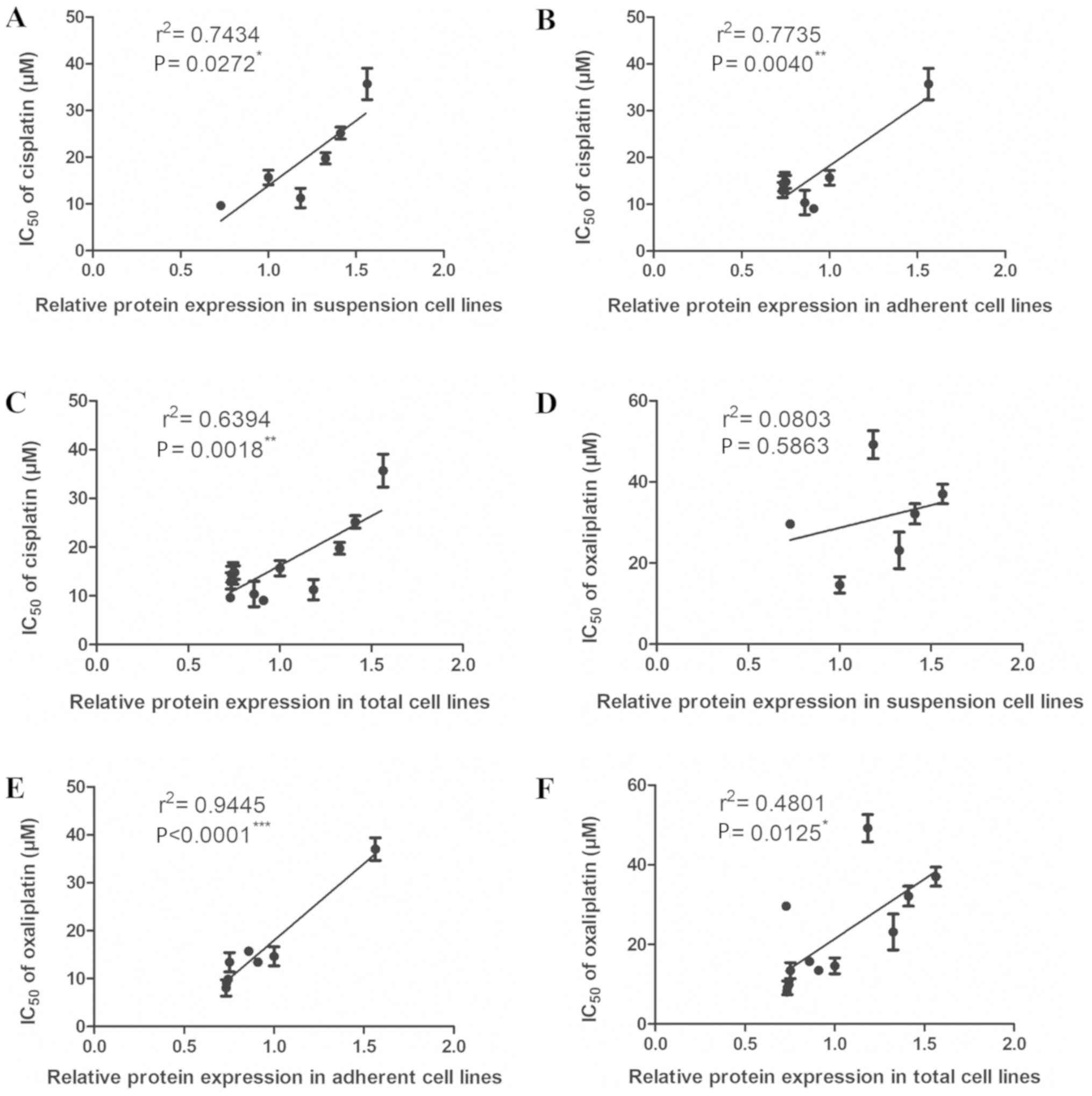
To investigate the effects of BRCA1 expression on
the chemosensitivity of gastric cancer cells to platinum agents,
correlation coefficients between BRCA1 protein expression level and
the IC50 of cisplatin or oxaliplatin were calculated
(Fig. 2). The suspension cell lines
demonstrated a significant positive correlation between BRCA1
protein expression level and the IC50 value of cisplatin
(P<0.05). In addition, significant positive correlations were
identified between the BRCA1 protein expression level and the
IC50 values of cisplatin (P<0.01) and oxaliplatin
(P<0.001) in the adherent cell lines.
Discussion
Gastric cancer was the third leading cause of
cancer-associated mortality worldwide in 2018 (27). Due to late detection and diagnosis at
an advanced stage, patients with gastric cancer often have a poor
prognosis (28). In Korea, gastric
cancer was estimated to be the fourth leading cause of
cancer-associated mortality both in males and females in 2018
(29). As one of the major types of
cancer, gastric cancer remains a global health burden; therefore,
there is a requirement to identify markers that may improve
prognosis and treatment.
BRCA1 expression level is widely used to predict the
prognosis of breast cancer and ovarian cancer. BRCA1 expression is
lower in sporadic and inherited breast cancer (30), and >50% of epithelial ovarian
cancer cases exhibit a BRCA1-deficient status (8,31). In
addition to the identified association between BRCA1 expression and
breast and ovarian cancer, previous studies have reported that
BRCA1 expression is associated with the prognosis of gastric cancer
(14–16). A high TNM stage or poorly
differentiated tumor is associated with a BRCA1-negative status
(14–17). SNU484 and SNU601 cells, and all
suspension gastric cancer cell lines used in the present study are
poorly differentiated (32,33).
Treatment regimes involving cisplatin and
oxaliplatin have been widely used to treat gastric cancer (26). In general, the current study revealed
that the IC50 values of both platinum agents were higher
in the suspension cell lines (16.76±6.4 for cisplatin; 34.01±10.89
for oxaliplatin) compared with the adherent cell lines (12.68±2.75
for cisplatin; 11.63±3.00 for oxaliplatin). Previous clinical
studies have reported that poorly differentiated or advanced-stage
cancer cases are more likely to have a BRCA1-negative status
(14–17). The suspension cell lines used in the
present study were poorly differentiated and exhibited similar
properties to advanced-stage cancer, which suggests they would be
sensitive to platinum agents. However, the suspension cell lines
were identified to possess a higher resistance to cisplatin and
oxaliplatin compared with the adherent cell line. Considering a
secondary mutation that may restore BRCA1 function (34), further studies are required to assess
if the suspension cell lines could gain another mutation. When
correlation coefficients between BRCA1 expression and
IC50 values were determined, lower correlation
coefficients were revealed between BRCA1 expression levels and
IC50 values for the suspension cell lines compared with
the adherent cell lines, which indicates that BRCA1 influences
advanced-stage cancer cells to a lesser extent compared with cancer
cells at an earlier stage.
Cisplatin and oxaliplatin are understood to respond
to cancer cells via a similar mechanism, including the formation of
adducts to double strands of DNA (26). However, cisplatin and oxaliplatin
exhibit different effects on the DNA mismatch repair pathway.
Cisplatin-DNA adducts demonstrate a stronger affinity for the
mismatch repair proteins MSH2 and MutS compared with
oxaliplatin-DNA adducts; therefore, mismatch repair proteins are
more susceptible to cisplatin cytotoxicity (35,36).
When mismatch repair proteins bind to cisplatin-DNA adducts, the
cytotoxicity increases due to an enhancement of the apoptosis
pathway (37,38) and DNA translesion synthesis (39). If mismatch repair pathways are
deficient or mutated, cisplatin resistance typically occurs
(40). In gastric cancer,
hypermethylation of the promoter region of the mismatch repair
protein MLH1 has been reported (41), which leads to silencing of the MLH1
gene. Hypermethylation of the MLH1 promoter has been identified in
>50% of gastric cancer cases, which demonstrate a high level of
microsatellite instability (MSI-H) (42–47).
Furthermore, patients with MSI-H have been reported to have no MLH1
and MSH2 protein expression (43,48).
Downregulation of the mismatch repair gene alone does not promote
carcinogenesis (49); therefore,
additional alterations in the expression of other genes would be
required.
The BRCA1-associated genome surveillance complex
(BASC) is composed of numerous proteins, including BRCA1, MSH2,
MSH6, MutL homolg 1 (MLH1), ATM, bloom syndrome RecQ like helicase
and replication factor C, and the RAD50-MRE11-nibrin protein
complex. BRCA1 and MLH1 or BRCA1 and the MSH2-MSH6 heterodimer
interact with each other within the complex (50). In addition, a study investigating
hereditary nonpolyposis colon cancer, which increases the risk of
GC (51), revealed an interaction
between BRCA1 and the MSH2-MSH6 complex (52), which suggests BASC serves a role in
the pathogenicity of gastric cancer. Therefore, further studies
with a focus on mismatch repair proteins, including MSH2, MSH6 and
MLH1, are required to improve understanding regarding the
association between BRCA1 and the cytotoxicity of platinum
agents.
In conclusion, the present study revealed that the
expression level of BRCA1 is variable in different types of gastric
cancer. In addition, BRCA1 expression level in adherent gastric
cancer cells was identified to be correlated with the treatment
response to cisplatin and oxaliplatin. Furthermore, a correlation
was observed in the suspension cell lines for cisplatin. Therefore,
the current study suggests that BRCA1 may be used as a therapeutic
marker to predict the sensitivity for platinum based anticancer
agents in gastric cancer.
Acknowledgements
Not applicable.
Funding
The present study was supported by the National
Research Foundation funded by the Ministry of Science, ICT and
Future Planning (grant no. NRF-2015R1C1A2A01054457).
Availability of data and materials
The datasets used and/or analyzed in the current
study are available from the corresponding author upon reasonable
request.
Authors' contributions
HM conceived and designed the experiments. GK, JK
and SYH performed the experiments and collected the data. GK, IGH,
HSK and HM analyzed the data and prepared the manuscript.
Ethics approval and consent to
participate
Not applicable.
Patient consent for publication
Not applicable.
Competing interests
The authors declare that they have no competing
interests.
References
|
1
|
Rosen EM, Fan S, Pestell RG and Goldberg
ID: BRCA1 gene in breast cancer. J Cell Physiol. 196:19–41. 2003.
View Article : Google Scholar : PubMed/NCBI
|
|
2
|
Roy R, Chun J and Powell SN: BRCA1 and
BRCA2: Different roles in a common pathway of genome protection.
Nat Rev Cancer. 12:68–78. 2011. View
Article : Google Scholar : PubMed/NCBI
|
|
3
|
Joo WS, Jeffrey PD, Cantor SB, Finnin MS,
Livingston DM and Pavletich NP: Structure of the 53BP1 BRCT region
bound to p53 and its comparison to the Brca1 BRCT structure. Genes
Dev. 16:583–593. 2002. View Article : Google Scholar : PubMed/NCBI
|
|
4
|
Mullan PB, Quinn JE and Harkin DP: The
role of BRCA1 in transcriptional regulation and cell cycle control.
Oncogene. 25:5854–5863. 2006. View Article : Google Scholar : PubMed/NCBI
|
|
5
|
Streff H, Profato J, Ye Y, Nebgen D,
Peterson SK, Singletary C, Arun BK and Litton JK: Cancer incidence
in first- and second-degree relatives of BRCA1 and BRCA2 mutation
carriers. Oncologist. 21:869–874. 2016. View Article : Google Scholar : PubMed/NCBI
|
|
6
|
Linger RJ and Kruk PA: BRCA1 16 years
later: Risk-associated BRCA1 mutations and their functional
implications. FEBS J. 277:3086–3096. 2010. View Article : Google Scholar : PubMed/NCBI
|
|
7
|
Wilson CA, Ramos L, Villaseñor MR, Anders
KH, Press MF, Clarke K, Karlan B, Chen JJ, Scully R, Livingston D,
et al: Localization of human BRCA1 and its loss in high-grade,
non-inherited breast carcinomas. Nat Genet. 21:236–240. 1999.
View Article : Google Scholar : PubMed/NCBI
|
|
8
|
Sun C, Li N, Yang Z, Zhou B, He Y, Weng D,
Fang Y, Wu P, Chen P, Yang X, et al: miR-9 regulation of BRCA1 and
ovarian cancer sensitivity to cisplatin and PARP inhibition. J Natl
Cancer Inst. 105:1750–1758. 2013. View Article : Google Scholar : PubMed/NCBI
|
|
9
|
Pennington KP, Walsh T, Harrell MI, Lee
MK, Pennil CC, Rendi MH, Thornton A, Norquist BM, Casadei S, Nord
AS, et al: Germline and somatic mutations in homologous
recombination genes predict platinum response and survival in
ovarian, fallopian tube, and peritoneal carcinomas. Clin Cancer
Res. 20:764–775. 2014. View Article : Google Scholar : PubMed/NCBI
|
|
10
|
Esteller M, Silva JM, Dominguez G, Bonilla
F, Matias-Guiu X, Lerma E, Bussaglia E, Prat J, Harkes IC, Repasky
EA, et al: Promoter hypermethylation and BRCA1 inactivation in
sporadic breast and ovarian tumors. J Natl Cancer Inst. 92:564–569.
2000. View Article : Google Scholar : PubMed/NCBI
|
|
11
|
Moskwa P, Buffa FM, Pan Y, Panchakshari R,
Gottipati P, Muschel RJ, Beech J, Kulshrestha R, Abdelmohsen K,
Weinstock DM, et al: miR-182-mediated downregulation of BRCA1
impacts DNA repair and sensitivity to PARP inhibitors. Mol Cell.
41:210–220. 2011. View Article : Google Scholar : PubMed/NCBI
|
|
12
|
Liu Z, Liu J, Segura MF, Shao C, Lee P,
Gong Y, Hernando E and Wei JJ: MiR-182 overexpression in
tumourigenesis of high-grade serous ovarian carcinoma. J Pathol.
228:204–215. 2012. View Article : Google Scholar : PubMed/NCBI
|
|
13
|
Alexandrov LB, Nik-Zainal S, Siu HC, Leung
SY and Stratton MR: A mutational signature in gastric cancer
suggests therapeutic strategies. Nat Commun. 6:86832015. View Article : Google Scholar : PubMed/NCBI
|
|
14
|
Zhang ZZ, Liu YJ, Yin XL, Zhan P, Gu Y and
Ni XZ: Loss of BRCA1 expression leads to worse survival in patients
with gastric carcinoma. World J Gastroenterol. 19:1968–1974. 2013.
View Article : Google Scholar : PubMed/NCBI
|
|
15
|
Chen W, Wang J, Li X, Li J, Zhou L, Qiu T,
Zhang M and Liu P: Prognostic significance of BRCA1 expression in
gastric cancer. Med Oncol. 30:4232013. View Article : Google Scholar : PubMed/NCBI
|
|
16
|
Kim JW, Cho HJ, Kim M, Lee KH, Kim MA, Han
SW, Oh DY, Lee HJ, Im SA, Kim TY, et al: Differing effects of
adjuvant chemotherapy according to BRCA1 nuclear expression in
gastric cancer. Cancer Chemother Pharmacol. 71:1435–1443. 2013.
View Article : Google Scholar : PubMed/NCBI
|
|
17
|
Chen XR, Zhang WZ, Lin XQ and Wang JW:
Genetic instability of BRCA1 gene at locus D17S855 is related to
clinicopathological behaviors of gastric cancer from Chinese
population. World J Gastroenterol. 12:4246–4249. 2006. View Article : Google Scholar : PubMed/NCBI
|
|
18
|
Wang K, Xu L, Pan L, Xu K and Li G: The
functional BRCA1 rs799917 genetic polymorphism is associated with
gastric cancer risk in a Chinese Han population. Tumour Biol.
36:393–397. 2015. View Article : Google Scholar : PubMed/NCBI
|
|
19
|
Shim HJ, Yun JY, Hwang JE, Bae WK, Cho SH,
Lee JH, Kim HN, Shin MH, Kweon SS, Lee JH, et al: BRCA1 and XRCC1
polymorphisms associated with survival in advanced gastric cancer
treated with taxane and cisplatin. Cancer Sci. 101:1247–1254. 2010.
View Article : Google Scholar : PubMed/NCBI
|
|
20
|
Wang D and Lippard SJ: Cellular processing
of platinum anticancer drugs. Nat Rev Drug Discov. 4:307–320. 2005.
View Article : Google Scholar : PubMed/NCBI
|
|
21
|
Borst P, Rottenberg S and Jonkers J: How
do real tumors become resistant to cisplatin? Cell Cycle.
7:1353–1359. 2008. View Article : Google Scholar : PubMed/NCBI
|
|
22
|
Galluzzi L, Vitale I, Michels J, Brenner
C, Szabadkai G, Harel-Bellan A, Castedo M and Kroemer G: Systems
biology of cisplatin resistance: Past, present and future. Cell
Death Dis. 5:e12572014. View Article : Google Scholar : PubMed/NCBI
|
|
23
|
Zimmermann T, Zeizinger M and Burda JV:
Cisplatin interaction with cysteine and methionine, a theoretical
DFT study. J Inorg Biochem. 99:2184–2196. 2005. View Article : Google Scholar : PubMed/NCBI
|
|
24
|
Circu ML and Aw TY: Reactive oxygen
species, cellular redox systems, and apoptosis. Free Radic Biol
Med. 48:749–762. 2010. View Article : Google Scholar : PubMed/NCBI
|
|
25
|
Martinez-Balibrea E, Martínez-Cardús A,
Ginés A, Ruiz de Porras V, Moutinho C, Layos L, Manzano JL, Bugés
C, Bystrup S, Esteller M and Abad A: Tumor-related molecular
mechanisms of oxaliplatin resistance. Mol Cancer Ther.
14:1767–1776. 2015. View Article : Google Scholar : PubMed/NCBI
|
|
26
|
Ajani JA: Evolving chemotherapy for
advanced gastric cancer. Oncologist. 10 Suppl 3:S49–S58. 2005.
View Article : Google Scholar
|
|
27
|
Bray F, Ferlay J, Soerjomataram I, Siegel
RL, Torre LA and Jemal A: Global cancer statistics 2018: GLOBOCAN
estimates of incidence and mortality worldwide for 36 cancers in
185 countries. CA Cancer J Clin. 68:394–424. 2018. View Article : Google Scholar : PubMed/NCBI
|
|
28
|
Carcas LP: Gastric cancer review. J
Carcinog. 13:142014. View Article : Google Scholar : PubMed/NCBI
|
|
29
|
Jung KW, Won YJ, Oh CM, Kong HJ, Cho H,
Lee JK, Lee DH and Lee KH: Prediction of cancer incidence and
mortality in Korea, 2016. Cancer Res Treat. 48:451–457. 2016.
View Article : Google Scholar : PubMed/NCBI
|
|
30
|
Mueller CR and Roskelley CD: Regulation of
BRCA1 expression and its relationship to sporadic breast cancer.
Breast Cancer Res. 5:45–52. 2003. View
Article : Google Scholar : PubMed/NCBI
|
|
31
|
McMillen BD, Aponte MM, Liu Z, Helenowski
IB, Scholtens DM, Buttin BM and Wei JJ: Expression analysis of
MIR182 and its associated target genes in advanced ovarian
carcinoma. Mod Pathol. 25:1644–1653. 2012. View Article : Google Scholar : PubMed/NCBI
|
|
32
|
Park JG, Frucht H, LaRocca RV, Bliss DP
Jr, Kurita Y, Chen TR, Henslee JG, Trepel JB, Jensen RT, Johnson
BE, et al: Characteristics of cell lines established from human
gastric carcinoma. Cancer Res. 50:2773–2780. 1990.PubMed/NCBI
|
|
33
|
Park JG, Yang HK, Kim WH, Chung JK, Kang
MS, Lee JH, Oh JH, Park HS, Yeo KS, Kang SH, et al: Establishment
and characterization of human gastric carcinoma cell lines. Int J
Cancer. 70:443–449. 1997. View Article : Google Scholar : PubMed/NCBI
|
|
34
|
Swisher EM, Sakai W, Karlan BY, Wurz K,
Urban N and Taniguchi T: Secondary BRCA1 mutations in BRCA1-mutated
ovarian carcinomas with platinum resistance. Cancer Res.
68:2581–2586. 2008. View Article : Google Scholar : PubMed/NCBI
|
|
35
|
Fink D, Nebel S, Aebi S, Zheng H, Cenni B,
Nehmé A, Christen RD and Howell SB: The role of DNA mismatch repair
in platinum drug resistance. Cancer Res. 56:4881–4886.
1996.PubMed/NCBI
|
|
36
|
Zdraveski ZZ, Mello JA, Farinelli CK,
Essigmann JM and Marinus MG: MutS preferentially recognizes
cisplatin-over oxaliplatin-modified DNA. J Biol Chem.
277:1255–1260. 2002. View Article : Google Scholar : PubMed/NCBI
|
|
37
|
Nehmé A, Baskaran R, Aebi S, Fink D, Nebel
S, Cenni B, Wang JY, Howell SB and Christen RD: Differential
induction of c-Jun NH2-terminal kinase and c-Abl kinase in DNA
mismatch repair-proficient and -deficient cells exposed to
cisplatin. Cancer Res. 57:3253–3257. 1997.PubMed/NCBI
|
|
38
|
Nehmé A, Baskaran R, Nebel S, Fink D,
Howell SB, Wang JY and Christen RD: Induction of JNK and c-Abl
signalling by cisplatin and oxaliplatin in mismatch
repair-proficient and -deficient cells. Br J Cancer. 79:1104–1110.
1999. View Article : Google Scholar : PubMed/NCBI
|
|
39
|
Vaisman A, Varchenko M, Umar A, Kunkel TA,
Risinger JI, Barrett JC, Hamilton TC and Chaney SG: The role of
hMLH1, hMSH3, and hMSH6 defects in cisplatin and oxaliplatin
resistance: Correlation with replicative bypass of platinum-DNA
adducts. Cancer Res. 58:3579–3585. 1998.PubMed/NCBI
|
|
40
|
Martin LP, Hamilton TC and Schilder RJ:
Platinum resistance: The role of DNA repair pathways. Clin Cancer
Res. 14:1291–1295. 2008. View Article : Google Scholar : PubMed/NCBI
|
|
41
|
Strathdee G, MacKean MJ, Illand M and
Brown R: A role for methylation of the hMLH1 promoter in loss of
hMLH1 expression and drug resistance in ovarian cancer. Oncogene.
18:2335–2341. 1999. View Article : Google Scholar : PubMed/NCBI
|
|
42
|
Yamamoto H, Perez-Piteira J, Yoshida T,
Terada M, Itoh F, Imai K and Perucho M: Gastric cancers of the
microsatellite mutator phenotype display characteristic genetic and
clinical features. Gastroenterology. 116:1348–1357. 1999.
View Article : Google Scholar : PubMed/NCBI
|
|
43
|
Wu MS, Lee CW, Shun CT, Wang HP, Lee WJ,
Chang MC, Sheu JC and Lin JT: Distinct clinicopathologic and
genetic profiles in sporadic gastric cancer with different mutator
phenotypes. Genes Chromosomes Cancer. 27:403–411. 2000. View Article : Google Scholar : PubMed/NCBI
|
|
44
|
Bacani J, Zwingerman R, Di Nicola N,
Spencer S, Wegrynowski T, Mitchell K, Hay K, Redston M, Holowaty E,
Huntsman D, et al: Tumor microsatellite instability in early onset
gastric cancer. J Mol Diagn. 7:465–477. 2005. View Article : Google Scholar : PubMed/NCBI
|
|
45
|
Leung SY, Yuen ST, Chung LP, Chu KM, Chan
AS and Ho JC: hMLH1 promoter methylation and lack of hMLH1
expression in sporadic gastric carcinomas with high-frequency
microsatellite instability. Cancer Res. 59:159–164. 1999.PubMed/NCBI
|
|
46
|
Pinto M, Oliveira C, Machado JC, Cirnes L,
Tavares J, Carneiro F, Hamelin R, Hofstra R, Seruca R and
Sobrinho-Simões M: MSI-L gastric carcinomas share the hMLH1
methylation status of MSI-H carcinomas but not their
clinicopathological profile. Lab Invest. 80:1915–1923. 2000.
View Article : Google Scholar : PubMed/NCBI
|
|
47
|
Fleisher AS, Esteller M, Wang S, Tamura G,
Suzuki H, Yin J, Zou TT, Abraham JM, Kong D, Smolinski KN, et al:
Hypermethylation of the hMLH1 gene promoter in human gastric
cancers with microsatellite instability. Cancer Res. 59:1090–1095.
1999.PubMed/NCBI
|
|
48
|
Scartozzi M, Galizia E, Freddari F,
Berardi R, Cellerino R and Cascinu S: Molecular biology of sporadic
gastric cancer: Prognostic indicators and novel therapeutic
approaches. Cancer Treat Rev. 30:451–459. 2004. View Article : Google Scholar : PubMed/NCBI
|
|
49
|
Eshleman JR and Markowitz SD: Mismatch
repair defects in human carcinogenesis. Hum Mol Genet. 5:1489–1494.
1996. View Article : Google Scholar : PubMed/NCBI
|
|
50
|
Wang Y, Cortez D, Yazdi P, Neff N, Elledge
SJ and Qin J: BASC, a super complex of BRCA1-associated proteins
involved in the recognition and repair of aberrant DNA structures.
Genes Dev. 14:927–939. 2000.PubMed/NCBI
|
|
51
|
Caldas C, Carneiro F, Lynch HT, Yokota J,
Wiesner GL, Powell SM, Lewis FR, Huntsman DG, Pharoah PD, Jankowski
JA, et al: Familial gastric cancer: Overview and guidelines for
management. J Med Genet. 36:873–880. 1999.PubMed/NCBI
|
|
52
|
Wang Q, Zhang H, Guerrette S, Chen J,
Mazurek A, Wilson T, Slupianek A, Skorski T, Fishel R and Greene
MI: Adenosine nucleotide modulates the physical interaction between
hMSH2 and BRCA1. Oncogene. 20:4640–4649. 2001. View Article : Google Scholar : PubMed/NCBI
|