Introduction
Melanoma is an invasive and malignant type of tumor,
which mainly occurs in the skin and oral mucosa (1). The number of cases of mortality
associated with cutaneous melanoma in the United States in 2017
reached 9,730, and it accounted for 1.6% of all cases of
cancer-associated mortality (2).
Despite decades of research efforts and development of antimelanoma
drugs, the therapeutic outcome of melanoma is generally
unsatisfactory.
Early detection and monitoring of patients with
melanoma are particularly important for melanoma therapy. MicroRNAs
(miRNAs/miRs) are a class of small non-coding molecules, 18–25 nt
in length, which have emerged as a powerful novel tool for the
diagnosis and monitoring of patients with various types of cancer
(3,4). Additionally, our previous studies
demonstrated that expression levels of miR-125b are markedly
downregulated in melanoma tissues and cell lines, and that miR-125b
is able to inhibit the proliferation and invasion of melanoma cells
(5,6). However, the functional role of miR-125b
in melanoma tissues, and its association with the survival and
prognosis of patients with melanoma, remain unclear.
The majority of miRNA samples in studies are derived
from fresh tissues. However, since it is difficult to obtain fresh
tissues due to limited sources, medical and research institutions
have commonly used the formalin fixation plus paraffin embedding
method to preserve pathological tissues. A number of previous
studies identified that miRNAs in formalin-fixed paraffin-embedded
(FFPE) tissues could still provide reliable expression profiles, as
they exhibited a high correlation with those in fresh tissues
(7); even tissues that have been
preserved for >30 years can still provide reliable miRNA
expression data (8). These
characteristics of FFPE miRNAs are beneficial for improving the
utilization of the largest bank of specimens to explore the
development, progression and prognosis of human tumors.
The aim of the present study was to detect the
expression of miR-125b in previously confirmed FFPE melanoma
tissues, and to analyze the associations among miR-125b expression
and the metastasis and prognosis of melanoma. This was performed in
an attempt to provide novel predictors and interventional targets
to improve the early screening level and efficacy of diagnosis and
treatment of this rare but devastating human malignant disease.
Materials and methods
Sample collection and grouping
The present study included 29 FFPE melanoma tissue
specimens and 16 FFPE intradermal nevus (IDN) tissue specimens,
which were obtained from patients who underwent radical resection
at The First Affiliated Hospital of Nanchang University (Nanchang,
Jiangxi, China) between January 2014 and May 2017. All specimens
were fixed with 20% neutral-buffered formalin for 24 h at room
temperature (25–27°C) and stored following paraffin embedding. The
thickness of paraffin sections was 5–10 µm. The inclusion criteria
for patients with melanoma were: i) Patients who had not received
radiotherapy or chemotherapy prior to surgery; ii) patients without
other co-existing cancer, heart disease and/or diabetes; and iii)
patients with complete clinicopathological data. FFPE tissue
specimens of all patients were prepared by the same technician, and
reviewed and confirmed by two pathologists. Clinicopathological
data of each patient and survival data were collected during the
follow-up period (6–32 months). The patients were divided into
three groups according to the 8th Edition of the American Joint
Committee on Cancer melanoma staging system (9): IDN (control; n=16), primary melanoma
(stage I and II; n=16) and metastatic melanoma (stage III and IV;
n=13). The 29 patients with melanoma were comprised of 13 males and
16 females, who ranged in age between 34 and 81 years, including 15
patients >60 years and 14 patients <60 years. The 16 patients
with IDN were comprised of 7 males and 9 females, who ranged in age
between 29 and 75 years, including 6 patients >60 years and 10
patients <60 years. The Ethics Committee of The First Affiliated
Hospital of Nanchang University reviewed and approved the present
study, and written informed consent was obtained from each patient
at each examination phase. The present study complied with the
principles of the Declaration of Helsinki.
miR-125b extraction
Expression levels of miR-125b in each group were
detected by reverse transcription-quantitative PCR (RT-qPCR).
Reagents, primers and instruments used in the present study
included miR-125b extraction reagent, the miRNA prep Pure FFPE kit,
miR-125b reverse transcription reagent, the miRcute Plus miRNA
First-Strand cDNA Synthesis kit, the miR-125b PCR reagent kit and
miRcute Plus miRNA qPCR Detection kit SYBR Green (all purchased
from Beijing Wanter Biopharmaceutical Co., Ltd.). U6 was used as
the internal control. FFPE tissues were sliced into 6–8 sections of
5–10 µm thickness (the first 2–3 sections were discarded) from
which RNA was extracted using TRIzol reagent (Thermo Fisher
Scientific, Inc.) according to the manufacturer's protocol. All
procedures were performed under ribozyme-free conditions. The total
RNA was measured for optical density, and subsequently stored in
RNA solution at −80°C.
RT-PCR
To each tube, 10 µl 2X miRNA RT Reaction Buffer, 4
µl total miRNA and 2 µl miRNA RT Enzyme mixed with 4 ml RNase-Free
ddH2O were added, totaling 20 µl. PCR reaction solution
of each sample was reverse transcribed under the conditions of 42°C
for 60 min, followed by 95°C for 3 min, and then the obtained cDNA
product was stored at −20°C.
qPCR
The reaction solution for PCR (20 µl) consisted of
10 µl 2X miRcute Plus miRNA Premix (with SYBR and ROX), 0.4 µl
Reverse Primer (10 µm), 2 µl 50X ROX Reference Dye and 7.6 µl
ddH2O. cDNA was added to the reaction solution and mixed
gently. qPCR was conducted under the following conditions: Initial
template denaturation at 95°C for 15 min for one cycle, 94°C for 20
sec for 5 cycles, 63–65°C for 30 sec for 5 cycles, 72°C for 34 sec
for 5 cycles, 94°C for 20 sec for 45 cycles of cyclic denaturation
and 60°C for 34 sec for 45 cycles of cyclic c annealing and
extension. Relative miR-125b expression was measured using the
2−ΔΔCq method (10). The
primers were as follows: miR-125b forward, 5′ddd-TGC CTC CCT GAG
ACC CTA ACT TGT GA-3′ and reverse, 5′-GTGCAGGGTCCGAGGT-3′; and U6
forward, 5′-CGCTTCGGCAGCACATATAC-3′ and reverse,
5′-AGGGGCCATGCTAATCTTCT-3′.
Statistical analysis
All experiments were repeated three times. Data were
analyzed using SPSS version 22.0 statistical software (IBM Corp.).
Results are expressed as the means ± standard deviation. One-way
ANOVA and a least significant difference post hoc test were used
when making comparisons among multiple groups. An
independent-sample t-test was used for comparisons between two
groups. Categorical variables were assessed by Fisher's exact test.
Receiver operating characteristic (ROC) curve analysis was used to
evaluate the value of diagnosis and prognostic prediction.
Kaplan-Meier survival curves were plotted to compare the survival
of the miR-125b low-expression group and the high-expression group.
A log-rank test was used to compare the survival curves of the two
groups. Cox regression analysis was used to compare the overall
survival in the miR-125b low- and high-expression groups. P<0.05
was considered to indicate a statistically significant.
Results
Comparison of miR-125b expression in
different groups
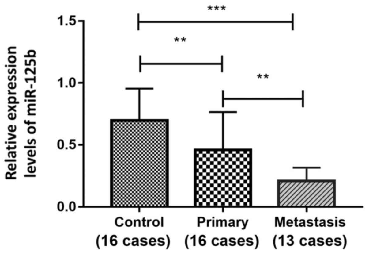
Expression levels of miR-125b in FFPE melanoma
tissues were detected by RT-qPCR. As shown in Fig. 1, the expression levels of miR-125b in
the primary melanoma group (n=16) and in the metastatic melanoma
group (n=13) were significantly lower than that in the IDN group
(n=16). Additionally, the expression level of miR-125b in the
metastatic melanoma group was significantly lower than that in the
primary melanoma group (0.21±0.11, 0.70±0.26 and 0.46±0.31, for
metastasis, IDN and primary groups, respectively; P<0.05).
Associations among miR-125b expression
and clinical features of patients with melanoma
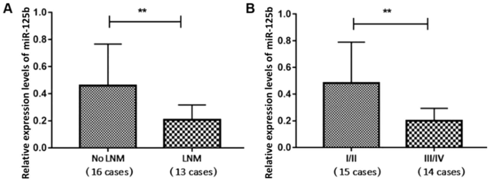
The clinicopathological features of the patients are
listed in Table I. The miR-125b
expression levels of the 16 patients with melanoma with no lymph
node metastasis (LNM) and the 13 patients with melanoma with LNM
were 0.46±0.31 and 0.21±0.11, respectively, revealing a significant
difference between the two groups (P<0.05; Fig. 2A). The expression levels of miR-125b
in the 15 cases of TNM stage I and II (I/II) melanoma and 14 cases
of TNM stage III and IV (III/IV) were 0.47±0.31 and 0.21±0.09,
respectively, revealing a significant difference between the two
groups (P<0.05; Fig. 2B).
Expression levels of miR-125b were associated with LNM and TNM
stage, but not with patient age, sex, tumor size or the location of
the lesion (acral vs. non-acral; Table
I; Fig. 2).
 | Table I.Associations between miR-125b
expression and the clinical features of patients with melanoma. |
Table I.
Associations between miR-125b
expression and the clinical features of patients with melanoma.
| Clinical
features | Number, n (%) | miR-125b expression
(mean ± standard deviation) | P-value |
|---|
| Sex |
|
| 0.443 |
| Male | 13 (44.83) | 0.30±0.18 |
|
|
Female | 16 (55.17) | 0.38±0.32 |
|
| Age, years |
|
| 0.874 |
| ≥60 | 15 (51.72) | 0.35±0.27 |
|
|
<60 | 14 (48.28) | 0.34±0.27 |
|
| Tumor diameter,
cm |
|
| 0.425 |
| ≥2 | 11 (37.93) | 0.29±0.29 |
|
|
<2 | 18 (62.07) | 0.38±0.26 |
|
| Lymph node
metastasis |
|
| 0.001a |
| No | 16 (55.17) | 0.46±0.31 |
|
| Yes | 13 (44.83) | 0.21±0.11 |
|
| TNM stage |
|
| 0.004a |
| I/II | 15 (51.72) | 0.47±0.31 |
|
|
III/IV | 14 (48.28) | 0.21±0.09 |
|
| Lesion location |
|
| 0.991 |
|
Acral | 17 (58.62) | 0.35±0.25 |
|
|
Non-acral | 12 (41.38) | 0.35±0.30 |
|
Diagnostic value of miR-125b
expression in melanoma
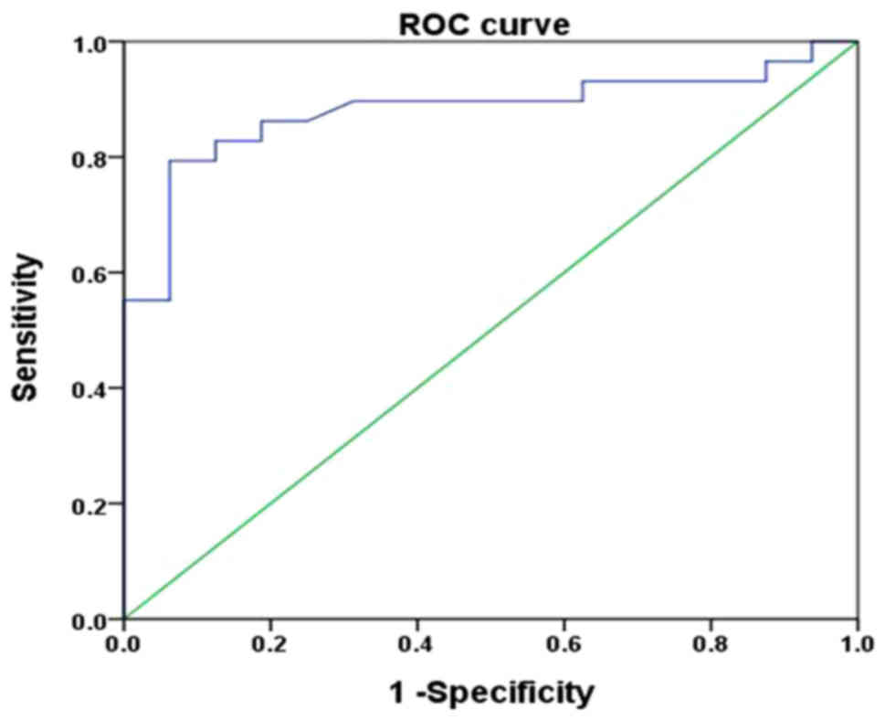
In the ROC curve analysis, the AUC was 0.880, with a
95% CI of 0.777–0.984 (P<0.001), with a sensitivity and
specificity of 79.3 and 93.7%, respectively, and a cutoff value of
0.73 (Fig. 3). These results
suggested that miR-125b may be used as a predictor of diagnosis for
patients with melanoma.
Association between miR-125b
expression and the prognosis of patients with melanoma
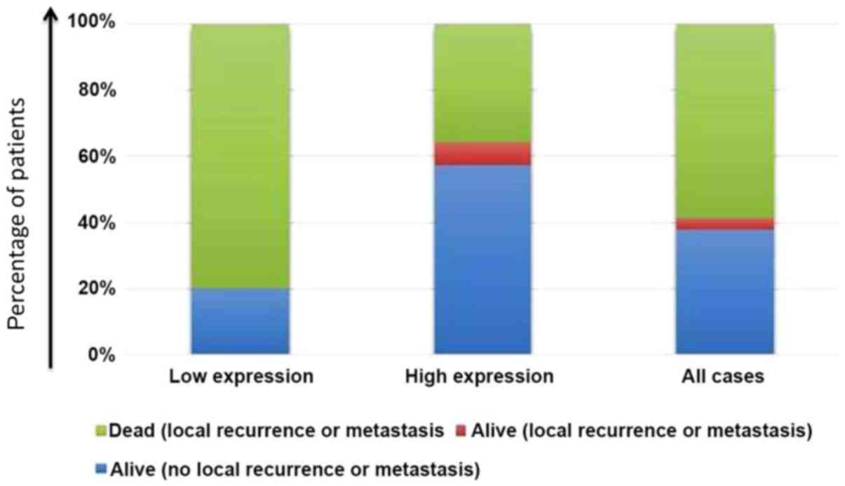
Using the median value (0.30) as the cut-off value,
the expression levels of miR-125b were used to classify the
patients into a high-expression group and a low-expression group.
No patients were lost to follow-up or died of other causes. The
mortality rate in the low-expression group was significantly higher
than that in the high-expression group (P<0.05; Table II, Fig.
4).
| Table II.Comparison of prognosis of patients in
the microRNA-125b low-expression and high-expression groups. |
Table II.
Comparison of prognosis of patients in
the microRNA-125b low-expression and high-expression groups.
| Patient
prognosis | Low expression | High expression | P-value |
|---|
| Alive (no local
recurrence or metastasis) | 3 | 8 | 0.034a |
| Alive (local
recurrence or metastasis) | 0 | 1 |
|
| Dead (local
recurrence or metastasis) | 12 | 5 |
|
| Dead (other diseases
or accidents) | 0 | 0 |
|
| Dead (lost to
follow-up) | 0 | 0 |
|
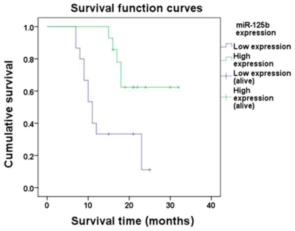
Kaplan-Meier analysis demonstrated that the 1- and
2-year cumulative survival rates in the high-expression group were
100 and 62.3% compared with 33.3 and 11.1% in the low-expression
group, respectively. A log-rank test identified a P-value of 0.005,
indicating that the survival time of patients in the low-expression
group was significantly shorter than that of patients in the
high-expression group (Fig. 5).
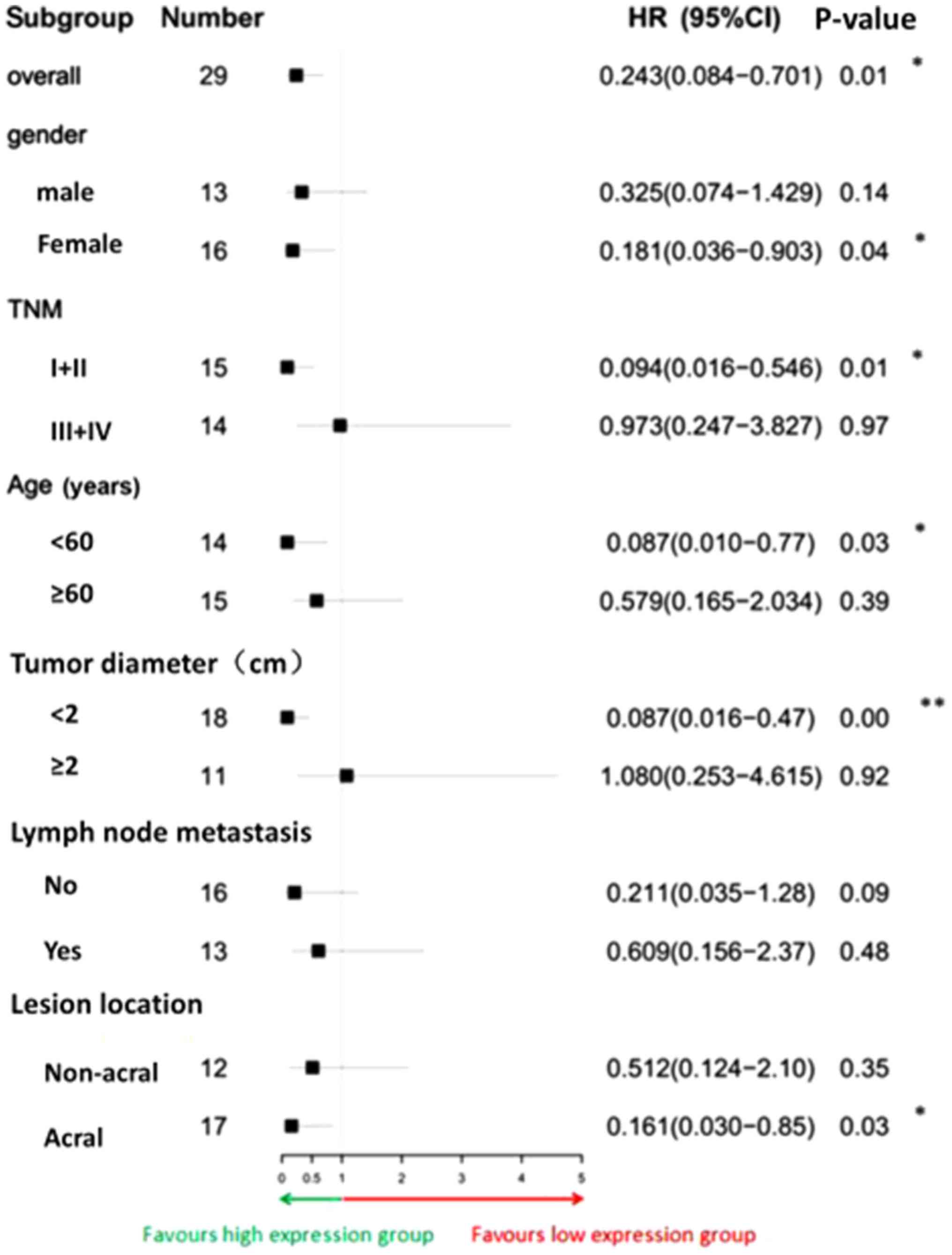
Survival of the patients was further analyzed by Cox
proportional hazards model (Fig. 6).
The overall risk of mortality of the miR-125b high expression group
was significantly lower than that of the low expression group (HR,
0.243; 95% CI, 0.084–0.701). In the five subgroups of female sex,
TNM stage I/II, age <60 years, tumor diameter <2 cm and tumor
located at the extremities, the risk of mortality in the high
expression group was significantly lower than that in the low
expression group (P<0.05). The associations between clinical
features and expression of miR-125b and overall survival of
patients with melanoma were analyzed by univariate and multivariate
Cox regression analysis. In univariate analysis, lesion location,
lymph node metastasis and miR-125b expression were significantly
associated with overall survival of patients. However, further
multivariate analysis revealed that lesion location and lymph node
metastasis could not enter the Cox regression equation. The results
of this indicated that only miR-125b expression was an independent
predictor of overall survival in patients with melanoma (HR, 0.252;
95% CI, 0.087–0.729; Table
III).
| Table III.Univariate and multivariate Cox
regression analyses of miR-125b expression, clinicopathological
features and overall survival. |
Table III.
Univariate and multivariate Cox
regression analyses of miR-125b expression, clinicopathological
features and overall survival.
|
| Univariate
analysis | Multivariate
analysis |
|---|
|
|
|
|
|---|
| Clinicopathological
feature | HR (95% CI) | P-value | HR (95% CI) | P-value |
|---|
| Sex |
| 0.979 |
|
|
| Male | Reference |
|
|
|
|
Female | 1.013
(0.390–2.628) |
|
|
|
| Age, years |
| 0.080 |
|
|
|
<60 | Reference |
|
|
|
| ≥60 | 2.471
(0.898–6.797) |
|
|
|
| Tumor diameter,
cm |
| 0.124 |
|
|
|
<2 | Reference |
|
|
|
| ≥2 | 2.116
(0.814–5.501) |
|
|
|
| Lymph node
metastasis |
| 0.012 |
| 0.266 |
| No | Reference |
|
|
|
|
Yes | 3.822
(1.339–10.912) |
|
|
|
| TNM stage |
| 0.053 |
|
|
|
I/II | Reference |
|
|
|
|
III/IV | 2.703
(0.985–7.413) |
|
|
|
| Lesion
location |
| 0.041 |
| 0.185 |
|
Non-acral | Reference |
|
|
|
|
Acral | 0.364
(0.138–0.960) |
|
|
|
| miR-125b
expression |
| 0.010 |
| 0.011 |
| Low
expression | Reference |
| Reference |
|
| High
expression | 0.243
(0.084–0.707) |
| 0.252
(0.087–0.729) |
|
Discussion
The lifetime risk of melanoma has been estimated to
be 1 in 75 and rising each year with >60,000 Americans
developing melanoma in 2010 (11).
The case-fatality rate is extremely high in patients with
metastatic melanoma (12). Risk
factors of melanoma include excessive ultraviolet light exposure,
photosensitive skin, improper management of pigmented nevi that may
induce malignant alterations, the existence of large numbers of
abnormally developed nevi and a family history of skin cancer. It
is necessary to identify effective approaches for the early
detection and treatment of high-risk populations of melanoma.
With the advantages of efficiency and accuracy,
various biomarkers have been the most valued tool in the diagnosis
and management of melanoma. miR-125b is an miRNA that serves an
essential role in tumor progression. Previous studies have
demonstrated that miR-125b may be a cancer suppressor gene of
multiple tumors, and suggested that miR-125b may be associated with
the development and progression of various types of cancer,
including prostate, oral and ovarian cancer (13–15).
Kappelmann et al (16) first
reported that the expression level of miR-125b in melanoma cells
obtained from primary and metastatic sources was lower than that in
normal human melanocytes. Further experiments demonstrated that the
downregulatory mechanism of miR-125b in melanoma cells inhibited
cell proliferation, which suggested that miR-125b may be a
potential tumor suppressor in melanoma. Our previous studies
(5,6)
demonstrated that the expression of miR-125b was able to inhibit
the proliferation and invasion of melanoma cells. Based on the
aforementioned research regarding RNA-protein-cell interactions
(5,6), the present study aimed to identify the
functional role of miR-125b in melanoma tissue and its association
with the survival and prognosis of patients with melanoma.
In the present study, expression levels of miR-125b
were detected in FFPE tissues from 29 patients with melanoma and 16
patients with IDN as the control. The results of the present study
revealed that the expression levels of miR-125b in the former were
significantly lower than those in in the latter. This result,
together with previous findings (5,6),
demonstrated that miR-125b served an anti-oncogenic role in the
occurrence and development of melanoma.
Analysis of the associations between miR-125b
expression and the clinical features of patients with melanoma
identified that the expression levels of miR-125b in patients with
LNM were significantly lower than those in patients with primary
melanoma without LNM and distant metastases (P<0.05). In
addition, the expression levels of miR-125b in patients with high
TNM stages were lower than those in patients with low TNM stages.
There were no significant associations among the expression levels
of miR-125b and sex, age, tumor size or lesion location
(P>0.05). This suggested that miR-125b expression was closely
associated with LNM and the TNM stage of melanoma. Glud et
al (17) detected the expression
levels of miR-125b in 28 paired patients with T2N0M0 and T2N1M0 by
RT-qPCR, and revealed that the expression levels of miR-125b in the
T2N1M0 patients were significantly downregulated compared with the
expression levels in the T2N0M0 patients (P<0.05), which is
consistent with the findings of the present study, further
indicating that miR-125b is involved in LNM and the TNM stage of
melanoma.
Expression levels of miR-125b in melanoma cell lines
are lower than those in melanocytes (16). Therefore, it may be possible to use
miR-125b expression as a diagnostic marker of melanoma. In the
present study, ROC curves were generated according to the
expression levels of miR-125b in patients with melanoma, by using
sensitivity as the ordinate and 1-specificity as the horizontal
ordinate, to explore the association between sensitivity and
specificity, knowing that the larger the AUC value, the more
accurate the diagnosis would be (18). The AUC value reported in the present
study was 0.880, with a 95% CI of 0.777–0.984 (P<0.001), a
sensitivity of 79.3% and a specificity of 93.7%, which indicated
that miR-125b may be a valuable marker for the diagnosis of
melanoma. However, larger sample size studies are required to
confirm if miR-125b could be used as a diagnostic indicator for
melanoma. Pathological evaluation remains the gold standard for the
diagnosis of melanoma.
It is important to predict the prognosis of patients
with melanoma. Since miR-125b has an impact on the proliferation
and invasion of melanoma cells, the association between the level
of miR-125b expression and the prognosis of patients with melanoma
may be explored. The 1- and 2-year survival rates of patients with
melanoma with high expression levels of miR-125b were 100 and
62.3%, respectively, compared with 33.3 and 11.1% in those patients
with low expression, indicating that the survival time of patients
in the low-expression group was significantly shorter than that of
patients in the high-expression group. The clinical outcomes of
patients with low or high expression levels of miR-125b were
statistically analyzed, and the results revealed that the
proportion of deaths in the high-expression group was significantly
lower than that in the low-expression group. These findings
suggested that low expression levels of miR-125b increased the risk
of mortality and reduced the survival time of patients with
melanoma. Univariate and multivariate Cox regression analysis
revealed that the expression of miR-125b was an independent
predictor for the overall survival of patients with melanoma. There
was a direct link between the level of miR-125b expression and the
survival of patients with melanoma.
The present study demonstrated that the expression
levels of miRNA-125b in melanoma tissues were decreased,
particularly in metastatic melanoma tissue. The abnormal miRNA-125b
expression was associated with lymph node metastasis and TNM stage
and its low expression was closely associated with the prognosis of
patients with melanoma. As miR-125b expression is relatively stable
in melanoma, it is relatively easy to obtain a specimen and the
methods for miRNA extraction/use are easy to follow. Therefore,
miR-125b may be used as an independent factor to predict the
prognosis of patients with melanoma, and it is a potential
diagnostic biomarker and therapeutic target of melanoma.
Acknowledgements
Not applicable.
Funding
This study was supported by the Jiangxi Province Key
Research and Development Program (grant no. 20161BBG70162), the
Jiangxi Province Science Foundation for Distinguished Young
Scholars (grant no. 20171BCB23090) and the Jiangxi Province Natural
Science Foundation (grant no. 20171ACB21053).
Availability of data and materials
All data generated or analyzed during this study are
included in this published article.
Authors' contributions
JZ, JY and QJ designed the study and performed the
experiments. HL, SN and SL collected the data. YX and CZ analyzed
the data. JY and QJ prepared the manuscript. All authors read and
approved the final manuscript.
Ethics approval and consent to
participate
The Ethics Committee of the First Affiliated
Hospital of Nanchang University reviewed and approved the present
study, and written informed consent was obtained from each patient
at each examination phase. The present study complied with the
principles of the Declaration of Helsinki.
Patient consent for publication
Not applicable.
Competing interests
The authors declare that they have no competing
interests.
References
|
1
|
Myles ZM, Buchanan N, King JB, Singh S,
White A, Wu M and Ajani U: Anatomic distribution of malignant
melanoma on the non-Hispanic black patient, 1998–2007. Arch
Dermatol. 148:797–801. 2012. View Article : Google Scholar : PubMed/NCBI
|
|
2
|
NIH National Cancer Institute:
Surveillance, Epidemiology, and End Results Program, Cancer Stat
Facts: Melanoma of the Skin (EB/OL). https://seer.cancer.gov/statfacts/html/melanhtml2017
|
|
3
|
Golabchi K, Soleimani-Jelodar R, Aghadoost
N, Momeni F, Moridikia A, Nahand JS, Masoudifar A, Razmjoo H and
Mirzaei H: MicroRNAs in retinoblastoma: Potential diagnostic and
therapeutic biomarkers. J Cell Physiol. 233:3016–3023. 2018.
View Article : Google Scholar : PubMed/NCBI
|
|
4
|
Gholamin S, Mirzaei H, Razavi SM,
Hassanian SM, Saadatpour L, Masoudifar A, ShahidSales S and Avan A:
GD2-targeted immunotherapy and potential value of circulating
microRNAs in neuroblastoma. J Cell Physiol. 233:866–879. 2018.
View Article : Google Scholar : PubMed/NCBI
|
|
5
|
Zhang J, Lu L, Xiong Y, Qin W, Zhang Y,
Qian Y, Jiang H and Liu W: MLK3 promotes melanoma proliferation and
invasion and is a target of microRNA-125b. Clin Exp Dermatol.
39:376–384. 2014. View Article : Google Scholar : PubMed/NCBI
|
|
6
|
Zhang J, Na S, Liu C, Pan S, Cai J and Qiu
J: MicroRNA-125b suppresses the epithelial-mesenchymal transition
and cell invasion by targeting ITGA9 in melanoma. Tumour Biol.
37:5941–5949. 2016. View Article : Google Scholar : PubMed/NCBI
|
|
7
|
Wang F, Wang L, Briggs C, Sicinska E,
Gaston SM, Mamon H, Kulke MH, Zamponi R, Loda M, Maher E, et al:
DNA degradation test predicts success in whole-genome amplification
from diverse clinical samples. J Mol Diagn. 9:441–451. 2007.
View Article : Google Scholar : PubMed/NCBI
|
|
8
|
von Ahlfen S, Missel A, Bendrat K and
Schlumpberger M: Determinants of RNA quality from FFPE samples.
PLoS One. 2:e12612007. View Article : Google Scholar : PubMed/NCBI
|
|
9
|
Amin MB, Edge SB, Greene FL, Byrd DR,
Brookland RK, Washington MK, Gershenwald JE, Compton CC, HessK R,
Sullivan DC, et al: AJCC cancer staging manual. 8th. Springer; New
York, NY: 2017, View Article : Google Scholar
|
|
10
|
Livak KJ and Schmittgen TD: Analysis of
relative gene expression data using real-time quantitative PCR and
the 2(-Delta Delta C(T)) method. Methods. 25:402–408. 2001.
View Article : Google Scholar : PubMed/NCBI
|
|
11
|
Wu S and Singh RK: Resistance to
chemotherapy and molecularly targeted therapies: Rationale for
combination therapy in malignant melanoma. Curr Mol Med.
11:553–563. 2011. View Article : Google Scholar : PubMed/NCBI
|
|
12
|
Garbe C, Peris K, Hauschild A, Saiag P,
Middleton M, Bastholt L, Grob JJ, Malvehy J, Newton-Bishop J,
Stratigos AJ, et al: Diagnosis and treatment of melanoma. European
consensus-based interdisciplinary guideline-update 2016. Eur J
Cancer. 63:201–217. 2016. View Article : Google Scholar : PubMed/NCBI
|
|
13
|
Fredsøe J, Rasmussen AKI, Thomsen AR,
Mouritzen P, Høyer S, Borre M, Ørntoft TF and Sørensen KD:
Diagnostic and prognostic MicroRNA biomarkers for prostate cancer
in cell-free urine. Eur Urol Focus. 4:825–833. 2018. View Article : Google Scholar : PubMed/NCBI
|
|
14
|
Chang SM and Hu WW: Long non-coding RNA
MALAT1 promotes oral squamous cell carcinoma development via
microRNA-125b/STAT3 axis. J Cell Physiol. 233:3384–3396. 2018.
View Article : Google Scholar : PubMed/NCBI
|
|
15
|
Yang L, Zhang X, Ma Y, Zhao X, Li B and
Wang H: Ascites promotes cell migration through the repression of
miR-125b in ovarian cancer. Oncotarget. 8:51008–51015.
2017.PubMed/NCBI
|
|
16
|
Kappelmann M, Kuphal S, Meister G,
Vardimon L and Bosserhoff AK: MicroRNA miR-125b controls melanoma
progression by direct regulation of c-Jun protein expression.
Oncogene. 32:2984–2991. 2013. View Article : Google Scholar : PubMed/NCBI
|
|
17
|
Glud M, Rossing M, Hother C, Holst L,
Hastrup N, Nielsen FC, Gniadecki R and Drzewiecki KT:
Downregulation of miR-125b in metastatic cutaneous malignant
melanoma. Melanoma Res. 20:479–484. 2010. View Article : Google Scholar : PubMed/NCBI
|
|
18
|
Ma H, Bandos AI and Gur D: On the use of
partial area under the ROC curve for comparison of two diagnostic
tests. Biom J. 57:304–320. 2015. View Article : Google Scholar : PubMed/NCBI
|