Introduction
Tumor growth is the basis for all aspects of cancer
development (1); therefore, the
inhibition of tumor cell proliferation is considered to be a
promising therapeutic strategy for cancer treatment (2). Mantle cell lymphoma (MCL), as a rare
subtype of non-Hodgkin lymphoma is associated with poor patient
prognosis even after active treatment (3). Following great efforts to improve the
treatment of MCL, a series of novel drugs have been developed, the
uses of which have resulted in markedly improved patient outcomes
(4,5). However, the median survival time of
patients with MCL remains at 10 years, thus further improvements
are required (6).
The reprogramming of glucose metabolism is
frequently observed in cancer cells (7), and accelerated glucose metabolism is
able to distinguish cancer cells from normal cells (8). The initiation of glucose metabolism
requires the uptake of glucose into cells, in which glucose
transporter 1 (GLUT1) facilitates the transport of glucose across
the mammalian plasma membrane (9).
Overexpression of GLUT1 is believed to contribute to the
proliferation of cancer cells (10,11). The
recently identified long non-coding RNA GATA6 antisense RNA 1
(lncRNA GATA6-AS) plays a pivotal role in endothelial-mesenchymal
transition (12). Preliminary
microarray data, prior to the present study, identified
downregulation of lncRNA GATA6-AS in patients with MCL. In the
present study it was revealed that lncRNA GATA6-AS may inhibit
cancer cell proliferation in MCL by downregulating GLUT1.
Materials and methods
Human samples and cell lines
Plasma samples were derived from the blood of 47
patients with MCL (patient group) and 42 healthy volunteers
(control group), who were admitted to the Jilin Central Hospital
(Jilin, China) between January 2015 and May 2018. All healthy
volunteers possessed normal physiological conditions, as determined
by systemic physiological examination. The inclusion criteria for
patients were as follows: i) Patients with MCL at stage I or II,
confirmed by histopathological testing; and ii) patients fully
understood the experimental protocol and signed informed content.
Exclusion criteria: i) Patients with MCL in addition to another
disease/s; and ii) patients who had received treatment up to 3
months before admission. The patient group was composed of 25 males
and 22 females, aged between 26 and 67 years (mean age, 46.8±4.8
years). The control group was composed of 22 females and 20 males,
and aged between 25 and 66 years, (mean age, 45.9±4.4 years). Both
groups possessed a similar age and gender distribution. The present
study was approved by the Ethics committee of Jilin Central
Hospital, and all participants gave written informed consent to
participate.
JVM-2 and Z-138 human MCL cell lines were provided
by the American Type Culture Collection (ATCC). ATCC-formulated
RPMI-1640 medium (ATCC; cat. no. 30–2001) supplemented with 10%
fetal bovine serum (ATCC; cat. no. 30-2020) was used to culture
cells under normal conditions (37°C; 5% CO2).
Reverse transcription-quantitative
polymerase chain reaction (RT-qPCR)
The Monarch® Total RNA Miniprep kit (New
England BioLabs, Inc.) was used to extract total RNA from cells and
patient samples, and the High-Capacity cDNA RT kit (Thermo Fisher
Scientific, Inc.) was used to synthesize cDNA (both according to
the manufacturer's protocol) using following conditions: 25°C for 5
min, 55°C for 20 min and 80°C for 5 min. The SuperScript III
Platinum One-Step RT-qPCR kit (Thermo Fisher Scientific, Inc.) was
used to prepare all PCR reactions. The primer sequences were as
follows: lncRNA GATA6-AS forward, 5′-ATGCGCTTTTTGCCCTGAAG-3′, and
reverse, 5′-AGGTCAGCTGGGGAATGTTG-3′; β-actin forward,
5′-GACCTCTATGCCAACACAGT-3′, and reverse,
5′-AGTACTTGCGCTCAGGAGGA-3′. Thermocylcing conditions: 95°C for 2
min, followed by 40 cycles of 95°C for 15 sec and 56°C for 30 sec.
RNA expression levels were quantified using the 2−ΔΔCq
method (13), and normalized to
β-actin.
Cell transfection
lncRNA GATA6-AS and GLUT1 expression vectors were
designed and synthesized by Shanghai GenePharma Co., Ltd. Cells
were cultured to 80–90% confluence and Lipofectamine®
2000 reagent (Invitrogen; Thermo Fisher Scientific, Inc.) was used
to transfect cancer cells with the appropriate vectors (15 nM).
Cells transfected with empty vector were used as the negative
control (NC), and untransfected cells were used as the control (C).
lncRNA GATA6-AS and GLUT1 expression was detected 12 h
post-transfection. The overexpression rates of lncRNA GATA6-AS and
GLUT1 reached 180% (180–200%) prior to subsequent
experimentation.
Glucose uptake assay
Following transfection, cells were harvested and
counted. Subsequently, 3×10⁵ cells were washed with PBS
and resuspended in 2 ml Krebs-Ringer-HEPES (KRH) buffer, which was
composed of 25 mM HEPES (pH 7.4), 120 mM NaCl, 1.3 mM
CaCl2, 1.2 mM MgSO4, 1.3 mM
KH2PO4 and 5 mM KCl. To initiate glucose
uptake, 1 µCi of [3H]-2-deoxyglucose (PerkinElmer, Inc.) was added
and cells were cultured at 37°C for 30 min. Cells were then washed
in ice-cold KRH buffer to terminate glucose uptake. Liquid
scintillation spectrometry was used to measure radioactivity, and
the [3H]-2-deoxyglucose content of the cells was expressed as
disintegrations per minute.
In vitro cell proliferation assay
Following transfection, the Cell Counting Kit-8
(CCK-8; Sigma-Aldrich; Merck KGaA) assay was used to assess cell
proliferation ability. Briefly, cells were harvested to prepare a
single cell suspension (3×10⁴ cells/ml), which was
transferred to a 96-well plate (100 µl/well). Cells were incubated
at 37°C, 5% CO2, and CCK-8 solution was added at 24, 48,
72 and 96-h time points. Optical density values were determined
using the Fisherbrand™ accuSkan™ GO UV/Vis
Microplate Spectrophotometer (Thermo Fisher Scientific, Inc.) at
450 nm.
Western blot analysis
Western blotting was performed using conventional
methods. RIPA solution (Sangon Biotech Co., Ltd.,) was used to
extract the total protein from cells, and the protein
concentrations were determined using a bicinchoninic acid assay kit
(Sangon Biotech Co., Ltd.). Electrophoresis was performed using 10%
SDS-PAGE gels with 35 µg protein per lane. Following gel transfer
to PVDF membranes, blocking was performed using PBS containing 5%
non-fat milk at room temperature for 2 h. Primary, rabbit
anti-human antibodies for GLUT1 (1:1,200; cat. no. ab15309) and
GAPDH (1:2,000; cat. no. ab8245) were purchased from Abcam, and
incubated with the membranes at 4°C for 12 h. A secondary, goat
anti-rabbit IgG-HRP antibody (1:1,000; cat. no. MBS435036) was
purchased from MyBioSource, Inc., and incubation was performed at
room temperature for 2 h. Signal development was performed using
ECL™ Select Western Blotting Detection Reagent
(Sigma-Aldrich; Merck KGaA) and ImageJ v1.46 software (National
Institutes of Health) was used to capture gel images.
Statistical analysis
The data were processed using GraphPad prism 6
(GraphPad Software, Inc.). All experiments were performed in
triplicate and the data are presented as the mean ± standard
deviation. Student's t-test was used for comparisons between two
groups, and one-way ANOVA followed by Tukey's test was used for
comparisons among multiple groups. Receiver operating
characteristic (ROC) curve analysis was performed to evaluate the
diagnostic value of plasma lncRNA GATA6-AS in MCL, with MCL
patients as true positive cases, and healthy volunteers as true
negative cases. P<0.05 was considered to indicate a
statistically significant difference.
Results
Downregulation of lncRNA GATA6-AS
distinguishes MCL patients from healthy controls
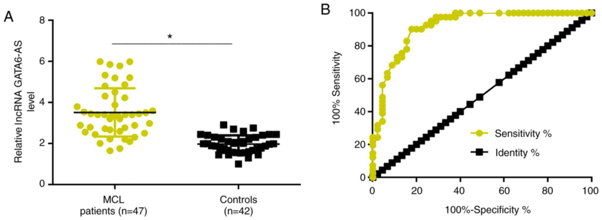
RT-qPCR was performed to detect the expression of
lncRNA GATA6-AS in the plasma of patients with MCL and healthy
controls. Compared with the healthy controls, plasma expression
levels of lncRNA GATA6-AS were significantly reduced in patients
with MCL (P<0.05; Fig. 1A). ROC
curve analysis was performed to evaluate the diagnostic value of
plasma lncRNA GATA6-AS in MCL. As revealed in Fig. 2B, the area under the curve was
0.9195, with a standard error of 0.02916 and 95% confidence
interval of 0.8623–0.9767.
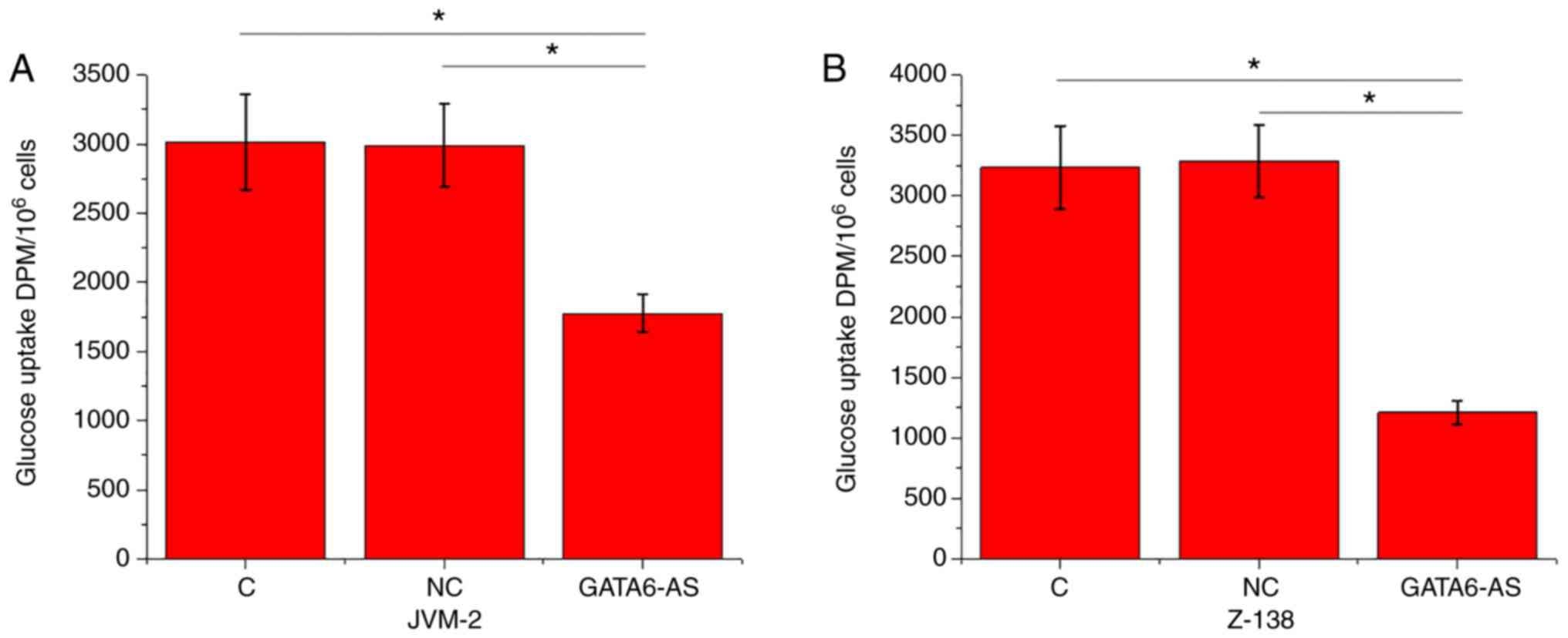
lncRNA GATA6-AS overexpression
inhibits glucose uptake in JVM-2 and Z-138 human MCL cells
Glucose uptake in JVM-2 and Z-138 human MCL cells
was detected using a glucose uptake assay. Compared with the
control and negative control groups, glucose uptake was
significantly reduced in JVM-2 (Fig.
2A) and Z-138 (Fig. 2B) human
MCL cells following lncRNA GATA6-AS overexpression (P<0.05).
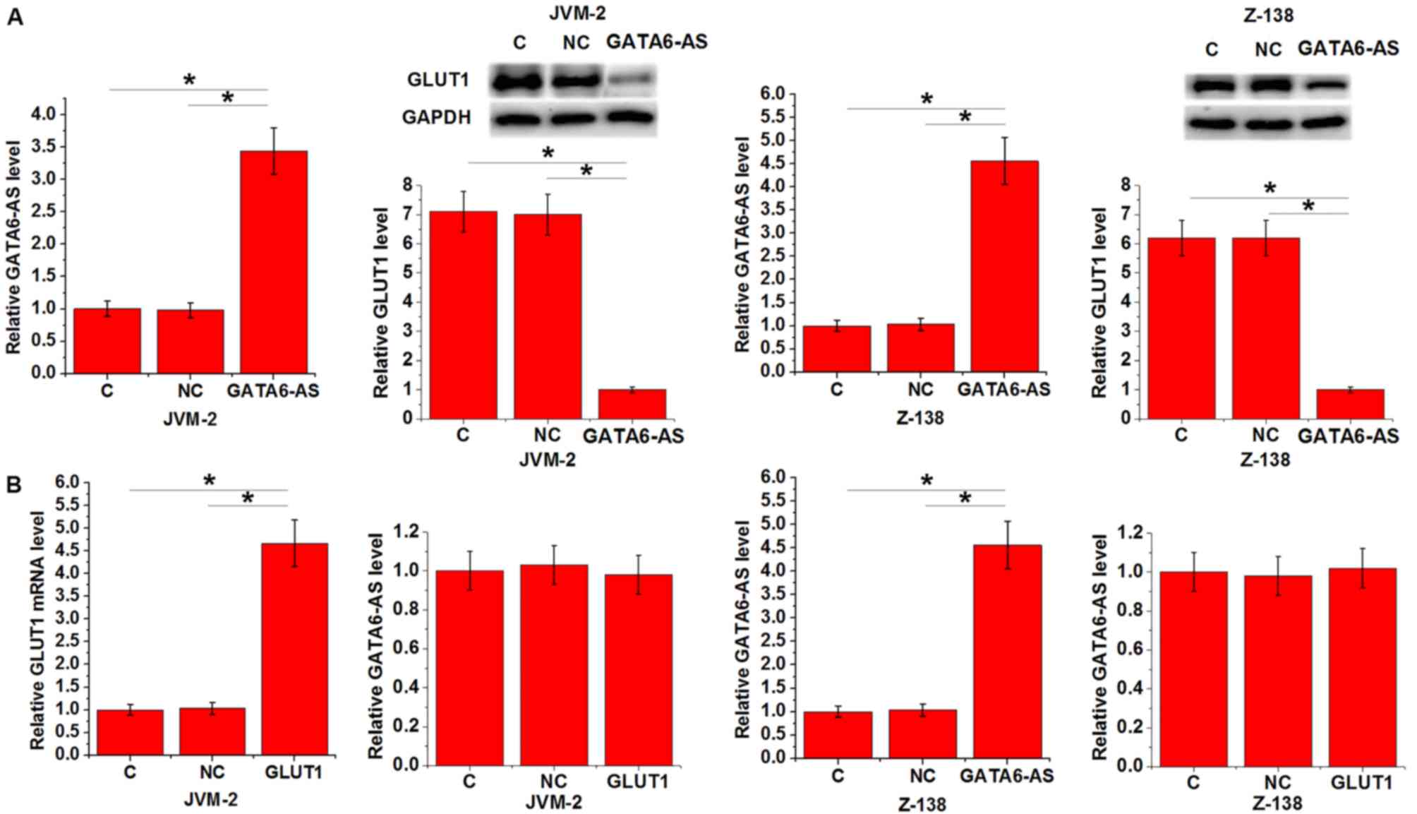
lncRNA GATA6-AS regulates GLUT1
expression in JVM-2 and Z-138 human MCL cells
GLUT1 is a one of the principal components of
glucose uptake. Therefore, the expression of GLUT1 was detected in
JVM-2 and Z-138 human MCL cell lines following lncRNA GATA6-AS
overexpression. Compared with control and negative control groups,
expression of GLUT1 was significantly reduced in both cell lines
following lncRNA GATA6-AS overexpression (P<0.05; Fig. 3A). By contrast, no significant
differences in the expression levels of lncRNA GATA6-AS were
observed following GLUT1 overexpression (P<0.05; Fig. 3B).
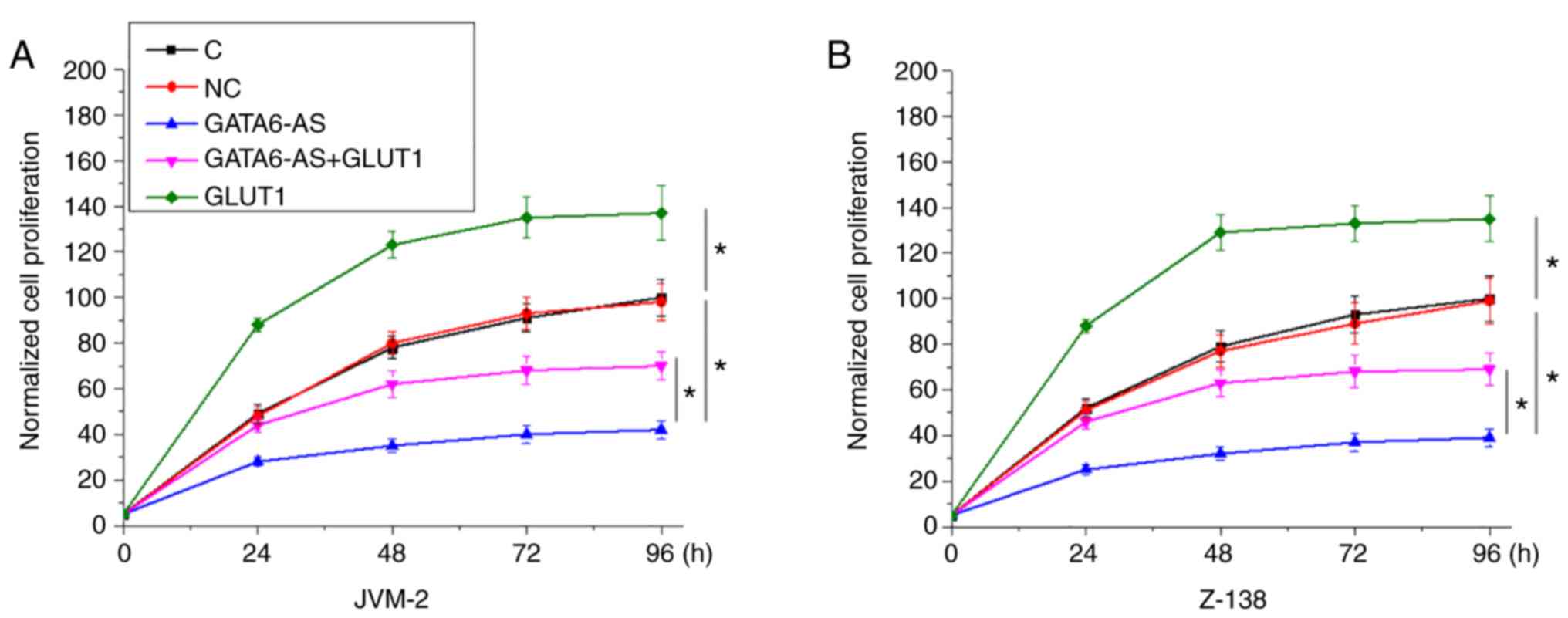
lncRNA GATA6-AS regulates GLUT1
involvement in the proliferation of JVM-2 and Z-138 human MCL
cells
Following transfection with lncRNA GATA6-AS and
GLUT1 expression vectors, the proliferation of JVM-2 and Z-138
human MCL cells was detected using a CCK-8 assay. Compared with
control and negative control groups, GATA6-AS overexpression
significantly inhibited, while GLUT1 overexpression significantly
promoted the proliferation of JVM-2 (Fig. 4A) and Z-138 (Fig. 4B) human MCL cells. Compared with
cells transfected with GATA6-AS expression vectors alone, cells
transfected with both GATA6-AS and GLUT1 expression vectors showed
significantly increased proliferation rates (P<0.05).
Discussion
lncRNA GATA6-AS has recently been identified as a
key component in endothelial-mesenchymal transition, while its
involvement in other physiological or pathological processes is
unknown. In the present study, a novel function of lncRNA GATA6-AS
was reported in MCL. This is the first report to indicate the
involvement of lncRNA GATA6-AS in cancer cell proliferation, which
is likely to be as a result of its interaction with GLUT1.
As a primary component of glucose uptake, GLUT1 is
involved in the regulation of cancer cell proliferation in
different types of human malignancies, including prostate cancer
(10) and triple-negative breast
cancer (11). The involvement of
GLUT1 in MCL still hasn't been well studied. In the present study,
GLUT1 overexpression resulted in significant promotion of MCL cell
proliferation. Therefore, GLUT1 may also serve an oncogenic role in
MCL by promoting cancer cell proliferation.
It has been reported that the development of MCL is
accompanied by changes in the expression pattern of a large set of
lncRNAs (14). However, the number
of studies addressing the functions of these lncRNAs in MCL is
limited (15). The findings of the
present study indicate that the inhibition of lncRNA GATA6-AS
expression may serve as a potential therapeutic target for the
treatment of MCL. It was also observed that lncRNA GATA6-AS was
downregulated in MCL, and that lncRNA GATA6-AS overexpression
inhibited the proliferation of MCL cell lines. This suggests that
in MCL, GATA6-AS is likely to be a tumor suppressor lncRNA, and
that its overexpression may be used therapeutically.
Early diagnosis is the key to successful
post-treatment survival in patients with cancer (16). The present study only included
patients in the early cancer stages of MCL (stage I and II). ROC
curve analysis revealed that downregulation of lncRNA GATA6-AS
effectively distinguished between patients and healthy controls.
Therefore, lncRNA GATA6-AS may have potential in the early
diagnosis of MCL.
GLUT1 is involved in cancer development and
progression through its interaction with different signaling
molecules, including lncRNAs (17,18). The
present study revealed that this involvement is likely to be
downstream of lncRNA GATA6-AS in the regulation of MCL cell
proliferation. However, as GLUT1 overexpression only attenuated,
not reversed the inhibitory effects of GATA6-AS overexpression on
cell proliferation, lncRNA GATA6-AS may interact with multiple
signaling molecules. Future studies will focus on the
identification of other downstream effectors of lncRNA
GATA6-AS.
Notably, GLUT 2–4 (class 1 GLUT), GLUT5 (class 2
GLUT) and GLUT6 (class 3 GLUT) failed to respond to lncRNA GATA6-AS
overexpression (preliminary data not shown), indicating a specific
interaction between GLUT1 and lncRNA GATA6-AS. In addition, no
potential targeting site of lncRNA GATA6 was identified on GLUT1;
therefore, it was hypothesized that the interactions between lncRNA
GATA6 and GLUT1 were mediated by other means, including
pathological factors. Future studies aim to detect the levels of
glycolytic intermediates to confirm the involvement of GLUT1 in
MLC. Furthermore, the interaction between GATA6AS with GATA6S in
MCL is also a topic for future investigation.
In conclusion, lncRNA GATA6-AS was downregulated in
MCL. Its overexpression may serve as a potential therapeutic
strategy for the treatment of MCL, by inhibiting cancer cell
proliferation through the downregulation of GLUT1.
Acknowledgements
Not applicable.
Funding
The present study was supported by the Jilin
Provincial Health Department in China (grant no. 2017ZC029), and
the Science and Technology Bureau project of Jilin City (grant no.
201830557).
Availability of data and materials
The datasets used and/or analyzed during the present
study are available from the corresponding author upon reasonable
request.
Authors' contributions
XW and PL designed the experiments. ZF performed the
experiments with the assistance of CM, MZ and CZ, who also analyzed
the data. ZF, XW and PL wrote the manuscript, which was revised by
all of the authors.
Ethics approval and consent to
participate
The present study was approved by the Ethics
Committee of Jilin Central Hospital, and all participants gave
written informed consent to participate.
Patient consent for publication
Not applicable.
Competing interests
The authors declare that they have no competing
interests.
References
|
1
|
Chang DW, Satterfield WC, Son D, Neto N,
Madewell JE, Raymond AK, Patrick CW Jr, Miller MJ, Costelloe CM and
Weber KL: Use of vascularized periosteum or bone to improve healing
of segmental allografts after tumor resection: An ovine rib model.
Plast Reconstr Surg. 123:71–78. 2009. View Article : Google Scholar : PubMed/NCBI
|
|
2
|
Sulciner ML, Serhan CN, Gilligan MM, Mudge
DK, Chang J, Gartung A, Lehner KA, Bielenberg DR, Schmidt B, Dalli
J, et al: Resolvins suppress tumor growth and enhance cancer
therapy. J Exp Med. 215:115–140. 2018. View Article : Google Scholar : PubMed/NCBI
|
|
3
|
Cheah CY, Seymour JF and Wang ML: Mantle
cell lymphoma. J Clin Oncol. 34:1256–1269. 2016. View Article : Google Scholar : PubMed/NCBI
|
|
4
|
Robak T, Huang H, Jin J, Zhu J, Liu T,
Samoilova O, Pylypenko H, Verhoef G, Siritanaratkul N, Osmanov E,
et al: Bortezomib-based therapy for newly diagnosed mantle-cell
lymphoma. N Engl J Med. 372:944–953. 2015. View Article : Google Scholar : PubMed/NCBI
|
|
5
|
Ruan J, Martin P, Shah B, Schuster SJ,
Smith SM, Furman RR, Christos P, Rodriguez A, Svoboda J, Lewis J,
et al: Lenalidomide plus rituximab as initial treatment for
mantle-cell lymphoma. N Engl J Med. 373:1835–1844. 2015. View Article : Google Scholar : PubMed/NCBI
|
|
6
|
Eskelund CW, Kolstad A, Jerkeman M, Räty
R, Laurell A, Eloranta S, Smedby KE, Husby S, Pedersen LB, Andersen
NS, et al: 15-year follow-up of the second nordic mantle cell
lymphoma trial (MCL2): Prolonged remissions without survival
plateau. Br J Haematol. 175:410–418. 2016. View Article : Google Scholar : PubMed/NCBI
|
|
7
|
Hay N: Reprogramming glucose metabolism in
cancer: Can it be exploited for cancer therapy? Nat Rev Cancer.
16:635–649. 2016. View Article : Google Scholar : PubMed/NCBI
|
|
8
|
Liberti MV and Locasale JW: The warburg
effect: How does it benefit cancer cells? Trends Biochem Sci.
41:211–218. 2016. View Article : Google Scholar : PubMed/NCBI
|
|
9
|
Olson AL and Pessin JE: Structure,
function, and regulation of the mammalian facilitative glucose
transporter gene family. Annu Rev Nutr. 16:235–256. 1996.
View Article : Google Scholar : PubMed/NCBI
|
|
10
|
Xiao H, Wang J, Yan W, Cui Y, Chen Z, Gao
X, Wen X and Chen J: GLUT1 regulates cell glycolysis and
proliferation in prostate cancer. Prostate. 78:86–94. 2018.
View Article : Google Scholar : PubMed/NCBI
|
|
11
|
Oh S, Kim H, Nam K and Shin I: Glut1
promotes cell proliferation, migration and invasion by regulating
epidermal growth factor receptor and integrin signaling in
triple-negative breast cancer cells. BMB Rep. 50:132–137. 2017.
View Article : Google Scholar : PubMed/NCBI
|
|
12
|
Neumann P, Jaé N, Knau A, Glaser SF,
Fouani Y, Rossbach O, Krüger M, John D, Bindereif A, Grote P, et
al: The lncRNA GATA6-AS epigenetically regulates endothelial gene
expression via interaction with LOXL2. Nat Commun. 9:2372018.
View Article : Google Scholar : PubMed/NCBI
|
|
13
|
Livak KJ and Schmittgen TD: Analysis of
relative gene expression data using real-time quantitative PCR and
the 2(-Delta Delta C(T)) method. Methods. 25:402–408. 2001.
View Article : Google Scholar : PubMed/NCBI
|
|
14
|
Hu G, Gupta SK, Troska TP, Nair A and
Gupta M: Long non-coding RNA profile in mantle cell lymphoma
identifies a functional lncRNA ROR1-AS1 associated with EZH2/PRC2
complex. Oncotarget. 8:80223–80234. 2017.PubMed/NCBI
|
|
15
|
Wang X, Sehgal L, Jain N, Khashab T,
Mathur R and Samaniego F: LncRNA MALAT1 promotes development of
mantle cell lymphoma by associating with EZH2. J Transl Med.
14:3462016. View Article : Google Scholar : PubMed/NCBI
|
|
16
|
Vose JM: Mantle cell lymphoma: 2015 update
on diagnosis, risk-stratification, and clinical management. Am J
Hematol. 90:739–745. 2015. View Article : Google Scholar : PubMed/NCBI
|
|
17
|
Zou ZW, Ma C, Medoro L, Chen L, Wang B,
Gupta R, Liu T, Yang XZ, Chen TT, Wang RZ, et al: LncRNA ANRIL is
up-regulated in nasopharyngeal carcinoma and promotes the cancer
progression via increasing proliferation, reprograming cell glucose
metabolism and inducing side-population stem-like cancer cells.
Oncotarget. 7:61741–61754. 2016. View Article : Google Scholar : PubMed/NCBI
|
|
18
|
Liu X and Gan B: lncRNA NBR2 modulates
cancer cell sensitivity to phenformin through GLUT1. Cell Cycle.
15:3471–3481. 2016. View Article : Google Scholar : PubMed/NCBI
|