Introduction
Breast cancer has been among the leading causes of
cancer-associated mortality among Chinese women in the past ten
years (1). The 5-year survival rates
vary from 100% (Stage I) to 22% (Stage IV) (2); thus, early diagnosis is crucial. Tumor
growth has been demonstrated to be largely dependent on
angiogenesis (3). The formation of
new blood vessels serves a pivotal role in the local growth,
invasion and distant metastasis of breast cancer (4). Therefore, this identifying this
distinct characteristic between malignant and benign breast lesions
may represent a supplementary method for improving diagnostic
performance and accuracy. Color Doppler flow imaging (CDFI) and
power Doppler flow imaging have been extensively applied in
clinical practice (5,6). However, such non-invasive methods
adjunct to grayscale ultrasonography (US) exhibit low sensitivity
in detecting microvascularity (7),
and are largely dependent on the mean Doppler frequency shift, with
inevitable loss of low-velocity blood flow information. By
contrast, superb microvascular imaging (SMI), an emerging Doppler
US method, suppresses clutter to delineate a wider range of blood
flow signals with a higher resolution. Therefore, SMI is capable of
detecting low-velocity and high-velocity flow, while CDFI is unable
to detect very low-flow states due to the different blood flow
extraction principles (8). Previous
trials have confirmed the higher sensitivity of SMI in depicting
central and peripheral vessels in hepatic lesions, compared with
CDFI (9,10).
The American College of Radiology first introduced
the Breast Imaging Reporting and Data System (BI-RADS) in 2003,
which was updated in 2013 (11).
This reporting system includes a wide range of US findings for
malignancy classifications, such as shape and echogenicity pattern
and posterior acoustic characteristics. The updated version added
subdivisions to category 4, taking into consideration the fact that
BI-RADS category 4 is the most controversial (12). The likelihood of malignancy in that
stratification ranges from 2–95% (13), as BI-RADS category 4 breast masses
are affected by a diverse range of clinical factors such as
abscess, hematoma and fat necrosis (14).
To the best of our knowledge, few studies have
applied the SMI technique to differentiate the malignancy among
BI-RADS 4 breast lesions. Therefore, the aim of the present study
was to explore the diagnostic value of SMI in differentiating
between malignant and benign BI-RADS 4 breast lesions compared with
conventional US.
Patients and methods
Clinical data
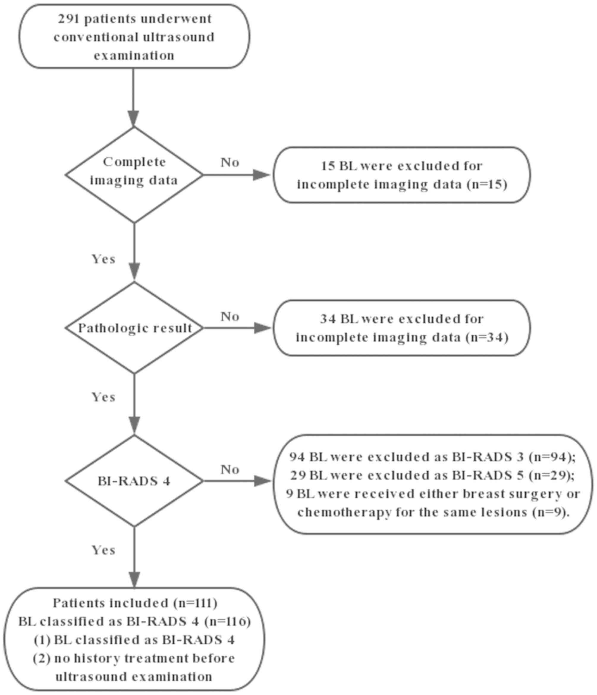
Between February 2016 and May 2018, 291 patients
were diagnosed with breast lesions detected by conventional US in
our hospital. In total, 116 lesions in 111 patients (age range,
16–64 years; mean age, 47.13±9.29 years) were recruited in this
prospective study. Among the 111 patients, 5 presented with 2
lesions. The flowchart of the selection process is presented in
Fig. 1 and the inclusion criteria
were as follows: i) Breast lesion detectable by US; ii) breast
lesion was classified as BI-RADS 4 category; and iii) no history of
treatment prior to US examination. The exclusion criteria were as
follows: i) Incomplete data (n=49); and ii) breast surgery or
chemotherapy for the same lesions (n=9). The mean diameter of the
lesions was 17.93±8.51 mm (range, 8.0–48.3 mm), while the mean
depth was 26.45±11.35 mm (range, 8.1–47.3 mm). All lesions were
pathologically confirmed by a US-guided core needle biopsy and/or
surgery according to standard clinical protocols. This prospective
study was approved by the Ethics Committee of Shanghai Pudong New
Area People's Hospital (Shanghai, China). All patients in the
present study were provided with information on all the
examinations and procedures and provided written informed consent
to participate in the study.
Equipment and methods
All patients initially underwent a grayscale US
examination, using a TOSHIBA Aplio 500 (Toshiba Medical System
Corporation, Tokyo, Japan) with high-frequency (14 MHz) line array
transducers. When a breast lesion was detected, the lesion size,
depth, shape and other US characteristics, including the margin,
echogenicity and posterior acoustic elements were recorded.
Grayscale US was followed by CDFI (frame rate 10–15 Hz) and SMI
(frame rate >50 Hz) to evaluate vascular quantity, morphology
and distribution. The velocity scope of SMI was adapted to <2.5
cm per second. Gentle pressure was applied through the transducer
to prevent vessel collapsing. During the examination, patients were
placed in the supine position with the arms elevated and were
instructed to breathe calmly.
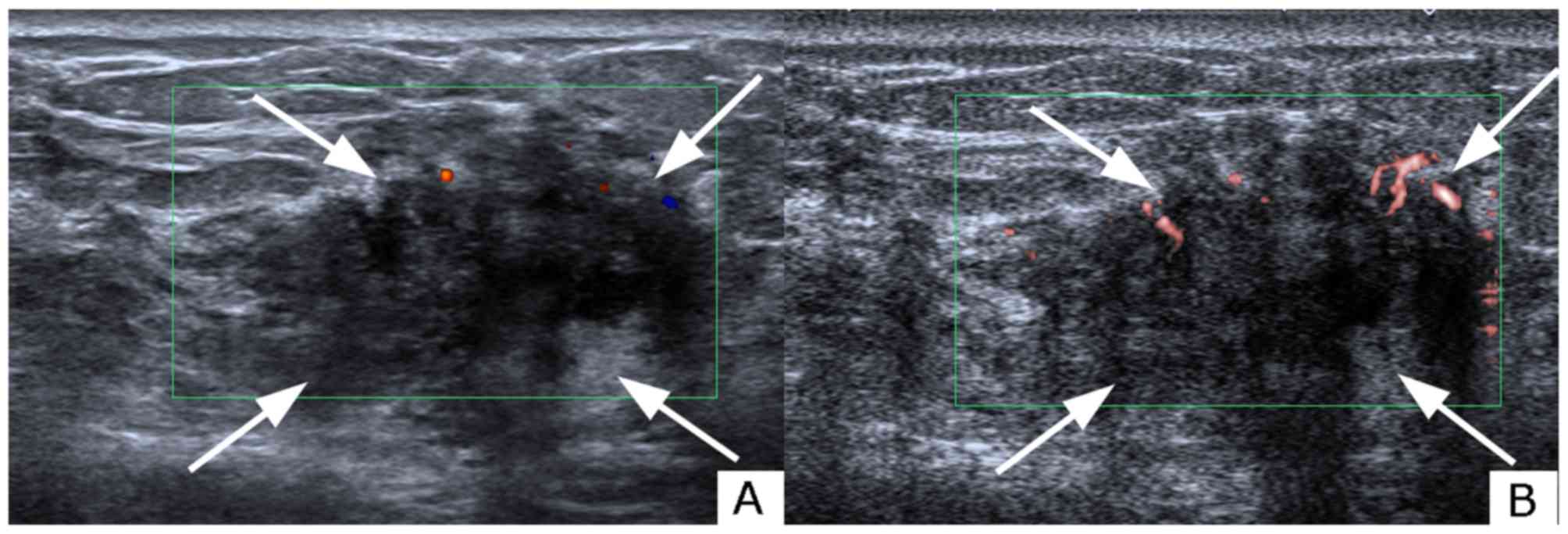
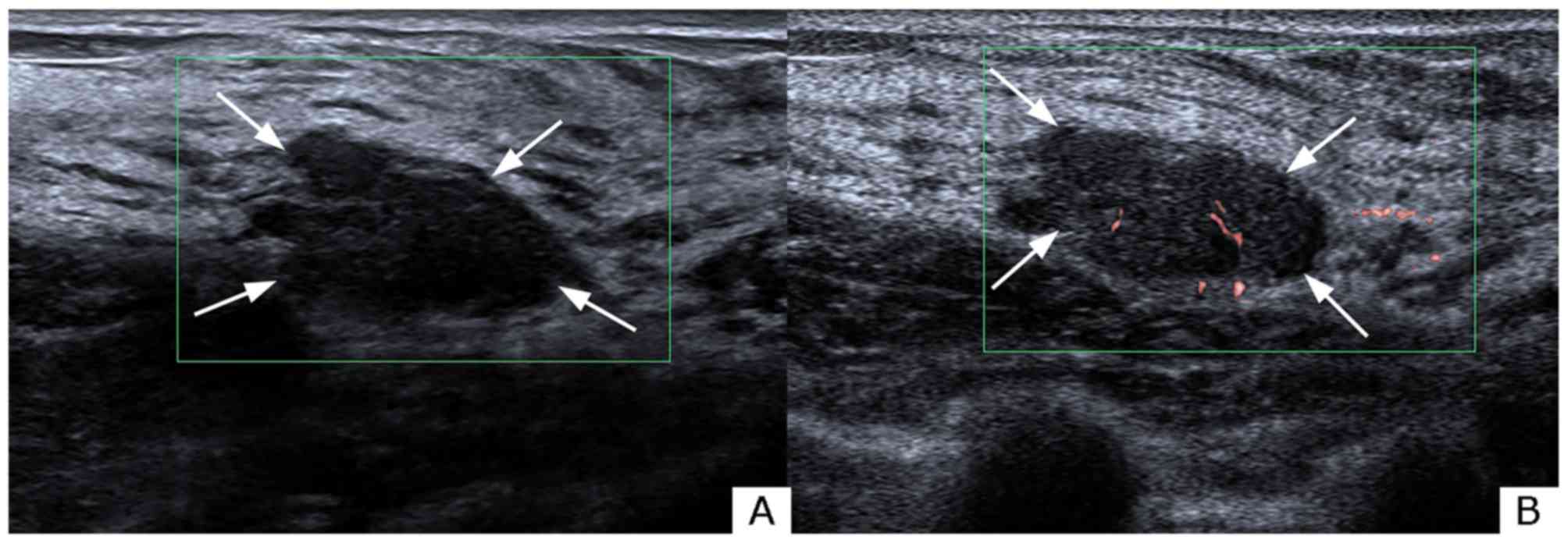
The same radiologist (YCZ) with >3 years of
experience in breast US and 1-year experience in SMI conducted all
examinations. The same imaging area for CDFI and SMI vascular blood
flow images were acquired as the reference area for the breast
tissue (Figs. 2 and 3). All images were recorded and transferred
to the hospital's internal online database. The images were
evaluated by two radiologists (YZ and SHD), who had 10- and 5-years
respective experience in CDFI and breast imaging, and 2 years in
SMI. A two-stage rating process was applied to the imaging of the
breast lesions. Each breast lesion was first rated according to
BI-RADS based on the aforementioned US characteristics; thereafter,
vascular quantity, morphology and distribution were rated based on
CDFI and SMI findings. Vascular quantity was graded according to
the Adler's classification (15) as
follows: i) Absent, grade 0; ii) minimal, grade 1; iii) moderate,
grade 2; or iv) marked, grade 3, dependent on the amount of blood
flow in the region of interest (15). Grade 0 referred to no blood flow
detected; minimal (grade 1) flow generally referred to 1 or 2
pixels containing flow (<0.1 cm in diameter); moderate (grade 2)
referred to a certain number of small vessels and/or a main vessel
and marked (grade 3) vascularity was defined as ≥4 vessels
visualized (15). Morphological
characteristics were evaluated using a classification that included
7 categories, including not applicable (N/A), linear, dot-like,
penetrating, branching, penetrating and branching, and shunt.
Vessel distribution was further divided into three categories,
peripheral, central, and both peripheral and central, respectively.
Based on these findings, the two aforementioned radiologists then
re-corrected BI-RADS stratification. If any disagreement occurred,
a third senior radiologist (QJ), with an experience of >15 years
in breast US and 2 years in SMI, was consulted. All radiologists
were blinded to the pathological findings.
Statistical analysis
The χ2 test or Fisher's exact tests were
applied for categorical variables, while an independent-samples
t-test was applied for the comparison of continuous variables.
Quantitative data are expressed as mean ± standard deviation when
normally distributed. The findings on new vessel formation from
CDFI and SMI were compared between the malignant and benign lesions
using χ2 test. A receiver operating characteristics
(ROC) curve was formulated to determine the diagnostic value of
CDFI and SMI. The areas under the curves (AUCs) of different
diagnostic modalities were compared using the χ2 test.
Statistical analysis was performed using the pathological results
as the diagnostic gold standard. P<0.05 was considered to
indicate statistically significant differences. Data analysis was
performed using SPSS 25.0 software (IBM Corp., Armonk, NY,
USA).
Results
US characteristics
Of the 116 breast lesions examined, 30 (25.9%)
lesions were pathologically confirmed as malignant. The malignant
lesions were further categorized as invasive ductal carcinomas
(n=17), ductal carcinoma in situ (n=6), tubular carcinomas
(n=2), invasive lobular carcinomas (n=2) and mucinous carcinomas
(n=3). Among the 86 benign lesions, the most common benign
pathological result was fibrocystic change (n=24), followed by
fibroadenoma (n=23) and ductal hyperplasia (n=18). The pathological
results of the breast masses are summarized in Table I. Conventional US characteristics,
such as irregular shape and non-circumscribed margins [malignant,
93.3% (28/30); benign, 77.9% (67/86)], were more commonly
identified in malignant breast masses when compared with benign
breast lesions (P<0.05; Table
II).
 | Table I.Pathological results of all BI-RADS 4
category breast lesions, [n (%)]. |
Table I.
Pathological results of all BI-RADS 4
category breast lesions, [n (%)].
| Lesions | n (%) |
|---|
| Malignant | 30 (25.9) |
| Invasive
ductal carcinoma | 17 (56.7) |
| Ductal
carcinoma in situ | 6 (20.0) |
| Tubular
carcinoma | 2 (6.7) |
| Invasive
lobular carcinoma | 2 (6.7) |
| Mucinous
carcinoma | 3 (10.0) |
| Benign | 86 (74.1) |
|
Papilloma | 7 (8.1) |
|
Fibroadenoma | 23 (26.7) |
|
Fibrocystic change | 24 (27.9) |
| Ductal
hyperplasia | 18 (20.9) |
|
Sclerosing adenosis | 3 (3.5) |
| Columnar
cell lesions | 11 (12.8) |
| Table II.Conventional ultrasound
characteristics of benign and malignant breast lesions, [n
(%)]. |
Table II.
Conventional ultrasound
characteristics of benign and malignant breast lesions, [n
(%)].
| Characteristic | Malignant (n=30) | Benign (n=86) | Overall | P-value |
|---|
| Mean age (year) | 52.43±8.12 | 45.16±8.96 | 47.13±9.29 | 0.014a |
| Size (diameter,
mm) | 28.24±9.54 | 14.34±4.05 | 17.93±8.51 | 0.014a |
| Depth (mm) | 30.11±13.06 | 25.18±10.47 | 26.45±11.35 |
<0.001a |
| Position |
|
|
| 0.924 |
|
Left | 14 (46.7) | 41 (47.7) | 55 (47.4) |
|
|
Right | 16 (53.3) | 45 (52.3) | 61 (52.6) |
|
| Shape |
|
|
|
<0.001a |
|
Oval | 5 (16.7) | 60 (69.8) | 65 (56.0) |
|
|
Round | 4 (13.3) | 14 (16.3) | 18 (15.5) |
|
|
Irregular | 21 (70.0) | 12 (14.0) | 33 (28.4) |
|
| Margin |
|
|
| 0.001a |
|
Circumscribed | 2 (6.7) | 19 (22.1) | 21 (18.1) |
|
|
Indistinct | 2 (6.7) | 26 (30.2) | 28 (24.1) |
|
|
Angular | 2 (6.7) | 5 (5.8) | 7 (6.0) |
|
|
Microlobulated | 20 (66.7) | 30 (34.9) | 50 (43.1) |
|
|
Spiculated | 4 (13.3) | 6 (7.0) | 10 (8.6) |
|
| Orientation |
|
|
| 0.032a |
|
Parallel | 16 (53.3) | 64 (74.4) | 80 (69.0) |
|
|
Non-parallel | 14 (46.7) | 22 (25.6) | 36 (31.0) |
|
| Posterior acoustic
features |
|
|
|
<0.001a |
|
None | 9 (30.0) | 57 (66.3) | 66 (56.9) |
|
|
Enhancement | 3 (10.0) | 6 (7.0) | 9 (7.8) |
|
|
Shadowing | 7 (23.3) | 23 (26.7) | 30 (25.9) |
|
|
Combined pattern | 11 (36.7) | 0 (0.0) | 11 (9.5) |
|
| Echo pattern |
|
|
| 0.060 |
|
Hypoechoic | 15 (50.0) | 54 (62.8) | 69 (59.5) |
|
|
Isoechoic | 4 (13.3) | 16 (18.6) | 20 (17.2) |
|
|
Hyperechoic | 6 (20.0) | 1 (1.2) | 7 (6.0) |
|
|
Complex | 5 (16.7) | 15 (17.4) | 20 (17.2) |
|
Evaluation of microvasculature
parameters in breast lesions using CDFI and SMI
The vascular quantity was evaluated using Adler's
classification, as shown in Table
III. CDFI and SMI exhibited a noticeable variance between
malignant and benign masses (P<0.001). CDFI identified 21 breast
masses as avascular, while SMI revealed absent vascularity in 17 of
those masses, which indicated that SMI was more efficient in
distinguishing microvessels. Based on the identification of the
microvasculature, SMI detected 80.00% of malignant lesions that
contained ≥4 vessels, while CDFI only identified 56.67% of
malignant breast lesions with rich blood flow signals. In
comparison, the majority of benign lesions were avascular (CDFI:
22.1%; SMI: 18.6%) or hypo-vascular (CDFI: 53.5%; SMI: 52.3%;
Table III). Avascular lesions were
rarely identified in malignant breast lesions; under CDFI
examination, 2 out of 30 malignant lesions exhibited this feature,
whereas SMI detected one malignant lesion with no vascularity. In
addition, the morphology of the vessels differed significantly
different between CDFI and SMI (P<0.01). Penetrating and
branching vessels were detected in malignant breast lesions using
CDFI and SMI (53.33 and 10.00%, respectively). However, SMI
exhibited higher sensitivity in detecting complex morphological
characteristics in benign breast masses, while CDFI was unable to
display penetrating, branching and shunt vessels in benign breast
masses. In terms of vessel distribution, malignant breast lesions
more frequently exhibited peripheral and both peripheral and
central distribution compared with central distribution alone,
whereas benign masses more frequently exhibited central and both
peripheral and central distribution. However, there was no
significant difference between benign and malignant breast lesions
in terms of vessel distribution using either CDFI (P=0.269) or SMI
(P=0.220) (Table III).
| Table III.Vascularity findings of malignant and
benign breast lesions using CDFI and SMI, [n (%)]. |
Table III.
Vascularity findings of malignant and
benign breast lesions using CDFI and SMI, [n (%)].
|
| CDFI | SMI |
|---|
|
|
|
|
|---|
| Variable | Malignant
(n=30) | Benign (n=86) | P-value | Malignant
(n=30) | Benign (n=86) | P-value |
|---|
| Adler
classification |
|
| 0.009 |
|
|
<0.001a |
| Grade
0 | 2 (6.7) | 19 (22.1) |
| 1 (3.3) | 16 (18.6) |
|
| Grade
1 | 5 (16.7) | 25 (29.1) |
| 2 (6.7) | 14 (16.3) |
|
| Grade
2 | 6 (20.0) | 21 (24.4) |
| 3 (10.0) | 31 (36.0) |
|
| Grade
3 | 17 (56.7) | 21 (24.4) |
| 24 (80.0) | 25 (29.1) |
|
| Morphology |
|
| <0.001 |
|
|
<0.001a |
|
N/A | 2 (6.7) | 19 (22.1) |
| 1 (3.3) | 16 (18.6) |
|
|
Linear | 14 (46.7) | 44 (51.2) |
| 1 (3.3) | 49 (57.0) |
|
|
Dot-like | 7 (23.3) | 23 (26.7) |
| 2 (6.7) | 17 (19.8) |
|
| Penetrating | 2 (6.7) | 0 (0.0) |
| 2 (6.7) | 2 (2.3) |
|
| Branching | 2 (6.7) | 0 (0.0) |
| 1 (3.3) | 2 (2.3) |
|
| Penetrating &
Branching | 3 (10.0) | 0 (0.0) |
| 16 (53.3) | 0 (0.0) |
|
| Penetrating &
Branching & Shunt | 0 (0.0) | 0 (0.0) |
| 7 (23.3) | 0 (0.0) |
|
| Distribution |
|
| 0.269 |
|
| 0.220 |
|
N/A | 2 (6.7) | 19 (22.1) |
| 1 (3.3) | 16 (18.6) |
|
|
Peripheral | 12 (40.0) | 33 (38.4) |
| 4 (13.3) | 10 (11.6) |
|
|
Central | 3 (10.0) | 6 (7.0) |
| 5 (16.7) | 15 (17.4) |
|
|
Both | 13 (43.3) | 28 (32.6) |
| 20 (66.7) | 45 (52.3) |
|
Risk of malignancy grading and
diagnostic performance of US, US + CDFI and US + SMI
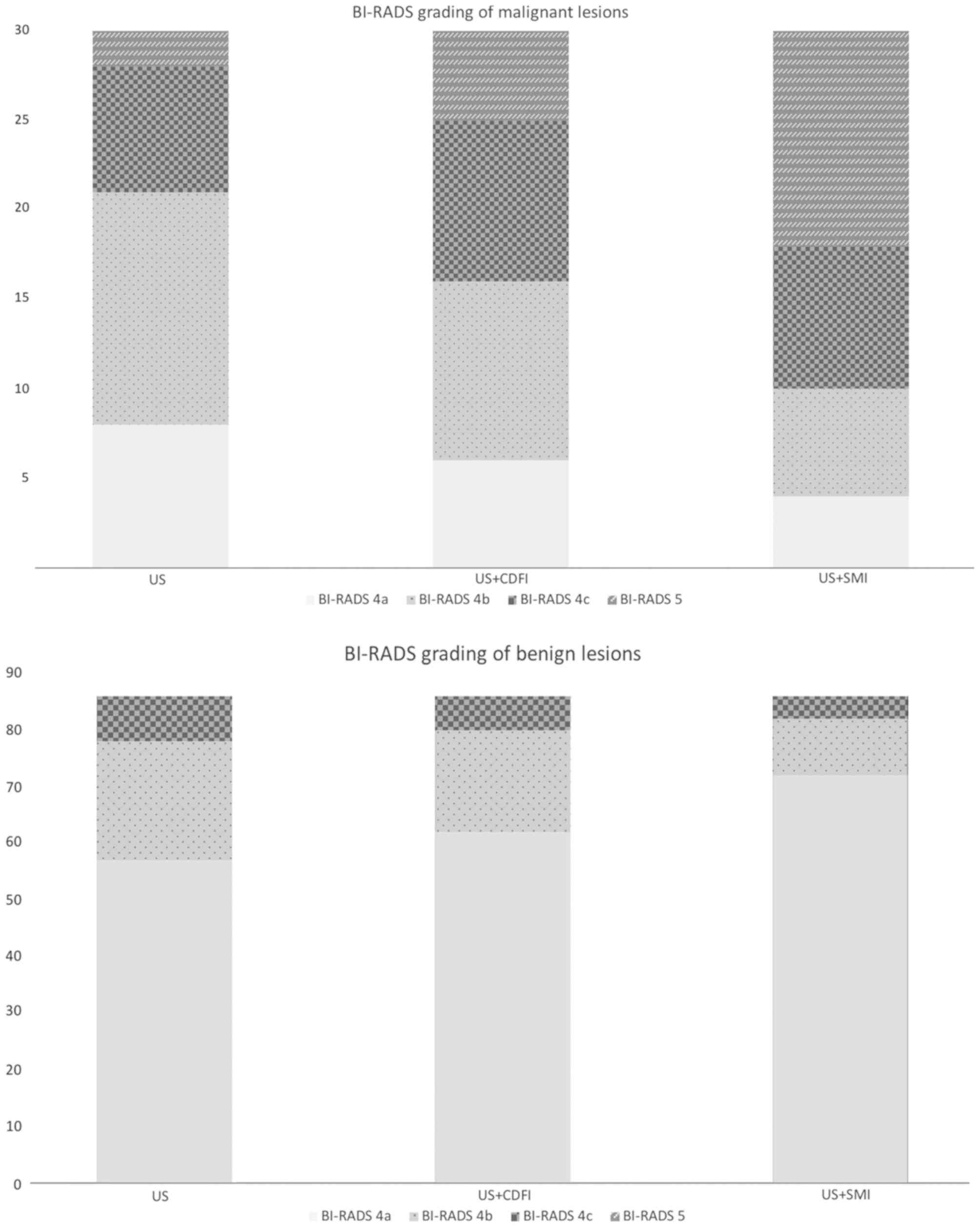
In terms of subdivision of the BI-RADS category 4 of
breast lesions, none were downgraded by either CDFI or SMI. A total
of 5 breast neoplasms were upgraded to category 5 with CDFI, while
12 breast neoplasms were upgraded to category 5 following SMI
examination (Fig. 4). Regarding the
86 benign lesions, 72, 62 and 57 were graded as BI-RADS 4a under US
+ SMI, US + CDFI, and US examination alone. None of the benign
lesions were graded as BI-RADS 5 under any of the three
examinations. When breast lesions rated as BI-RADS 4a were
considered as benign, and the remaining breast lesions rated as
BI-RADS 4b, 4c and 5 as malignant, the sensitivity, specificity and
accuracy rate for SMI were 86.67, 83.72 and 84.48%, respectively;
the sensitivity, specificity and accuracy rate for CDFI were 80.00,
72.09 and 74.14% (Table IV). The
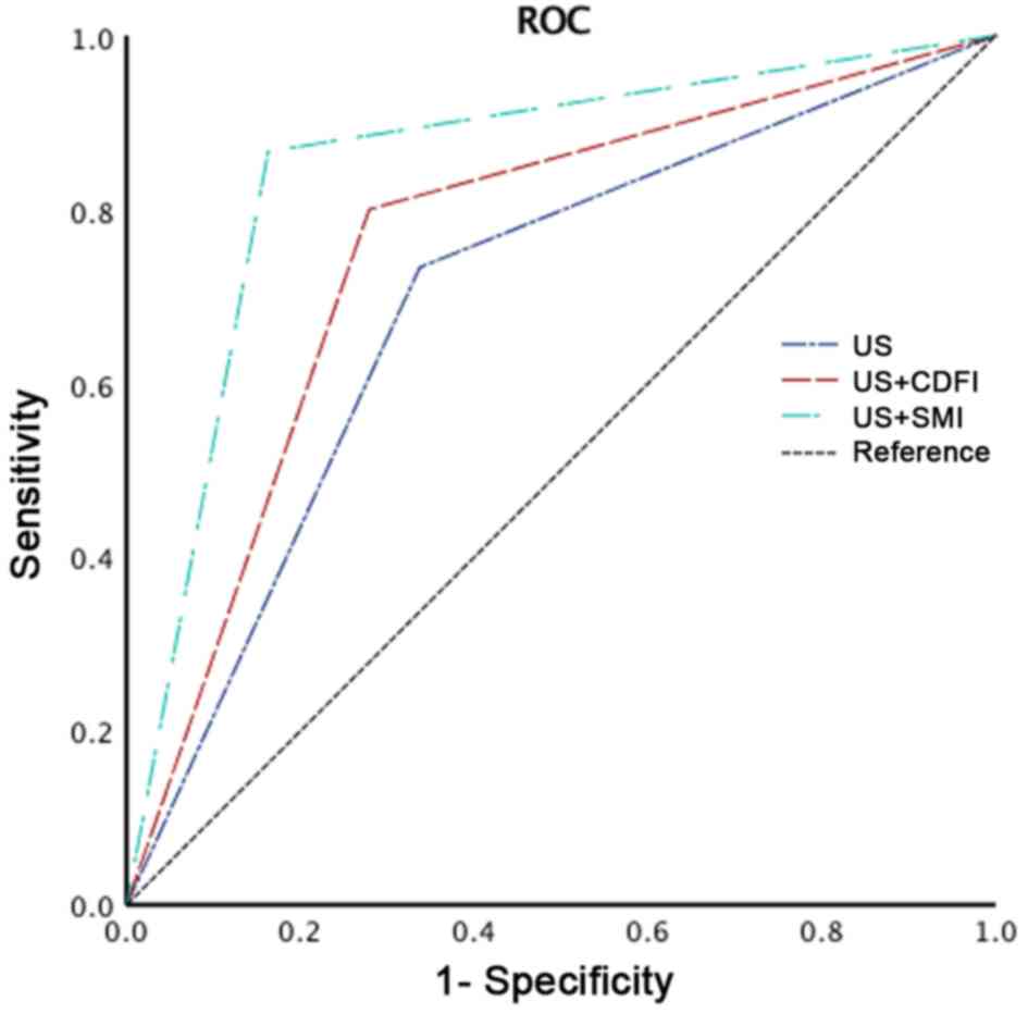
AUC values for US, US + CDFI and US + SMI were 0.698 [95%
confidence interval (CI): 0.589–0.807], 0.760 (95% CI: 0.660–0.860)
and 0.852 (95% CI: 0.768–0.936), respectively (Fig. 5). There was a significant difference
in the AUC value between US + CDFI and US + SMI (P<0.001).
| Table IV.Comparison of the diagnostic
performance of US, US+CDFI and US+SMI. |
Table IV.
Comparison of the diagnostic
performance of US, US+CDFI and US+SMI.
| Variable | Sensitivity
(%) | Specificity
(%) | Accuracy (%) | AUC | 95% CI | P-value |
|---|
| US | 73.33 | 66.28 | 68.10 | 0.698 | 0.589–0.807 |
|
| US + CDFI | 80.00 | 72.09 | 74.14 | 0.760 | 0.660–0.860 | <0.001 |
| US + SMI | 86.67 | 83.72 | 84.48 | 0.852 | 0.768–0.936 |
|
Discussion
US has been widely applied as a first-line
diagnostic technique in differentiating malignant and benign tumors
based on the evaluation of angiogenesis and the growth of irregular
vascular structures (16,17). Compared with magnetic resonance
imaging and contrast-enhanced US, Doppler US has the advantages of
being simpler, more cost-effective and more risk-free. CDFI, one of
the most widely used US techniques, provides valuable data for
evaluating blood flow, but with limitation in detecting vessels
<0.1 mm, as CDFI is generally associated with data loss due to
movement artifacts attributed to the single-dimension filter
(18). Due to the advances in US
techniques, SMI is a pioneering technique that has the ability to
visualize lower-speed bloodstream without motion artifacts
(7). This advantage has been widely
reported in the analysis of microvascular flow within thyroid
nodules (19), testicular (20) and hepatic tumors (21). Multiple studies support the efficacy
of SMI in specifically visualizing minute vessels and slow speed
blood flow, with high resolution and fewer motion artifacts. After
observing 123 breast masses, Ma et al (22) concluded that SMI (83.7%) achieved a
better visualization of vascularity compared with CDFI (74.8%). The
study of Zhu et al (23)
evaluated microvascular blood flow in 123 breast lesions in 121
patients and demonstrated that the improved visualization of the
microvasculature, including low-flow vessels, with SMI (87.80%)
when compared with CDFI (78.05%). These studies support the
findings of the present study, which demonstrated that SMI detected
the presence of vessels in 99 breast masses (85.3%), while CDFI
only detected blood flow in 95 masses (81.90%). Among avascular
breast masses, only one was pathologically proven to be malignant.
In the present study, malignant breast lesions tended to have ≥4
vessels based on the Adler's classification. Specifically, 80.00%
of the malignant breast lesions were rated as grade 3 using SMI,
whereas CDFI classified only 56.67% of such lesions as grade 3.
Therefore, malignant breast neoplasms displayed more enriched flow
signals. This finding was consistent with the nature of neoplastic
angiogenesis, as malignant and benign lesions exhibit distinct
degrees of neo-vasculature development (24). Vascularity growth is a closely
associated with neoplastic proliferation (25).
In addition to vessel quantity, the present study
also observed vascular morphology and distribution as potential
indicators of malignancy. A high correlation between breast cancer
angiogenesis, vascular morphology and distribution has been
reported (26). One of the critical
characteristics of malignant lesions is the presence of penetrating
and complex branching patterns (27). Xiao et al (28) indicated that penetrating, spiculated
or radially arranged vessels are more likely to be identified in
malignant breast lesions. Zhan et al (29), demonstrated that SMI depicted an
increase in the median number of penetrating vessels when compared
with CDFI. Similarly, in the present study, no shunt vessels were
detected by CDFI, whereas 7 malignant breast lesions with shunt
vessels were identified by SMI. With regards to the distribution of
vascularity, there was no statistically significant difference
between CDFI and SMI. Therefore, our findings re-confirmed that
both the number and morphological characteristics of vessels are
key to differentiating breast malignancies (30). The overgrowth of immature capillaries
from the vessels surrounding the lesions may explain the findings
of the present study.
In the present study, the risk of malignancy was
based on the BI-RADS system. SMI was demonstrated to be superior in
terms of sensitivity (86.67%, 26/30), specificity (83.72%, 72/86)
and accuracy rate (84.48%, 98/116). However, the increased efficacy
of SMI in extracting microvascular information may lead to an
increase in false positive diagnoses. For example, 1 fibroadenoma
and 1 papilloma were observed to be delineated with penetrating and
branching vessels, respectively under SMI examination. When
integrating with other vascular characteristics, the two benign
breast lesions were upgraded from 4a to 4c and 4b, respectively.
Furthermore, calcifications in the hyperechoic area may result in
the misdiagnosis of true negative cases.
There were certain limitations to the present study.
Firstly, the study was only conducted in one center with limited
pathological groups. Second, all the examinations were conducted by
one radiologist and, consequently, there was no interpretation of
inter-observer differences. Third, the inclusion of samples may be
biased, as BI-RADS category 3 breast masses were not included in
the present study, although none of the examined breast masses were
downgraded to category 3 neither by CDFI or SMI. Therefore, further
research should include larger samples, from multiple centers and
include the full scale of BI-RADS categories.
In summary, the present study compared SMI with CDFI
to evaluate vascular quantity, morphology and distribution for
differentiating between malignant and benign BI-RADS 4 category
breast lesions. SMI was able to overcome the shortcomings of CDFI
in detecting low-velocity blood flow due to motion artifacts. Our
findings demonstrated that SMI is superior to CDFI in identifying
and characterizing vascular details further. We also reported that,
as an adjunct to grayscale US, SMI exhibited notable diagnostic
performance in distinguishing between malignant and benign BI-RADS
category 4 breast lesions.
Acknowledgements
Not applicable.
Funding
The present study was funded by Important Weak
Subject Construction Project of Pudong Health and Family Planning
Commission of Shanghai (Shanghai, China; grant no. PWzbr
2017-10).
Availability of data and materials
The datasets used and/or analyzed during the present
study are available from the corresponding author on reasonable
request.
Authors' contributions
YCZ designed the study. YZ, SHD and QJ collected and
analyzed the data. DMZ, JS and XRS contributed the collection of
samples and provided intellectual input. YCZ was a major
contributor in writing the manuscript. All authors read and
approved the final manuscript.
Ethics approval and consent to
participate
The Ethics Committee of Shanghai Pudong New Area
People's Hospital (Shanghai, China) approved the prospective study.
All enrolled patients were notified of the examinations and
procedure, and written informed consents was provided by all
patients.
Patient consent for publication
Not applicable.
Competing interests
The authors declare that they have no competing
interests.
Glossary
Abbreviations
Abbreviations:
|
US
|
ultrasonography
|
|
CDFI
|
color Doppler flow imaging
|
|
SMI
|
superb microvascular imaging
|
|
BI-RADS
|
Breast Imaging Reporting and Data
System
|
|
US-FNA
|
ultrasound-guided fine needle
aspiration
|
|
ROC
|
receiver-operating characteristic
|
|
AUC
|
area under the curve
|
References
|
1
|
Li T, Mello-Thoms C and Brennan PC:
Descriptive epidemiology of breast cancer in China: Incidence,
mortality, survival and prevalence. Breast Cancer Res Treat.
159:395–406. 2016. View Article : Google Scholar : PubMed/NCBI
|
|
2
|
Rezaee A, Buck A, Raderer M, Langsteger W
and Beheshti M: Chapter 3-breast cancer BT-PET/CT in cancer: An
interdisciplinary approach to individualized imaging. Elsevier.
43–63. 2018.
|
|
3
|
Folkman J: Angiogenesis in cancer,
vascular, rheumatoid and other disease. Nat Med. 1:27–31. 1995.
View Article : Google Scholar : PubMed/NCBI
|
|
4
|
Stratman AN, Yu JA, Mulligan TS, Butler
MG, Sause ET and Weinstein BM: Chapter 24-blood vessel formation,
editor(s): Sally a. Moody, Principels of developmental genetics
(Second edition). Academic Press; pp. 421–449. 2015
|
|
5
|
Lee SH, Chung J, Choi HY, Choi SH, Ryu EB,
Ko KH, Koo HR, Park JS, Yi A, Youk JH, et al: Evaluation of
screening US-detected breast masses by combined use of elastography
and color doppler US with B-mode US in women with dense breasts: A
multicenter prospective study. Radiology. 285:660–669. 2017.
View Article : Google Scholar : PubMed/NCBI
|
|
6
|
Kook SH, Park HW, Lee YR, Lee YU, Pae WK
and Park YL: Evaluation of solid breast lesions with power Doppler
sonography. J Clin Ultrasound. 27:231–237. 1999. View Article : Google Scholar : PubMed/NCBI
|
|
7
|
Park AY, Seo BK, Woo OH, Jung KS, Cho KR,
Park EK, Cha SH and Cha J: The utility of ultrasound superb
microvascular imaging for evaluation of breast tumour vascularity:
Comparison with colour and power Doppler imaging regarding
diagnostic performance. Clin Radiol. 73:304–311. 2018. View Article : Google Scholar : PubMed/NCBI
|
|
8
|
Zhu YC, Zhang Y, Deng SH and Jiang Q: A
prospective study to compare superb microvascular imaging with
grayscale ultrasound and color doppler flow imaging of vascular
distribution and morphology in Thyroid nodules. Med Sci Monit.
24:9223–9231. 2018. View Article : Google Scholar : PubMed/NCBI
|
|
9
|
Dubinsky TJ, Revels J, Wang S, Toia G,
Sonneborn R, Hippe DS and Erpelding T: Comparison of Superb
Microvascular Imaging with color flow and power doppler imaging of
small hepatocellular carcinomas. J Ultrasound Med. 37:2915–2924.
2018. View Article : Google Scholar : PubMed/NCBI
|
|
10
|
He MN, Lv K, Jiang YX and Jiang TA:
Application of superb microvascular imaging in focal liver lesions.
World J Gastroenterol. 23:7765–7775. 2017. View Article : Google Scholar : PubMed/NCBI
|
|
11
|
Mercado CL: BI-RADS update. Radiol Clin
North Am. 52:481–487. 2014. View Article : Google Scholar : PubMed/NCBI
|
|
12
|
Levy L, Suissa M, Chiche JF, Teman G and
Martin B: BIRADS ultrasonography. Eur J Radiol. 61:202–211. 2007.
View Article : Google Scholar : PubMed/NCBI
|
|
13
|
Strigel RM, Burnside ES, Elezaby M, Fowler
AM, Kelcz F, Salkowski LR and DeMartini WB: Utility of BI-RADS
assessment category 4 subdivisions for screening breast MRI. AJR Am
J Roentgenol. 208:1392–1399. 2017. View Article : Google Scholar : PubMed/NCBI
|
|
14
|
Raza S, Goldkamp AL, Chikarmane SA and
Birdwell RL: US of breast masses categorized as BI-RADS 3, 4, and
5: Pictorial review of factors influencing clinical management.
Radiographics. 30:1199–1213. 2010. View Article : Google Scholar : PubMed/NCBI
|
|
15
|
Adler DD, Carson PL, Rubin JM and
Quinn-Reid D: Doppler ultrasound color flow imaging in the study of
breast cancer: Preliminary findings. Ultrasound Med Biol.
16:553–559. 1990. View Article : Google Scholar : PubMed/NCBI
|
|
16
|
Durand MA and Hooley RJ: Implementation of
whole-breast screening ultrasonography. Radiol Clin North Am.
55:527–539. 2017. View Article : Google Scholar : PubMed/NCBI
|
|
17
|
Niell BL, Freer PE, Weinfurtner RJ, Arleo
EK and Drukteinis JS: Screening for breast cancer. Radiol Clin
North Am. 55:1145–1162. 2017. View Article : Google Scholar : PubMed/NCBI
|
|
18
|
Thomas L and Szabo: Chapter 11-Doppler
modes in diagnostic ultrasound imaging: Inside out (Second
edition). Academic Press; pp. 431–500. 2014
|
|
19
|
Lu R, Meng Y, Zhang Y, Zhao W, Wang X, Jin
M and Guo R: Superb microvascular imaging (SMI) compared with
conventional ultrasound for evaluating thyroid nodules. BMC Med
Imaging. 17:652017. View Article : Google Scholar : PubMed/NCBI
|
|
20
|
Durmaz MS and Sivri M: Comparison of
superb micro-vascular imaging (SMI) and conventional Doppler
imaging techniques for evaluating testicular blood flow. J Med
Ultrason. 45:443–452. 2001. View Article : Google Scholar
|
|
21
|
Lee DH, Lee JY and Han JK: Superb
microvascular imaging technology for ultrasound examinations:
Initial experiences for hepatic tumors. Eur J Radiol. 85:2090–2095.
2016. View Article : Google Scholar : PubMed/NCBI
|
|
22
|
Ma Y, Li G, Li J and Ren WD: The
diagnostic value of superb microvascular imaging (SMI) in detecting
blood flow signals of breast lesions: A preliminary study comparing
SMI to color doppler flow imaging. Medicine (Baltimore).
94:e15022015. View Article : Google Scholar : PubMed/NCBI
|
|
23
|
Zhu YC, Zhang Y, Deng SH and Jiang Q:
Diagnostic performance of superb microvascular imaging (SMI)
combined with shear-Wave elastography in evaluating breast lesions.
Med Sci Monit. 24:5935–5942. 2018. View Article : Google Scholar : PubMed/NCBI
|
|
24
|
Less JR, Skalak TC, Sevick EM and Jain RK:
Microvascular architecture in a mammary carcinoma: Branching
patterns and vessel dimensions. Cancer Res. 51:265–273.
1991.PubMed/NCBI
|
|
25
|
Williams RG: The vascularity of normal and
neoplastic grafts in vivo. Cancer Res. 11:139–144. 1951.PubMed/NCBI
|
|
26
|
Kopeć M and Abramczyk H: Angiogenesis-a
crucial step in breast cancer growth, progression and dissemination
by Raman imaging. Spectrochim Acta Part A Mol Biomol Spectrosc.
198:338–345. 2018. View Article : Google Scholar
|
|
27
|
Raza S and Baum JK: Solid breast lesions:
Evaluation with power Doppler US. Radiology. 203:164–168. 1997.
View Article : Google Scholar : PubMed/NCBI
|
|
28
|
Xiao XY, Chen X, Guan XF, Wu H, Qin W and
Luo BM: Superb microvascular imaging in diagnosis of breast
lesions: A comparative study with contrast-enhanced
ultrasonographic microvascular imaging. Br J Radiol.
89:201605452016. View Article : Google Scholar
|
|
29
|
Zhan J, Diao XH, Jin JM, Chen L and Chen
Y: Superb microvascular imaging-A new vascular detecting
ultrasonographic technique for avascular breast masses: A
preliminary study. Eur J Radiol. 85:915–921. 2016. View Article : Google Scholar : PubMed/NCBI
|
|
30
|
Yen PL, Wu HK, Tseng HS, Kuo SJ, Huang YL,
Chen HT and Chen DR: Vascular morphologic information of
three-dimensional power Doppler ultrasound is valuable in the
classification of breast lesions. Clin Imaging. 36:267–271. 2012.
View Article : Google Scholar : PubMed/NCBI
|