Introduction
Non-small cell lung cancer (NSCLC) is a
heterogeneous class of tumors that accounts for >85% of all
newly diagnosed lung cancer cases (1). Despite the efforts that have been made
on the development of treatments for NSCLC, outcomes for patients
with this disease are generally poor due to the existence of
distant tumor metastasis in more than two out of three cases
(2). Cancer metastasis is a major
challenge for the treatment of almost all types of human
malignancies (3), and the prevention
of metastasis remains a major task in clinical practice.
Epithelial-mesenchymal transition (EMT) serves a
pivotal role in the metastasis of tumors (4). It has been well established that
activation of Akt signaling is involved in EMT (5), and inhibition of Akt signaling is
considered to be a promising target for the treatment of various
types of malignancy including NSCLC (6). Akt signaling in some cases achieves its
biological roles through interactions with long non-coding RNAs
(lncRNAs) (7,8). lncRNAs are a group of noncoding RNAs of
>200 nucleotides in length that have roles in cancer biology
(9). Long intergenic non-coding RNA
for kinase activation (LINK-A) is a recently identified lncRNA with
oncogenic effects in triple-negative breast cancer (TNBC) (10). It has also been reported that LINK-A
may induce oncogenic effects in TNBC through the activation of Akt
signaling (11). However, the
interaction between LINK-A and Akt signaling in other diseases
remains unknown. In the present study, the role of LINK-A lncRNA in
the metastasis of NSCLC through the activation of Akt signaling was
investigated.
Materials and methods
Patients
The present study included 134 patients with NSCLC
who were diagnosed and treated in the Navy General Hospital
People's Liberation Army (PLA) (Beijing, China) between January
2014 and March 2016. These patients included 72 males and 62
females, with an age range of 28–71 years, and a mean age of 47±7.2
years. Inclusion criteria were as follows: i) Patients were
pathologically diagnosed with NSCLC and undergoing first time
treatment; ii) patients were educated to above primary high school
level; iii) patients and their families were willing to
participate; iv) the patients completed the whole treatment
procedure in the Navy General Hospital PLA; v) the patients
completed 2 years of follow-up; and vi) only a single type of
metastasis was identified in cases of metastatic NSCLC. Exclusion
criteria were as follows: i) NSCLS was combined with other types of
malignancy; ii) the patients were treated prior to admission; iii)
the patients were transferred to the Navy General Hospital PLA
during treatment; or vi) the patients died of other causes during
follow-up. A total of 45 healthy controls (age range, 28–70 years;
mean age, 46±7.1 years) were enrolled at the physical health center
of the same hospital. The control group included 31 males and 14
females. No significant differences in age and sex were found
between two groups. The Ethics Committee of the Navy General
Hospital PLA approved the present study and all subjects provided
written informed consent.
Grouping and specimen collection
Among the 134 NSCLC patients, brain metastasis (BRM)
was found in 32 cases, bone metastasis (BOM) in 44 patients, liver
metastasis (LM) in 26 cases and no metastasis (NM) in the remaining
32 patients. Blood was extracted from the patients and the healthy
controls on the day of admission. Blood was also extracted from
patients on the day of discharge. Plasma was separated from blood
by centrifuging blood samples in EDTA tubes at 1,200 × g for 14 min
at room temperature. Plasma samples were stored in liquid nitrogen
before use.
Cell lines, cell culture and
transfection
The present study included 2 human NSCLC cell lines,
NCI-H1993 (H1993) and NCI-H1581 (H1581), and a normal human lung
cell line, HBEC3-KT (ATCC). RPMI-1640 medium containing 10% FBS
(Sangon Biotech Co., Ltd.) was used to culture the cells at 37°C
with 5% CO2. Full length LINK-A cDNA was obtained by PCR
using Phusion® High-Fidelity DNA Polymerase (New England
BioLabs, Inc.). The primer sequences used were: Forward,
5′-TGGAATTCAAGCTGTGGGTG-3′ and reverse,
5′-GCATTTTTATTTTAATTGAGGA-3′. Thermocycling conditions were as
follows: 95°C for 5 min; followed by 30 cycles of 95°C for 30 sec,
57°C for 30 sec and 72°C for 30 sec; and 72°C for 10 min. The cDNA
was inserted into a linearized pIRES2-EGFP vector (Clontech
Laboratories, Inc.) to produce an LINK-A expression vector.
Lipofectamine® 2000 reagent (cat. no. 11668-019;
Invitrogen; Thermo Fisher Scientific, Inc.) and 10 nM vector was
transfected into 5×10⁵ cells. Untransfected cells were
used as a control and cells transfected with empty vectors were
used as the negative control. Expression of LINK-A, determined by
RT-qPCR as aforementioned, at 200% of the control (200–220% in the
three cell lines) was achieved at 24 h post-transfection prior to
subsequent experiments. For Akt activator and inhibitor treatment,
cells were treated with Akt activator SC79 (5 and 10 µg/ml; cat.
no. SML0749; Sigma-Aldrich; Merck KGaA) or Akt inhibitor (SH-6; 50
µM; cat. no. sc-205974; Santa Cruz Biotechnology) at 37°C for 24 h
prior to use in subsequent experiments.
Reverse transcription-quantitative PCR
(RT-qPCR)
TRIzol® reagent (Thermo Fisher
Scientific, Inc.) was used to extract total RNA from plasma and
in vitro cultivated NCI-H1993 and NCI-H1581 cells. Reverse
transcription was performed to synthesize cDNA using a
High-Capacity cDNA Reverse Transcription kit (Applied Biosystems;
Thermo Fisher Scientific, Inc.) and oligo(dT) as the primer.
Reaction conditions were as follows: 25°C for 5 min, 50°C for 20
min and 75°C for 10 min. The KAPA Taq PCR kit (Kapa Biosystems;
Roche Diagnostics) was used to prepare samples for PCR and the
thermocycling conditions were as follows: 95°C for 5 min, followed
by 40 cycles of 95°C for 30 sec, 57°C for 30 sec and 72°C for 30
sec, and then 72°C for 10 min. The PCR product was checked by
agraose gel electrophoresis with ethidium bromide staining at room
temperature for 20 min. The gel was visulized using Gel Doc™ EZ Gel
Documentation System (Bio-Rad Laboratories, Inc.). SYBR Green PCR
Master mix (Thermo Fisher Scientific Inc.) was used in qPCR samples
and the following primers were used: Human LINK-A forward,
5′-TTCCCCCATTTTTCCTTTTC-3′ and reverse, 5′-CTCTGGTTGGGTGACTGGTT-3′;
and β-actin forward, 5′-GACCTCTATGCCAACACAGT-3′ and reverse,
5′-AGTACTTGCGCTCAGGAGGA-3′. qPCR was performed using the CFX96
Touch™ Real-Time PCR Detection system (Bio-Rad Laboratories, Inc.).
The thermocycling parameters for qPCR were: 95°C for 45 sec,
followed by 40 cycles of 95°C for 12 sec and 57°C for 28 sec. Data
were processed using the 2−ΔΔCq method (12) and LINK-A expression was normalized to
the lowest value of the endogenous control β-actin.
Transwell migration and invasion
assays
The three cell lines were added to RPMI-1640 medium
(Thermo Fisher Scientific, Inc.) containing 1% fetal bovine serum
(FBS; Sigma-Aldrich; Merck KGaA) and cell suspensions were prepared
at a density of ~4×10⁴ cells/ml. For the invasion assay,
the upper Transwell chambers were precoated with Matrigel (cat. no.
356234; EMD Millipore), and for the migration assay, the Transwell
chambers were uncoated. The upper Transwell chamber was filled with
0.1 ml cell suspension, and the lower chamber was filled with
RPMI-1640 medium supplemented with 20% FBS to induce cell migration
or invasion. The cells were incubated for 24 h at 37°C. The cells
were removed from the membranes using cotton swabs and the invading
and migrating cells were stained with 0.5% crystal violet
(Sigma-Aldrich; Merck KGaA) for 15 min at room temperature. Cell
migration and invasion were normalized to cell proliferation rate
at 24 h, which was performed using a Cell Counting Kit-8
(Sigma-Aldrich; Merck KGaA) by measuring optical density at 450 nm,
and data were expressed as a percentage of the control group.
Western blot analysis
Total protein was extracted using RIPA buffer
(Thermo Fisher Scientific, Inc.), and bicinchoninic acid assay
(BCA-1; Sigma-Aldrich; Merck KGaA) was performed to determine the
protein concentration. SDS-PAGE using a 10% gel was performed with
20 µg denatured protein per well. Following transfer of protein to
PVDF membranes, blocking was performed by incubation with skimmed
milk (5%) in PBS at room temperature for 1 h. The membranes were
then incubated with rabbit anti-human primary antibodies against
Akt (1:1,200; cat. no. ab126811; Abcam), phosphorylated (p-)Akt
(phospho T308; 1:1,400; cat. no. ab38449; Abcam), P13K (1:1,400;
cat. no. ab227204; Abcam), p-PI3K (phospho Y607; 1:1,400; cat. no.
ab182651; Abcam) and GAPDH (1:1,400; cat. no. ab9485; Abcam) at 4°C
overnight. Membranes were washed with TBST (0.3% Tween-20) and
incubated with horseradish peroxidase-conjugated immunoglobulin G
secondary antibody (1:1,000; cat. no. MBS435036; MyBioSource, Inc.)
for 3 h at room temperature. ECL™ Western Blotting Analysis System
(Sigma-Aldrich; Merck KGaA) was used to develop signals. Grey
values of Akt and p-Akt bands were normalized to GAPDH using ImageJ
v 1.46 software (National Institutes of Health).
Statistical analysis
All statistical analyses were performed using
GraphPad Prism 6 software (GraphPad Software, Inc.). Data are
presented as the mean ± standard deviation and were compared using
Student's t-test for comparisons between 2 groups or one-way
analysis of variance followed by Fisher's least significant
difference test to compare >2 groups. Receiver operating
characteristic (ROC) curve analysis was performed to evaluate the
diagnostic values of plasma LINK-A for different types of NSCLC,
using values from patients with NSCLC as true-positive cases and
healthy controls as true-negative cases. Kaplan-Meier plotter
(http://kmplot.com/analysis) was used to
plot survival curves, which were compared by log-rank test.
Comparisons of mortality rates were performed by χ2
test. P<0.05 was considered to indicate a statistically
significant difference.
Results
Plasma levels of LINK-A in healthy
controls and patients with different types of NSCLC
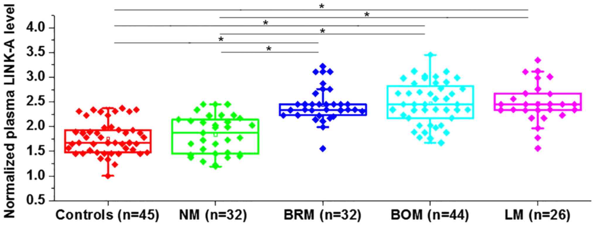
Differential expression indicates the involvement of
a certain gene in a disease. The expression of LINK-A was detected
in the plasma of healthy controls and in patients with
non-metastatic NSCLC and different types of metastatic NSCLC. The
expression level of LINK-A in the plasma was significantly higher
in the patients with three types of metastatic NSCLC (BRM, BOM and
LM) compared with that in patients with NM NSCLC and healthy
controls (Fig. 1). However, no
significant differences in plasma level of LINK-A were found
between healthy controls and patients with NM NSCLC. Data were
normalized to the sample with the lowest expression level.
Therefore, upregulation of LINK-A may specifically participate in
metastatic NSCLC.
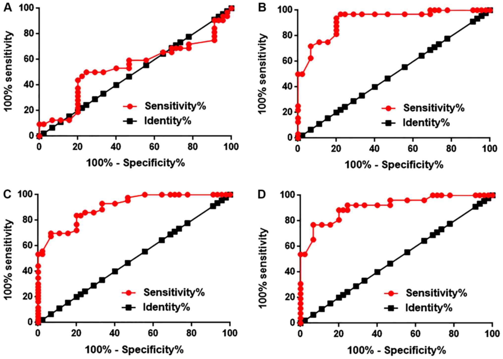
Diagnostic values of plasma levels of
LINK-A for different types of NSCLC
ROC curve analysis was performed to evaluate the
diagnostic values of plasma LINK-A for different types of NSCLC.
The area under the curve (AUC) of diagnosis of NM NSCLC was 0.5293,
with a standard error of 0.07086 and a 95% confidence interval of
0.4007 to 0.6785 (P=0.5558; Fig.
2A). The AUC of BRM NSCLC was 0.9212, with a standard error of
0.03087 and a 95% confidence interval of 0.8607 to 0.9817
(P<0.0001; Fig. 2B). The AUC of
BOM NSCLC was 0.9044, with a standard error of 0.02994 and a 95%
confidence interval of 0.8457 to 0.9631 (P<0.0001; Fig. 2C). The AUC of LM NSCLC was 0.9120,
with a standard error of 0.03587 and a 95% confidence interval of
0.8414 to 0.9825 (P<0.0001; Fig.
2D). Therefore, plasma LINK-A may serve as a diagnostic marker
for metastatic NSCLC, but not NM NSCLC.
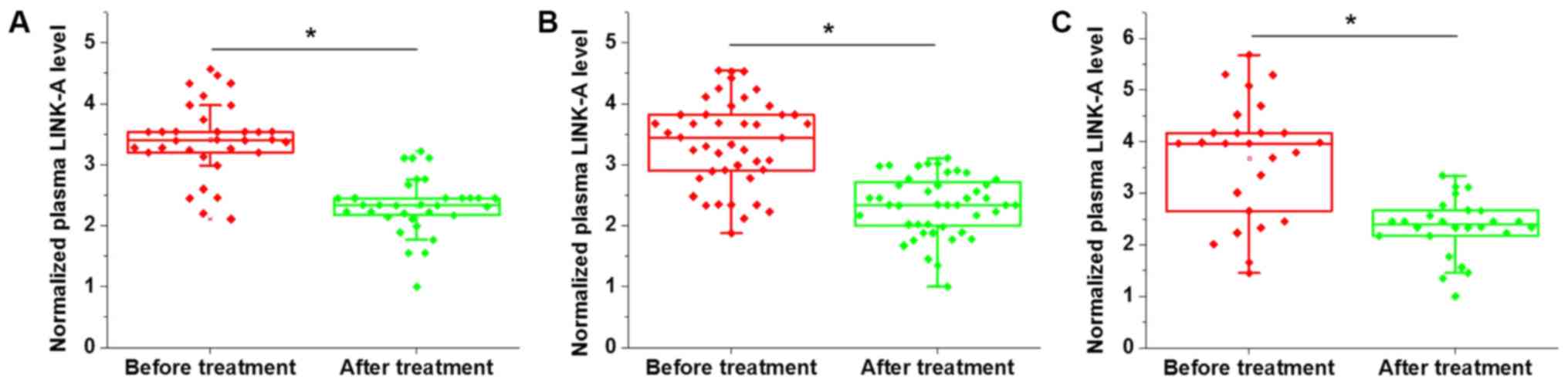
Comparison of plasma levels of LINK-A
prior to treatment and following discharge in patients with three
types of metastatic NSCLC
Comparison of plasma levels of LINK-A prior to
treatment and following discharge in patients with three types of
metastatic NSCLC indicated that, compared with the pre-treatment
levels, the plasma level of LINK-A were significantly decreased in
patients with BRM (Fig. 3A), BOM
(Fig. 3B) and LM (Fig. 3C) NSCLC. Therefore, plasma LINK-A may
serve as a marker for the treatment of metastatic NSCLC. Data were
normalized to the sample with the lowest expression level.
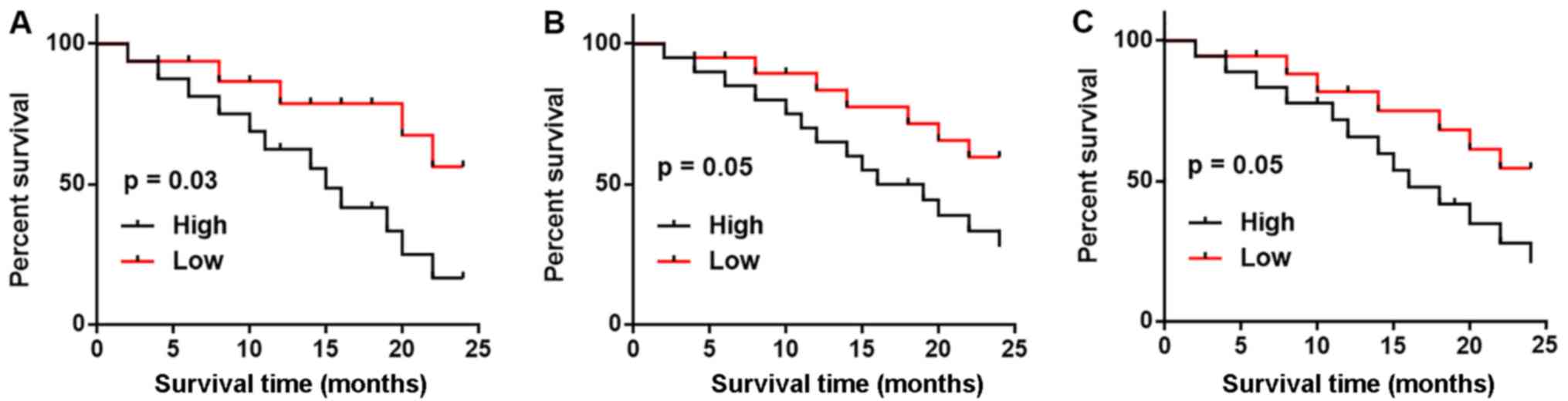
Comparison of 2-year mortality and
progression-free survival rates between patients with high and low
plasma levels of LINK-A
All patients with three types of metastatic NSCLC
were followed-up for two years and the survival conditions were
recorded. According to the median plasma level of LINK-A measured
on the day of admission, patients with each type of metastatic
NSCLC were divided into high and low groups. As shown in Table I, patients with high plasma levels of
LINK-A had higher mortality and lower progression-free survival
rates compared with patients with low plasma levels of LINK-A in
all three subtypes of metastatic NSCLC. Kaplan-Meier curve was used
to plot survival curves and these were compared by log-rank test.
As shown in Fig. 4, patients with
high LINK-A expression levels exhibited notably worse survival
rates compared with patients with low LINK-A expression levels in
BRM (Fig. 4A), BOM (Fig. 4B) and LM (Fig. 4C); however, the difference was only
significant in patients with BRM.
 | Table I.Comparison of 2-year MR and PFS rates
between patients with high and low plasma levels of LINK-A. |
Table I.
Comparison of 2-year MR and PFS rates
between patients with high and low plasma levels of LINK-A.
| Type of NSCLC | LINK-A
expression | Deceased, n | MR, % | Cases of PFS, n | PFS rate, % |
|---|
| Brain-metastatic | High (n=16) | 13 | 81.25 | 1 | 6.25 |
|
| Low (n=16) | 7 | 43.75 | 3 | 18.75a |
| Bone-metastatic | High (n=22) | 14 | 63.64 | 2 | 9.09 |
|
| Low (n=22) | 8 | 36.36 | 4 | 18.18a |
| Liver-metastatic | High (n=13) | 9 | 69.23 | 0 | 0.00 |
|
| Low (n=13) | 6 | 46.15 | 2 | 15.38a |
Effects of LINK-A overexpression on
Akt and cell migration and invasion
The present results indicate that LINK-A may be
involved in the metastasis of NSCLC. Activation of Akt plays
pivotal roles in tumor metastasis (13). Therefore, the potential interaction
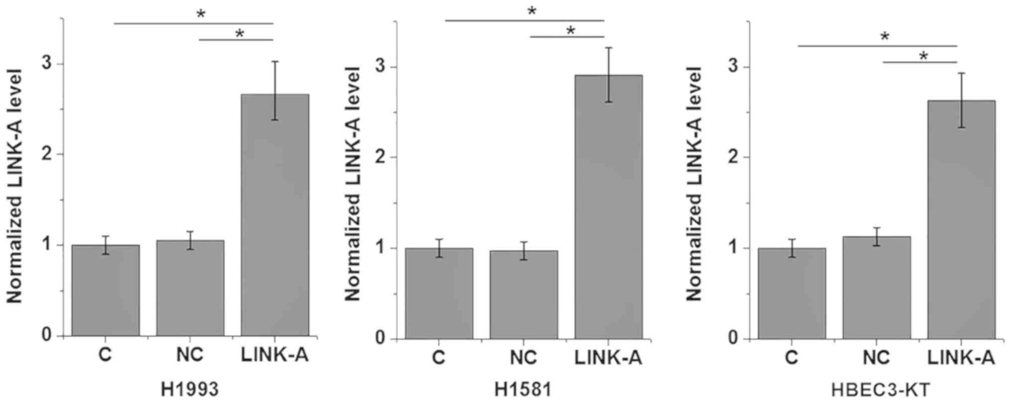
between LINK-A and Akt in NSCLC was explored by transfecting a
LINK-A expression vector into cells of human NSCLC and normal lung
tissue cell lines. LINK-A was overexpressed at 24 h
post-transfection (Fig. 5). As shown
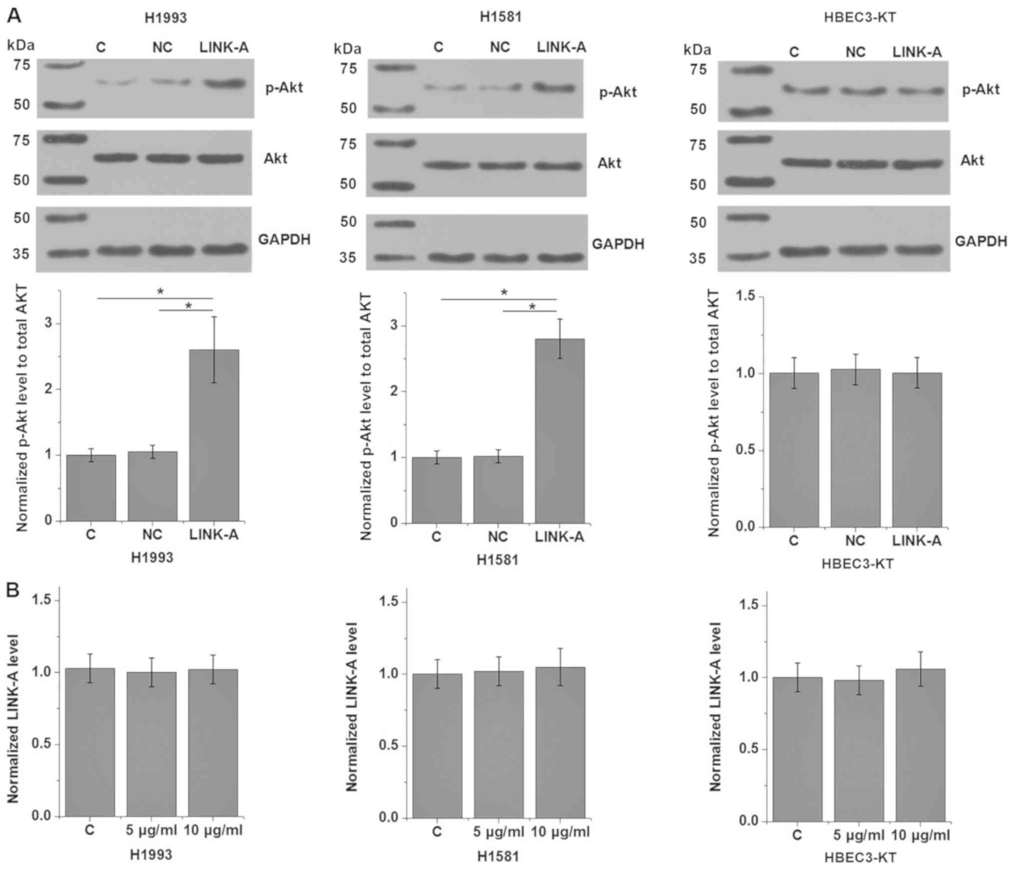
in Fig. 6A, LINK-A overexpression
significantly promoted the phosphorylation of Akt (P<0.05), but
not the expression of Akt in the cells of the human NSCLC H1993 and
H1581 cell lines compared with the control group (P<0.05). No
effect on p-Akt was observed on cells of the normal human lung
HBEC3-KT cell line compared with the control group. LINK-A
overexpression exhibited no significant effects on PI3K and p-PI3K
levels (data not shown), therefore PI3K was not further analyzed.
By contrast, Akt activator SC79 treatment (5 and 10 µg/ml) had no
significant effect on LINK-A expression (Fig. 6B). The involvement of LINK-A in NSCLC
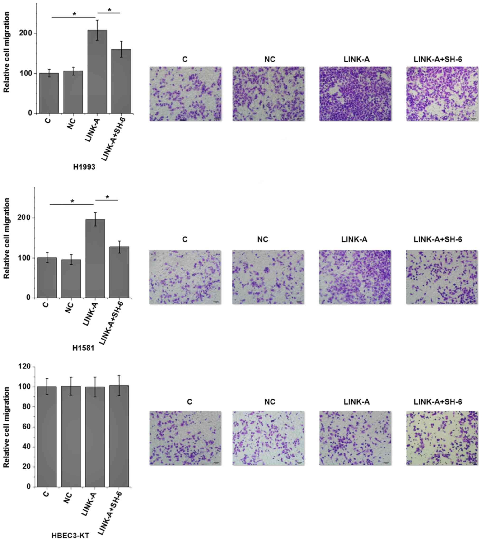
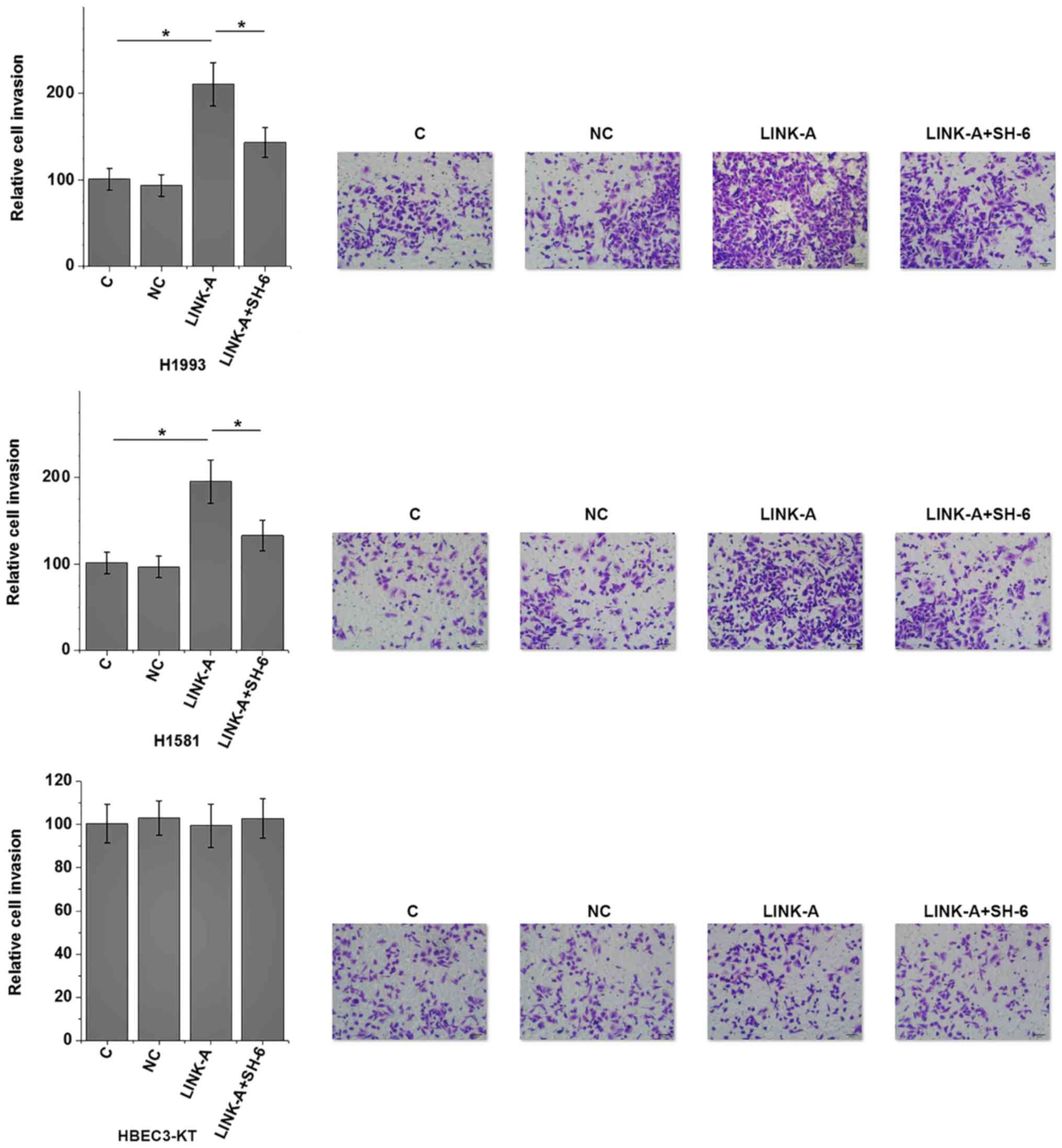
was further tested by in vitro cell migration and invasion
assays. Results showed that LINK-A overexpression significantly
promoted the migration (Fig. 7) and
invasion (Fig. 8) of the NSCLC H1993
and H1581 cell lines (P<0.05), but not the normal human lung
HBEC3-KT cell line (P>0.05). In addition, treatment with an Akt
inhibitor (SH-6; 50 µM) significantly reduced the enhancing effect
of LINK-A overexpression on cell migration (Fig. 7) and invasion (Fig. 8) in the NSCLC cell lines, but not the
normal human lung cell line. Therefore, LINK-A may mediate NSCLC
cell migration and invasion by functioning as an upstream activator
of Akt signaling. In addition, SH-6 alone is unlikely to affect the
migration and invasion of normal human lung HBEC3-KT cells, as both
LINK-A and LINK-A + SH-6 failed to significantly affect the
migration and invasion of these cells.
Discussion
In the present study, LINK-A was detected in the
plasma of patients with NSCLC and healthy controls. Upregulation of
LINK-A in metastatic NSCLC may promote the migration and invasion
of NSCLC cells through the activation of Akt signaling. The
interaction between LINK-A and Akt reported in the present study is
consistent with the role of this lncRNA in TNBC (11), indicating that different malignancies
may share similar pathological pathways.
Novel metastatic markers are required due to the
clinical value in estimating patient prognosis, and as potential
therapeutic targets (14). lncRNAs
serve critical roles in the pathogenesis of numerous aspects of
malignancies (9). Onset and
development of NSCLC is usually accompanied by changes in
expression of a large set of lncRNAs, and those lncRNAs show
upregulated or downregulated expression patterns that are
associated with the promotion or inhibition of disease progression
(15). In previous studies,
expression of lncRNAs SPRY4-IT1 and CCAT2 was increased in tumor
tissues compared with expression in paired healthy tissues in
patients with NSCLC (16,17), supporting their roles as oncogenic
lncRNAs in this disease. By contrast, lncRNA LOC285194 is
downregulated in NSCLC compared with that in healthy lung tissue,
indicating its role as a tumor suppression lncRNA (18). However, altered expression of these
lncRNAs has been demonstrated to correlate with tumor growth and
metastasis (14). In the present
study, upregulation of LINK-A was found in patients with metastatic
NSCLC, but not in patients with NM NSCLC. In addition, ROC curve
analysis also indicated that the plasma levels of LINK-A may be
used to effectively distinguish patients with metastatic NSCLC from
healthy controls, but not patients with NM NSCLC. Therefore, LINK-A
may be associated with the metastasis of NSCLC.
The process of disease treatment is accompanied by
changes in certain substances in the blood, and detecting those
substances may provide guidance for treatment (19). In the present study, plasma levels of
LINK-A were significantly lower on the day of discharge compared
with pre-treatment levels measured on the day of admission.
Therefore, plasma levels of LINK-A may reflect the treatment
outcomes of metastatic NSCLC. It was also observed that high plasma
LINK-A was associated with a higher mortality rate and lower
progression-free survival rate compared with the rates for patients
with low plasma LINK-A levels. Therefore, monitoring the changes in
plasma levels of LINK-A may provide guidance for the prognosis of
NSCLC.
It has been reported that LINK-A can directly
interact with phosphatidylinositol (3,4,5)-trisphosphate (PIP3) and AKT pleckstrin
homology domain at the single nucleotide level to promote the
interactions between AKT and PIP3, and subsequent enzyme activation
(11). In the present study,
activation of Akt was observed in NSCLC cells with LINK-A
overexpression, while an Akt activator failed to affect LINK-A
expression. In addition, LINK-A overexpression promoted NSCLC cell
migration and invasion, while an Akt inhibitor reduced these
effects of LINK-A overexpression on migration and invasion. In the
present study, LINK-A was not found to affect Akt expression, but
impacted Akt phosphorylation. These data indicate that LINK-A and
Akt signaling are associated in NSCLC. It is also worth noting that
LINK-A overexpression had no significant effects on normal human
lung cells. In this manner, LINK-A may serve as a potential
therapeutic target for the treatment of NSCLCL by inhibiting tumor
metastasis.
As the present study included 2 NSCLC cell lines and
a normal control cell line only, future studies with more cell
lines are required to further confirm the conclusions. The current
study also did not include LINK-A siRNA silencing due to the low
efficiency of knockdown, which will be resolved in future
studies.
In conclusion, LINK-A is overexpressed in the plasma
of patients with metastatic NSCLC. LINK-A may promote the migration
and invasion of NSCLC by activating Akt signaling. However, future
clinical studies are required to test the potential application of
LINK-A as a diagnostic marker and treatment target for NSCLC.
Acknowledgements
Not applicable.
Funding
No funding was received.
Availability of data and materials
The datasets generated and analyzed during the
present study are available from the corresponding author on
reasonable request.
Authors' contributions
JQL and TG designed experiments. JQL, WS and JL
performed experiments. XL and RZ collected and analyzed the data.
TG drafted the manuscript and all authors approved the
manuscript.
Ethics approval and consent to
participate
The present study was approved by The Ethics
Committee of the People's Liberation Army Navy General Hospital
(Beijing, China) and all subjects provided written informed
consent.
Patient consent for publication
Not applicable.
Competing interests
The authors declare that they have no competing
interests.
References
|
1
|
Gridelli C, Rossi A, Carbone DP, Guarize
J, Karachaliou N, Mok T, Petrella F, Spaggiari L and Rosell R:
Non-small-cell lung cancer. Nat Rev Dis Primers. 1:150092015.
View Article : Google Scholar : PubMed/NCBI
|
|
2
|
Brahmer J, Reckamp KL, Baas P, Crinò L,
Eberhardt WE, Poddubskaya E, Antonia S, Pluzanski A, Vokes EE,
Holgado E, et al: Nivolumab versus docetaxel in advanced
squamous-cell non-small-cell lung cancer. N Engl J Med.
373:123–135. 2015. View Article : Google Scholar : PubMed/NCBI
|
|
3
|
Gupta GP and Massagué J: Cancer
metastasis: Building a framework. Cell. 127:679–695. 2006.
View Article : Google Scholar : PubMed/NCBI
|
|
4
|
Chaffer CL, San Juan BP, Lim E and
Weinberg RA: EMT, cell plasticity and metastasis. Cancer Metastasis
Rev. 35:645–654. 2016. View Article : Google Scholar : PubMed/NCBI
|
|
5
|
Larue L and Bellacosa A:
Epithelial-mesenchymal transition in development and cancer: Role
of phosphatidylinositol 3′ kinase/AKT pathways. Oncogene.
24:7443–7454. 2005. View Article : Google Scholar : PubMed/NCBI
|
|
6
|
Wang S, Yan Y, Cheng Z, Hu Y and Liu T:
Sotetsuflavone suppresses invasion and metastasis in non-small-cell
lung cancer A549 cells by reversing EMT via the TNF-α/NF-κB and
PI3K/AKT signaling pathway. Cell Death Discov. 4:262018. View Article : Google Scholar : PubMed/NCBI
|
|
7
|
Xu S, Sui S, Zhang J, Bai N, Shi Q, Zhang
G, Gao S, You Z, Zhan C, Liu F and Pang D: Downregulation of long
noncoding RNA MALAT1 induces epithelial-to-mesenchymal transition
via the PI3K-AKT pathway in breast cancer. Int J Clin Exp Pathol.
8:4881–4891. 2015.PubMed/NCBI
|
|
8
|
Zhu Y, Zhang X, Qi L, Cai Y, Yang P, Xuan
G and Jiang Y: HULC long noncoding RNA silencing suppresses
angiogenesis by regulating ESM-1 via the PI3K/Akt/mTOR signaling
pathway in human gliomas. Oncotarget. 7:14429–14440.
2016.PubMed/NCBI
|
|
9
|
Cheetham SW, Gruhl F, Mattick JS and
Dinger ME: Long noncoding RNAs and the genetics of cancer. Br J
Cancer. 108:2419–2425. 2013. View Article : Google Scholar : PubMed/NCBI
|
|
10
|
Lin A, Li C, Xing Z, Hu Q, Liang K, Han L,
Wang C, Hawke DH, Wang S, Zhang Y, et al: The LINK-A lncRNA
activates normoxic HIF1α signalling in triple-negative breast
cancer. Nat Cell Biol. 18:213–224. 2016. View Article : Google Scholar : PubMed/NCBI
|
|
11
|
Lin A, Hu Q, Li C, Xing Z, Ma G, Wang C,
Li J, Ye Y, Yao J, Liang K, et al: The LINK-A lncRNA interacts with
ptdIns (3, 4, 5) P 3 to hyperactivate AKT and confer resistance to
AKT inhibitors. Nat Cell Biol. 19:238–251. 2017. View Article : Google Scholar : PubMed/NCBI
|
|
12
|
Livak KJ and Schmittgen TD: Analysis of
relative gene expression data using real-time quantitative PCR and
the 2(-Delta Delta C(T)) method. Methods. 25:402–408. 2001.
View Article : Google Scholar : PubMed/NCBI
|
|
13
|
Qiao M, Sheng S and Pardee AB: Metastasis
and AKT activation. Cell Cycle. 7:2991–2996. 2008. View Article : Google Scholar : PubMed/NCBI
|
|
14
|
Ramankulov A, Lein M, Johannsen M,
Schrader M, Miller K and Jung K: Plasma matrix metalloproteinase-7
as a metastatic marker and survival predictor in patients with
renal cell carcinomas. Cancer Sci. 99:1188–1194. 2008. View Article : Google Scholar : PubMed/NCBI
|
|
15
|
Zhou D, Xie M, He B, Gao Y, Yu Q, He B and
Chen Q: Microarray data re-annotation reveals specific lncRNAs and
their potential functions in non-small cell lung cancer subtypes.
Mol Med Rep. 16:5129–5136. 2017. View Article : Google Scholar : PubMed/NCBI
|
|
16
|
Sun M, Liu XH, Lu KH, Nie FQ, Xia R, Kong
R, Yang JS, Xu TP, Liu YW, Zou YF, et al: EZH2-mediated epigenetic
suppression of long noncoding RNA SPRY4-IT1 promotes NSCLC cell
proliferation and metastasis by affecting the
epithelial-mesenchymal transition. Cell Death Dis. 5:e12982014.
View Article : Google Scholar : PubMed/NCBI
|
|
17
|
Zhao Z, Wang J, Wang S, Chang H, Zhang T
and Qu J: LncRNA CCAT2 promotes tumorigenesis by over-expressed
Pokemon in non-small cell lung cancer. Biomed Pharmacother.
87:692–697. 2017. View Article : Google Scholar : PubMed/NCBI
|
|
18
|
Shi X, Chen Y, Chen J, Feng F, Li J and He
J: Antisense long noncoding RNA LOC285194 is downregulated in NSCLC
and associated with poor prognosis. Chest. 149:A2562016. View Article : Google Scholar
|
|
19
|
Matsuoka Y, Nishi D, Tanima Y, Itakura M,
Kojima M, Hamazaki K, Noguchi H and Hamazaki T: Serum pro-BDNF/BDNF
as a treatment biomarker for response to docosahexaenoic acid in
traumatized people vulnerable to developing psychological distress:
A randomized controlled trial. Transl Psychiatry. 5:e5962015.
View Article : Google Scholar : PubMed/NCBI
|