Introduction
Gastric carcinoma (GC) is the fifth most common
malignant tumor and the second most common cause of
cancer-associated mortality worldwide (1). Diagnosis and treatment methods for GC
have improved each year, but the prognosis for patients remains
poor. GC recurrence and metastasis remain the most prevalent causes
of poor long-term survival (2).
The growth and metastasis of tumors requires an
adequate blood supply. However, antivascular therapies have failed
to improve the survival time of patients with late-stage GC.
Additionally, it has been hypothesized that anti-angiogenesis
therapy may induce vasculogenic mimicry (VM) formation (3,4), a
bloody supply pattern formed without the participation of
endothelial cells. Therefore, VM may serve an important role in the
development of GC, and may be the main cause of anti-angiogenic
treatment failure.
VM was first described by Maniotis et al
(5). Currently, VM has been
demonstrated in numerous types of malignant solid tumor (6–9). VM
provides a blood supply to tumors at the early stages of
tumorigenesis, as well as a pathway for tumor metastasis.
Furthermore, preliminary research has demonstrated that patients
with GC with VM have shorter survival times and higher rates of
metastasis than patients with GC without VM (9).
N-myc downstream regulated gene 1 (NDRG1) serves an
inhibitory role in the migration, invasion and metastasis of
numerous different types of tumor by regulating
epithelial-mesenchymal transition (EMT) or downregulating the
erythroblastic leukemia viral oncogene homolog (ErbB) family of
receptors (10,11). However, whether the inhibitory effect
of NDRG1 involves EMT and human epidermal growth factor 2 (HER2) in
GC has not yet been fully elucidated. Furthermore, EMT has been
confirmed to serve a key role in VM formation in a previous,
preliminary study (12). At the same
time, the NDRG1 protein inhibits angiogenesis in a number of solid
tumors by inhibiting vascular endothelial growth factor (13). However, to the best of our knowledge,
no studies have reported the inhibition of angiogenesis by NDRG1 or
the function of NDRG1 in VM formation in GC.
The present study, therefore, investigated the role
of NDRG1 in VM formation in patients with GC. The results of the
present study may reveal a new functional role of NDRG1 in GC and
may provide insights into the development of anti-angiogenic GC
drugs.
Materials and methods
Patient samples
In total, 228 human GC tissue specimens were
collected from patients who underwent surgical resection between
January 2002 and December 2012 at the Tianjin Medical University
Cancer Institute and Hospital (Tianjin, China). The age range of
patients was 28–82 years. GC tumor tissue samples were collected
from patients who had not received chemotherapy, radiotherapy or
surgical treatment of prior to surgery. All GC specimens were
formalin fixed and paraffin embedded. The histopathological
diagnosis was confirmed by trained pathologists according to the
World Health Organization histological classification of stomach
tumors. Detailed pathological and clinical data, such as age, sex,
tumor size, histological differentiation, pathological stage,
Lauren type, and metastasis and recurrence, were collected for all
samples. Ethical approval was obtained from the Ethics Committee of
Tianjin Medical University (Tianjin, China). Written informed
consent was obtained from each patient. The privacy rights of human
subjects were maintained.
Immunohistochemical and histochemical
double-staining methods
All GC tissues were fixed in 10% neutral formalin
for 24 h at room temperature, and paraffin embedded. Specimens were
sectioned into 4 µm sections. Sections were deparaffinized with
xylene, rehydrated with a decreasing gradient of ethanol (100-80%),
and endogenous peroxidase activity was blocked with 3% hydrogen
peroxide in 100% methanol for 30 min at room temperature. The
sections were rehydrated and washed with PBS and pretreated with
0.01 M citrate buffer (pH 6.0) or EDTA buffer (pH 9.0) for 15 min
at 100°C in a microwave oven. Following blocking of non-specific
binding sites using normal goat serum (cat. no. ZLI-9022; OriGene
Technologies, Inc.) for 30 min at room temperature, the sections
were incubated overnight at 4°C with rabbit polyclonal anti-NDRG1
(1:400; ab124689; Abcam), mouse monoclonal anti-cluster of
differentiation (CD) 34 (1:550; Zm-0046; OriGene Technologies,
Inc.), rabbit polyclonal anti-vascular endothelial (VE)-cadherin
(1:400 dilution, ab33168, Abcam), mouse polyclonal anti-HER2 (1:200
dilution, ZA-0023, OriGene Technologies, Inc.), rabbit polyclonal
anti-Snail (1:200; ab180714; Abcam), rabbit polyclonal anti-Twist1
(1:100; Sc-15393; Santa Cruz Biotechnology, Inc.), rabbit
polyclonal anti-epithelial (E)-cadherin (1:100; Sc-7870; Santa Cruz
Biotechnology, Inc.) and rabbit polyclonal anti-vimentin (1:100;
ab92547; Abcam). The sections were then rinsed with PBS, and
super-sensitivity S-P IHC secondary antibodies kit (cat. no PV6001,
anti-Rabbit; cat. no. PV6002, anti-Mouse; OriGene Technologies,
Inc.) was applied to the sections at room temperature for 1 h. The
sections were stained with 3,3′-diaminobenzidine chromogen for
10–15 min at room temperature and washed with distilled water.
Following immunohistochemical staining for CD34, the
sections were washed with distilled water for 5 min and incubated
with periodic acid for 10 min and with Schiff reagent for 15 min.
Finally, all sections were counterstained with hematoxylin at 37°C
for 2 min, dehydrated by increasing gradient of ethanol (80–100%)
and mounted on coverslisps with Neutral gum (OriGene Technologies,
Inc.).
Gastric mucosa membrane adjacent to the GC tissues
was used as a positive control for PBS staining. PBS was used in
place of the primary antibodies as a negative control. Normal
tissue sections were used as a positive control according to the
manufacturer's protocol for each antibody supplier. All sections
were observed and analyzed under light microscope (magnification,
×400) by two pathologists. The expression of each marker was
assessed semi-quantitatively according to the number of cells that
stained positive and the intensity of immunostaining in individual
tumor cells (14). For HER2, only
immunoreactivity in the GC cell membrane was evaluated (15).
Periodic acid-Schiff (PAS)-Alcian blue
staining protocol
Tissue sections were deparaffinized as
aforementioned, washed with distilled water and then incubated for
10 min with Alcian blue buffer at room temperature. The sections
were then washed with distilled water for 5 min, incubated with
periodic acid for 10 min at room temperature and washed with
distilled water again. Sections were incubated with Schiff reagent
for 15 min at 37°C. Finally, all sections were counterstained with
hematoxylin in 37°C for 2 min, dehydrated by increasing gradient of
ethanol (80–100%) and mounted on coverslisps with Neutral gum. All
GC sections were classified as intestinal type or diffuse type
using the Alcian blue-periodic acid-Schiff method (16).
VM channel quantification
The characteristics of VM were assessed according to
whether: The wall of VM vessels was lined with GC cells; red cells
could be observed in the VM tube; or the GC cells in the VM wall
were CD34− and PAS+ based on CD34/PAS double
staining (17). VM channels in
immunohistochemistry stained sections were counted under light
microscope (magnification, ×400). In total, 5 fields were randomly
selected, and the average channel was defined as the number of VM
vessels in one section.
Database and analysis
The data of 442 patients with GC were collected from
The Cancer Genome Atlas (TCGA) (cancergenome.nih.gov). The overall gene expression in
patients with GC was analyzed using weighted gene co-expression
network analysis (WGCNA) (18). A
functional enrichment analysis of NDRG1 was performed using
Metascape ((http://metascape.org/) and the Gene Set
Enrichment Analysis (GSEA) (software.broadinstitute.org/gsea/login.jsp) on
the data from 415 patients with GC (19). GraphPad Prism version 6 (GraphPad
Software, Inc.) was used to present the data. The function of NDRG1
was analyzed using the ClueGO plug-in application in Cytoscape
2.8.3 (https://cytoscape.org). Kyoto
Encyclopedia of Genes and Genomes (https://www.genome.jp/kegg/) pathway enrichment
analysis was carried out and visualized using ClueGO and CluePedia
with P<0.05 as the cut-off value.
Statistical analysis
The data were analyzed using SPSS (version 16.0;
SPSS, Inc., Chicago, IL, USA). P<0.05 was considered to indicate
a statistically significant difference. Two-tailed Student's test
was used for comparison of ‘VM count’ in two independent groups.
‘VM count’ in different HER2 group was analyzed using ANOVA test
followed by Student-Newman-Keuls post-hoc test. The association
between two proteins expression or VM formation and the
clinicopathologic parameters was analyzed using two-tailed
χ2 test. The correlation analysis was performed using
Pearson's correlation or Spearman's correlation. Hazard and
survival analysis were calculated using the Kaplan-Meier method,
the log-rank method and multivariate Cox regression analysis.
Results
Correlation between NDRG1, VM and
clinicopathological factors
To identify any correlations between NDRG1 and VM in
patients with GC, a total of 228 GC specimens from the Tianjin
Medical University Cancer Institute and Hospital were studied via
immunohistochemistry and CD34/PAS double staining. Furthermore,
NDRG1, VM and clinicopathological parameters were analyzed to
identify any associations. The clinicopathological parameters of GC
specimens are presented in Table I.
In the GC specimens, tube cavities lined with PAS+,
CD34− GC cells and red blood cells within the cavities
were confirmed as VM channels (Fig.
1A). In total, 65 (28.51%) samples with VM were identified
among the 228 GC specimens. Channels positive for PAS and CD34 were
defined as endothelium-dependent blood vessels (EDVs) (Fig. 1A). The clinicopathological data of
patients with GC with NDRG1 or VM were compared with data of those
without NDRG1 or VM formation (Table
II). Notably, the results from the present study demonstrated
that NDRG1 expression and VM were correlated with tumor
histological differentiation, Tumor-Node-Metastasis (TNM) stage
(20), Lauren type, lymph node
metastasis, distant metastasis, recurrence and metastasis, and HER2
expression in GC (P<0.05; Table
II).
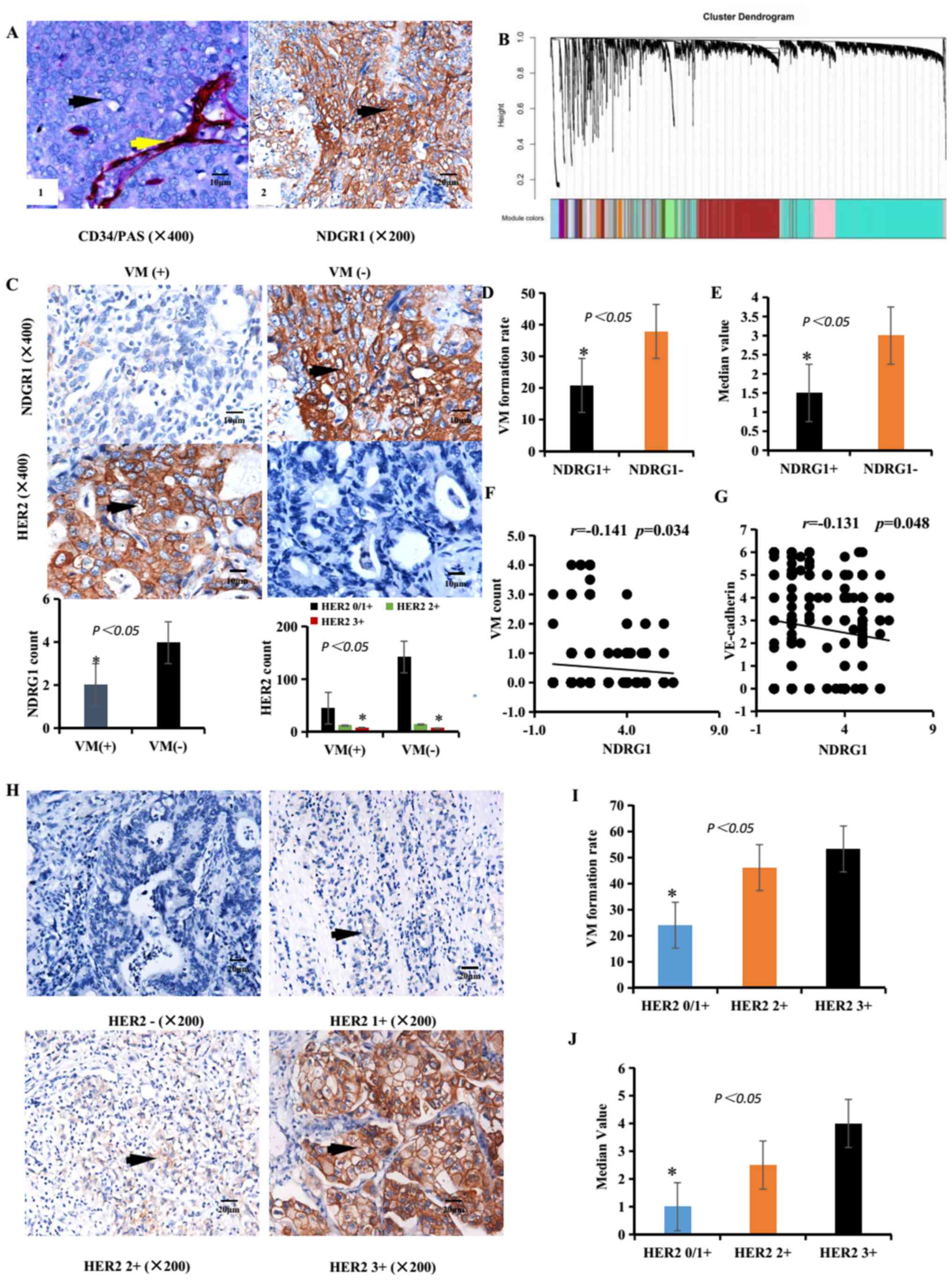 | Figure 1.Associations between NDRG1
expression, HER2 expression and VM in GC specimens. (A/left panel)
Identification of VM (black arrow, CD34-negative and PAS-positive
cells) and EDV (yellow arrow, CD34-positive and PAS-positive cells)
in GC specimens. Scale bar, 10 µm. Magnification, ×400. (A/right
panel) IHC staining of NDRG1 expression. Brown particles were
present in the cytoplasm and membranes of GC cells (black arrow).
Scale bar, 20 µm. Magnification, ×200. (B) Gene dendrogram obtained
by average linkage hierarchical clustering. Different gene modules
were obtained according to the segmentation results of the set
standard, which were represented by branches of the Dynamic Tree
Cut and different colors. A weighted correlation network analysis
revealed that overall gene expression data corresponded to an
overview of aggregation in the patients with GC. (C) IHC staining
of NDRG1 and HER2 expression in VM-positive (left images) and
VM-negative samples (right images) expression. Scale bar, 10 µm.
Magnification, ×400. (D) Rate of VM formation in different NDRG1
expression groups. (E) Median value of the VM count in different
NDRG1 expression groups. (F) Correlation between NDRG1 expression
and VM count using Pearson's correlation. (G) Correlation between
NDRG1 expression and VE-cadherin expression using Pearson's
correlation. (H) IHC staining of HER2 expression. Brown particles
were present in the membrane of GC cells (black arrow). Scale bar,
20 µm. Magnification, ×200. (I) Rate of VM formation in different
HER2 expression groups. (J) Median value of VM count in different
HER2 expression groups. *P<0.05. EDV Endothelium-dependent blood
vessels; GC, gastric carcinoma; HER2, human epidermal growth factor
receptor 2; IHC, immunohistochemistry; NDRG1, N-myc downstream
regulated gene 1; PAS, periodic acid-Schiff; VM, vasculogenic
mimicry. |
 | Table I.Clinical data of patients with
gastric cancer. |
Table I.
Clinical data of patients with
gastric cancer.
| Characteristic | Alive (n=92) | Deceased
(n=131) | Total
(n=228a) (%) |
|---|
| Sex |
|
Female | 27 | 44 | 74 (32.5) |
|
Male | 65 | 87 | 154 (67.6) |
| Age, years |
|
<60 | 50 | 53 | 105 (46.1) |
|
≥60 | 42 | 78 | 123 (54.0) |
| Tumor size,
cm3 |
|
<3 | 50 | 41 | 96 (42.1) |
| ≥3 | 42 | 90 | 132 (58.0) |
| Histological
differentiation |
|
I/II | 56 | 59 | 117 (51.3) |
|
III/IV | 36 | 72 | 111 (48.7) |
| TNM stage |
|
I/II | 56 | 41 | 99 (43.4) |
|
III/IV | 36 | 90 | 129 (56.6) |
| Lymphatic
metastasis |
|
Positive | 46 | 102 | 152 (66.7) |
|
Negative | 46 | 29 | 76 (33.3) |
| Distant
metastasis |
|
Positive | 11 | 72 | 85 (37.3) |
|
Negative | 81 | 59 | 143 (62.7) |
| Lauren type |
|
Intestinal | 56 | 65 | 123 (54.0) |
|
Diffuse | 36 | 66 | 105 (46.1) |
| Metastasis and
recurrence |
|
Positive | 8 | 95 | 105 (46.1) |
|
Negative | 84 | 36 | 123 (54.0) |
| Table II.Association between NDRG1 or VM and
the clinicopathological parameters with gastric carcinoma. |
Table II.
Association between NDRG1 or VM and
the clinicopathological parameters with gastric carcinoma.
|
| Tissue samples | Tissue samples |
|---|
|
|
|
|
|---|
| Variable | NDRG1 (n=125) | Non-NDRG1
(n=103) | χ2 | P-value | VM (n=65) | Non-VM (n=163) | χ2 | P-value |
|---|
| Age, years |
|
| 0.469 | 0.507 |
|
| 3.050 | 0.105 |
|
<60 | 55 | 50 |
|
| 24 | 81 |
|
|
|
≥60 | 70 | 53 |
|
| 41 | 82 |
|
|
| Sex |
|
| 0.534 | 0.465 |
|
| 0.941 | 0.352 |
|
Male | 87 | 67 |
|
| 47 | 107 |
|
|
|
Female | 38 | 36 |
|
| 18 | 56 |
|
|
| Tumor size,
cm3 |
|
| 0.408 | 0.523 |
|
| 0.165 | 0.767 |
|
<3 | 55 | 41 |
|
| 26 | 70 |
|
|
| ≥3 | 70 | 62 |
|
| 39 | 93 |
|
|
| Histological
differentiation |
|
| 8.353 | 0.005 |
|
| 4.660 | 0.040 |
|
I/II | 75 | 42 |
|
| 26 | 91 |
|
|
|
III/IV | 50 | 61 |
|
| 39 | 72 |
|
|
| TNM stage |
|
| 8.289 | 0.005 |
|
| 15.317 | <0.0001 |
|
I/II | 65 | 34 |
|
| 15 | 84 |
|
|
|
III/IV | 60 | 69 |
|
| 50 | 79 |
|
|
| Lauren type |
|
| 4.080 | 0.046 |
|
| 5.635 | 0.018 |
|
Intestinal | 75 | 48 |
|
| 27 | 96 |
|
|
|
Diffuse | 50 | 55 |
|
| 38 | 67 |
|
|
| Lymphatic
metastasis |
|
| 26.784 | <0.0001 |
|
| 11.018 | 0.001 |
|
Positive | 65 | 87 |
|
| 54 | 98 |
|
|
|
Negative | 60 | 16 |
|
| 11 | 65 |
|
|
| Distant
metastasis |
|
| 6.981 | 0.008 |
|
| 5.553 | 0.018 |
|
Positive | 37 | 48 |
|
| 32 | 53 |
|
|
|
Negative | 88 | 55 |
|
| 33 | 110 |
|
|
| Metastasis and
recurrence |
|
| 11.255 | 0.001 |
|
| 17.137 | <0.0001 |
|
Positive | 45 | 60 |
|
| 44 | 61 |
|
|
|
Negative | 80 | 43 |
|
| 21 | 102 |
|
|
| HER2 |
|
| 6.826 | 0.032 |
|
| 10.320 | 0.005 |
|
0/1+ | 110 | 77 |
|
| 45 | 142 |
|
|
| 2+ | 9 | 17 |
|
| 12 | 14 |
|
|
| 3+ | 6 | 9 |
|
| 8 | 7 |
|
|
Among the GC specimens, 125 (54.82%) samples were
positive for NDRG1 expression (Table
II and Fig. 1A). In
NDRG1-positive cases, 26 patients were identified as VM-positive,
and the VM formation rate was 20.80% (26/125). In the
NDRG1-negative cases, 39 patients were identified as VM-positive,
and the VM-positive rate was 37.86% (39/103). The overall
correlation patterns of genes across GC samples within TCGA was
analyzed using WGCNA (Fig. 1B). The
VM formation rates in NDRG1-negative and NDRG1-positive patients
were significantly different (χ2=8.068; P=0.005;
Fig. 1C and D and Table III). The median value of the VM
count in NDRG1-negative and NDRG1-positive samples was
significantly different (Fig. 1E).
The formation rate of VM in NDRG1-negative patients was higher than
that in NDRG1-positive patients. Furthermore, NDRG1 expression and
VM quantification were negatively correlated based on Pearson's
correlation (r=−0.141; P<0.05, Fig.
1F). NDRG1 expression and VE-cadherin expression were
negatively correlated based on Pearson's correlation (r=−0.131;
P<0.05, Fig. 1G).
| Table III.Association between NDRG1 and the
EMT-associated proteins. |
Table III.
Association between NDRG1 and the
EMT-associated proteins.
| Variable | NDRG1 (n=125) | Non-NDRG1
(n=103) | χ2 | P-value |
|---|
| VM |
|
| 8.068 | 0.005 |
|
Positive | 26 | 39 |
|
|
|
Negative | 99 | 64 |
|
|
| VE-cadherin |
|
| 4.803 | 0.036 |
|
Positive | 36 | 44 |
|
|
|
Negative | 89 | 59 |
|
|
| Twist1 |
|
| 6.739 | 0.011 |
|
Positive | 43 | 53 |
|
|
|
Negative | 82 | 50 |
|
|
| Snail |
|
| 8.419 | 0.004 |
|
Positive | 65 | 73 |
|
|
|
Negative | 60 | 30 |
|
|
| E-cadherin |
|
| 5.080 | 0.029 |
|
Positive | 85 | 55 |
|
|
|
Negative | 40 | 48 |
|
|
| Vimentin |
|
| 4.485 | 0.042 |
|
Positive | 30 | 38 |
|
|
|
Negative | 95 | 65 |
|
|
| HER2 |
|
| 6.826 | 0.032 |
|
0/1+ | 110 | 77 |
|
|
| 2+ | 9 | 17 |
|
|
| 3+ | 6 | 9 |
|
|
HER2 expression was rated 0/1+ in 187 (83.77%)
patients, 2+ in 26 (10.09%) patients and 3+ in 15 (6.14%) patients
(Table II). The expressions of HER2
are presented in Fig. 1H. In the
HER2 0/1+ group, VM-positivity was identified in 45 cases (24.1%);
in the HER2 2+ group, VM-positivity was identified in 12 cases
(46.2%); and in the HER2 3+ group, VM-positivity was identified in
8 cases (53.3%). There were considerable differences among the HER2
0/1+, HER2 2+ and HER2 3+ groups (χ2=10.320; P=0.005,
Table II and Fig. 1I). The HER2 2+ and HER2 3+ groups
demonstrated greater VM-positive rates than the HER2 0/1+ group
(P=0.017, χ2=5.683 and P=0.013, χ2=6.147,
respectively). The combination HER2 2+ and HER2 3+ cases
demonstrated a greater VM-positive rate than the HER2 0/1+ cases
(P=0.002, χ2=10.079). The median VM counts in the HER2
0/1+, HER2 2+ and HER2 3+ groups were significantly different
(Fig. 1J). Furthermore, HER2
expression and VM quantification was positively correlated based on
Spearman's correlation (r=0.153; P=0.021). Taken together, these
results of the present study indicate that NDRG1 expression may
weaken VM formation and that HER2 may promote VM formation.
Expression of NDRG1 is an indicator of
good prognosis in patients with GC
To evaluate the association between hazard ratio and
survival rate, Kaplan-Meier and multivariate Cox regression
analyses were performed. Using univariate Cox proportional hazards
regression analysis, pathological variables with VM, HER2, tumor
size, TNM stage, lymphatic metastasis, distant metastasis and
metastasis and recurrence were all identified to be significant
predictors of prognosis in patients with GC. When using the
multivariate Cox analysis only, HER2 and metastasis and recurrence
were significant independent risk factors in patients with GC, with
hazard ratios of 1.365 and 4.186, respectively. NDRG1 was the most
favorable factor in patients with GC with a hazard ratio of 0.66
(Table IV).
| Table IV.Univariate and multivariate Cox
regression analysis of variables associated with gastric cancer
mortality. |
Table IV.
Univariate and multivariate Cox
regression analysis of variables associated with gastric cancer
mortality.
|
| Univariate
analysis | Mutlivariate
analysis |
|---|
|
|
|
|
|---|
| Variable | HR | 95% CI | P-value | HR | 95% CI | P-value |
|---|
| Age, years | 1.008 | 0.992–1.024 | 0.306 |
|
|
|
| Sex, male =1 | 0.837 | 0.582–1.203 | 0.336 |
|
|
|
| VM, positive
=1 | 1.672 | 1.170–2.389 | 0.005 |
|
|
|
| NDRG1,
positive=1 | 0.659 | 0.467–0.929 | 0.017 | 0.660 | 0.462–0.941 | 0.022 |
| HER2,
positive=1 | 1.516 | 1.138–2.021 | 0.004 | 1.365 | 1.017–1.832 | 0.038 |
| Tumor size, ≥3
cm3=1 | 1.967 | 1.357–2.852 | <0.001 |
|
|
|
| Lauren type,
diffuse=1 | 1.396 | 0.990–1.969 | 0.057 |
|
|
|
| TNM stage,
III/IV=1 | 2.600 | 1.786–3.784 | <0.001 |
|
|
|
| Histological
differentiation, I/II=1 | 0.599 | 0.423–0.846 | 0.004 |
|
|
|
| Lymphatic
metastasis, positive=1 | 2.750 | 1.814–4.171 | <0.001 |
|
|
|
| Distant metastasis,
positive=1 | 2.980 | 2.095–4.240 | <0.001 |
|
|
|
| Metastasis and
recurrence, positive=1 | 5.326 | 3.568–7.949 | <0.001 | 4.186 | 2.421–7.238 | <0.001 |
A univariate survival analysis was performed using a
Kaplan-Meier survival analysis and the log-rank method. The
correlations between VM, NDRG1 protein expression, HER2 expression
and the survival rate of patients with GC following surgical
procedures [rate of follow-up, 97.81% (223/228)] were investigated.
Follow up was performed for all patients by clinic interview or
phone call. By the end of the follow-up time in January 2014, 5
patients or their family could not be contacted and were therefore
lost. Overall, 5 patients were lost during follow-up. The
cumulative survival function in patients with GC is presented in
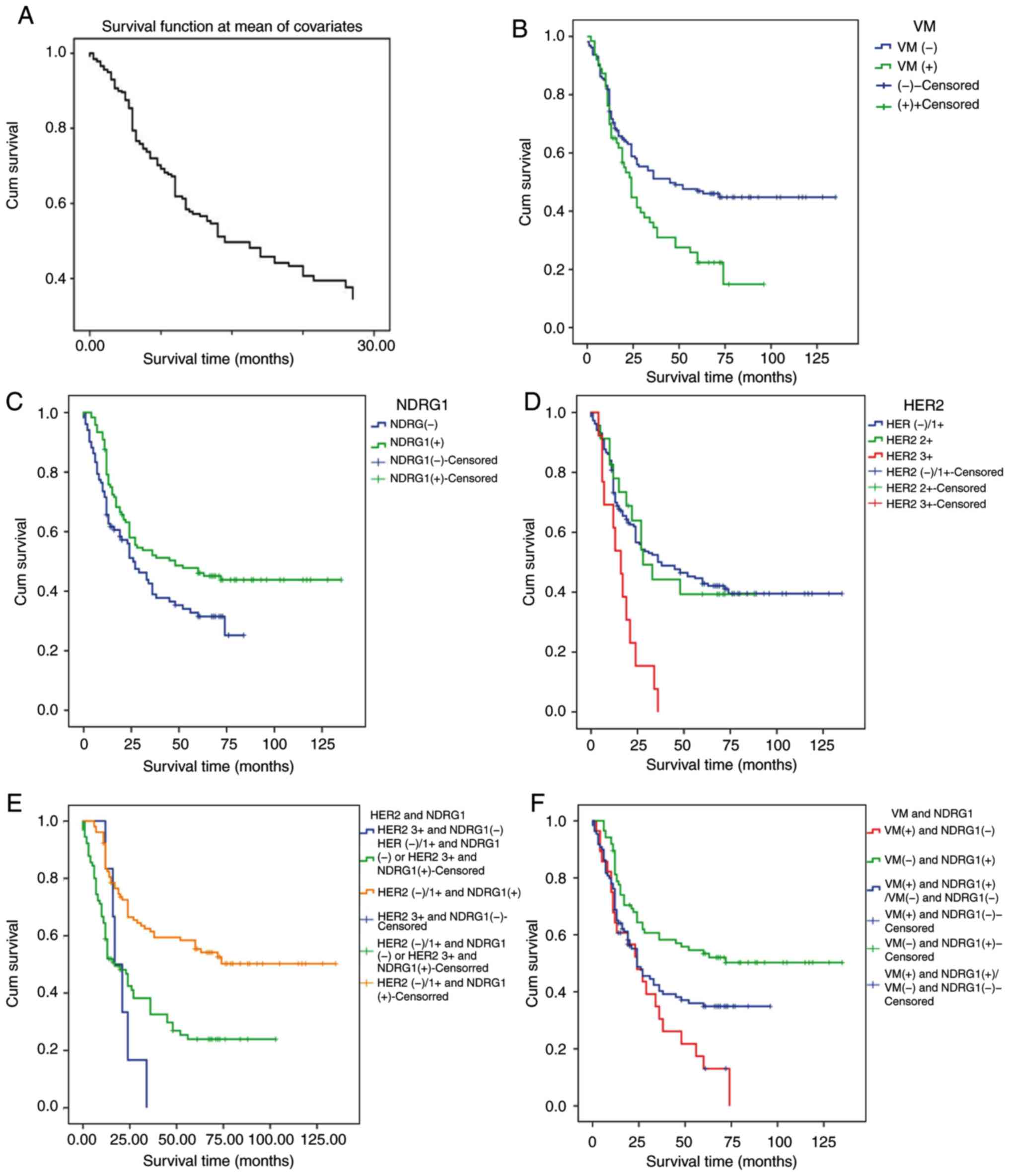
Fig. 2A. Patients without VM had a
significantly longer survival time compared with those with VM
(log-rank test, χ2=8.413; P=0.004; Fig. 2B). Patients with high NDRG1
expression levels had significantly longer survival time compared
with those with low NDRG1 expression levels (log-rank test,
χ2=5.925; P=0.015; Fig.
2C). Patients with HER2 3+ expression levels had a
significantly shorter survival time compared with those with HER2
2+ and HER2 0/1+ expression levels (log-rank test,
χ2=14.656; P=0.001; Fig.
2D). The results of the present study revealed that the overall
survival time was significantly lower in patients with HER2 3+ and
NDRG1-negative tumors compared with those with HER2 0/1+ and
NDRG1-positive tumors (log-rank test, χ2=25.96;
P<0.0001; Fig. 2E). The overall
survival time was significantly lower in patients with VM-positive
and NDRG1-negative samples compared with those with VM-negative and
NDRG1-positive samples (log rank test, χ2=14.058;
P=0.001; Fig. 2F). Therefore, the
results from the present study indicate that NDRG1 has a negative
association with the prognosis of patients with GC and HER2 has a
positive association with the prognosis of patients with GC.
NDRG1 protein may weaken VM formation
in patients with GC by decreasing EMT-associated markers and HER2
expression
To further clarify the role of NDRG1 in the decrease
in VM formation, the expression levels of VM-associated markers
(VE-cadherin), EMT-associated markers (Twist1, Snail, E-cadherin
and vimentin) and HER2 were detected via immunohistochemistry.
In the NDRG1-positive group, VE-cadherin expression
was positive in 45.00% (36/80), Twist1 expression was positive in
44.79% (43/96), Snail expression was positive in 47.10% (65/138),
E-cadherin expression was positive in 60.71% (85/140) and vimentin
expression was positive in 44.12% (30/68) of cases. In the
NDRG1-negative group, VE-cadherin expression was positive in 55%
(44/80), Twist1 expression was positive in 55.21% (53/96), Snail
expression was positive in 52.90% (73/138), E-cadherin expression
was positive in 39.29% (55/140) and vimentin expression was
positive in 55.88% (38/68) of cases. The expression of VE-cadherin
and EMT-associated markers in the NDRG1-negative and NDRG1-positive
groups was significantly different (Table III; Fig.
3B). These results suggest that NDRG1 suppresses the expression
of Twist1, Snail, VE-cadherin and vimentin, whereas NDRG1 enhances
the expression of E-cadherin (P<0.05).
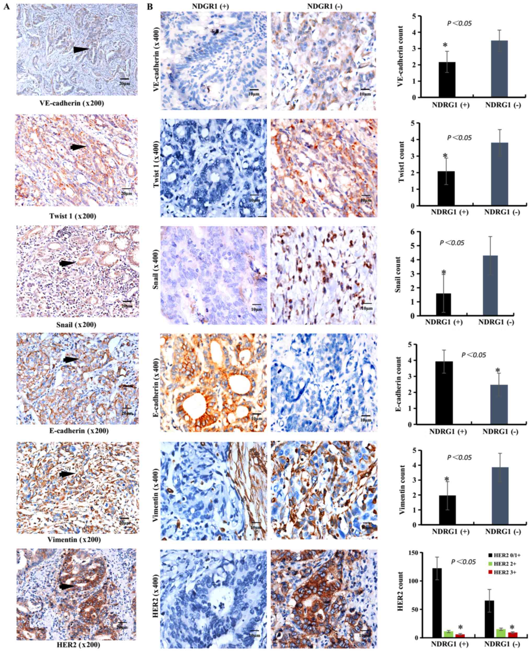 | Figure 3.IHC staining of VE-cadherin,
EMT-associated markers (Twist1, Snail, E-cadherin and vimentin) and
HER2 in GC samples. (A) IHC staining of VE-cadherin, Twist1, Snail,
E-cadherin and vimentin in GC samples. The brown particles were
present in the cytoplasm (VE-cadherin, Twist1, Snail and vimentin)
or membrane (VE-cadherin, E-cadherin and HER2) or nucleus (Snail)
of GC cells (black arrow). Scale bar, 20 µm. Magnification, ×200.
(B) IHC staining of VE-cadherin, Twist1, Snail, E-cadherin,
Vimentin and HER2 expression in NDRG1-positive (left images) and
NDRG1-negative (right images) samples. The positive rate of
VE-cadherin, Twist1, Snail, vimentin and HER2 in NDRG1-positive
were lower than that in NDRG1-negative. The positive rate of
E-cadherin in NDRG1-positive were higher than that in
NDRG1-negative. Scale bar, 10 µm. Magnification, ×400. *P<0.05.
IHC, immunohistochemistry; VE-cadherin, vascular
endothelial-cadherin; E-cadherin, epithelial-cadherin; EMT,
epithelial mesenchymal transition; HER2, human epidermal growth
factor 2; GC, gastric carcinoma; NDRG1, N-myc downstream regulated
gene 1. |
In addition, the functional enrichment analysis of
NDRG1 in 415 cases of GC from TCGA database was performed using
Metascape and GSEA. All 442 cases of GC within TCGA database are
presented in Table V.
| Table V.Clinical data of gastric carcinoma in
The Cancer Genome Atlas. |
Table V.
Clinical data of gastric carcinoma in
The Cancer Genome Atlas.
| Characteristic | Alive, (n=369) | Deceased, tumor
present, (n=58) | Deceased, no tumor,
(n=15) | Total, (n=442) |
|---|
| Sex, n (%) |
|
Female | 138 (37.4) | 16 (27.6) | 4 (27.0) | 158 (36.0) |
|
Male | 231 (63.0) | 42 (72.4) | 11 (73.3) | 284 (64.3) |
| Age, years |
| Mean (±
SD) | 65.4 (10.8) | 64.6 (10.1) | 73.3 (9.5) | 65.3 (10.5) |
| Median
(range) | 67 (30–90) | 66 (41–86) | 73 (47–90) | 66 (34–90) |
| Stage, n (%) |
| I | 3 (0.8) |
|
| 3 (0.7) |
| IA | 13 (3.5) |
| 2 (13.3) | 14 (3.2) |
| IB | 35 (9.5%) | 5 (8.6) | 3 (20.0) | 38 (8.6) |
| II | 32 (8.7) | 2 (3.4) |
| 29 (6.6) |
|
IIA | 38 (10.3) | 1 (1.7) | 1 (6.7) | 38 (8.6) |
|
IIB | 55 (15.0) | 1 (1.7) | 1 (6.7) | 55 (12.4) |
|
III | 2 (0.5) | 1 (1.7) |
| 2 (0.5) |
|
IIIA | 63 (17.1) | 11 (19.0) | 3 (20.0) | 68 (15.4) |
|
IIIB | 49 (13.3) | 11 (19.0) | 3 (20.0) | 62 (14.0) |
|
IIIC | 32 (8.7) | 6 (10.3) | 1 (6.7) | 38 (8.6) |
| IV | 24 (6.5) | 17 (29.3) |
| 38 (8.6) |
| Grade, n (%) |
| G1 | 12 (3.3) | 1 (1.7) |
| 10 (2.3) |
| G2 | 129 (35.0) | 24 (41.4) | 6 (40.0) | 138 (31.2) |
| G3 | 221 (59.9) | 31 (53.4) | 9 (60.0) | 240 (54.3) |
| GX | 7 (1.9) | 2 (3.4) |
| 7 (1.6) |
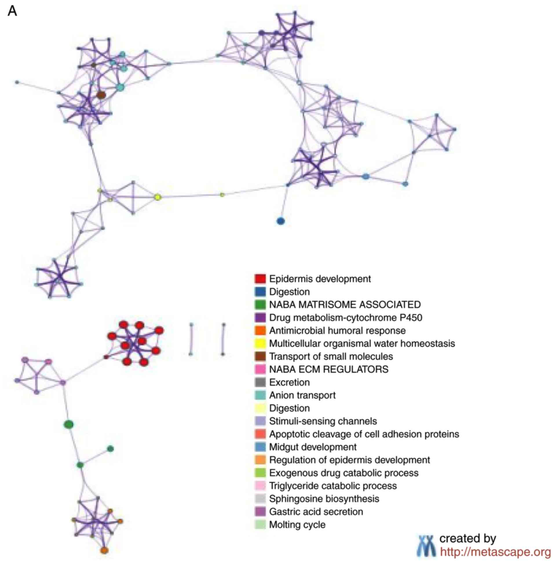
The Metascape analysis revealed that 242 genes
exhibited significantly increased expression (P<0.05; Fig. 4A), and 239 genes exhibited
significantly decreased expression in the 211 samples with high
NDRG1 expression (P<0.05; Fig.
4B). The GSEA analysis indicated that 47 pathways were
significantly upregulated (P<0.05; Fig. 4C) and 46 pathways were significantly
downregulated in the 211 samples with high NDRG1 expression
(P<0.05; Fig. 4D). P-values were
automatically generated by the Cytoscape software.
Compared with the two groups with high and low NDRG1
expression, the differentially expressed genes were focused
primarily on the pathways of development and differentiation of
epithelial cells. The results of the present study indicate that
NDRG1 serves an important role in maintaining the epithelial
phenotype of GC, and VM occurs more easily in GC with low NDRG1
expression.
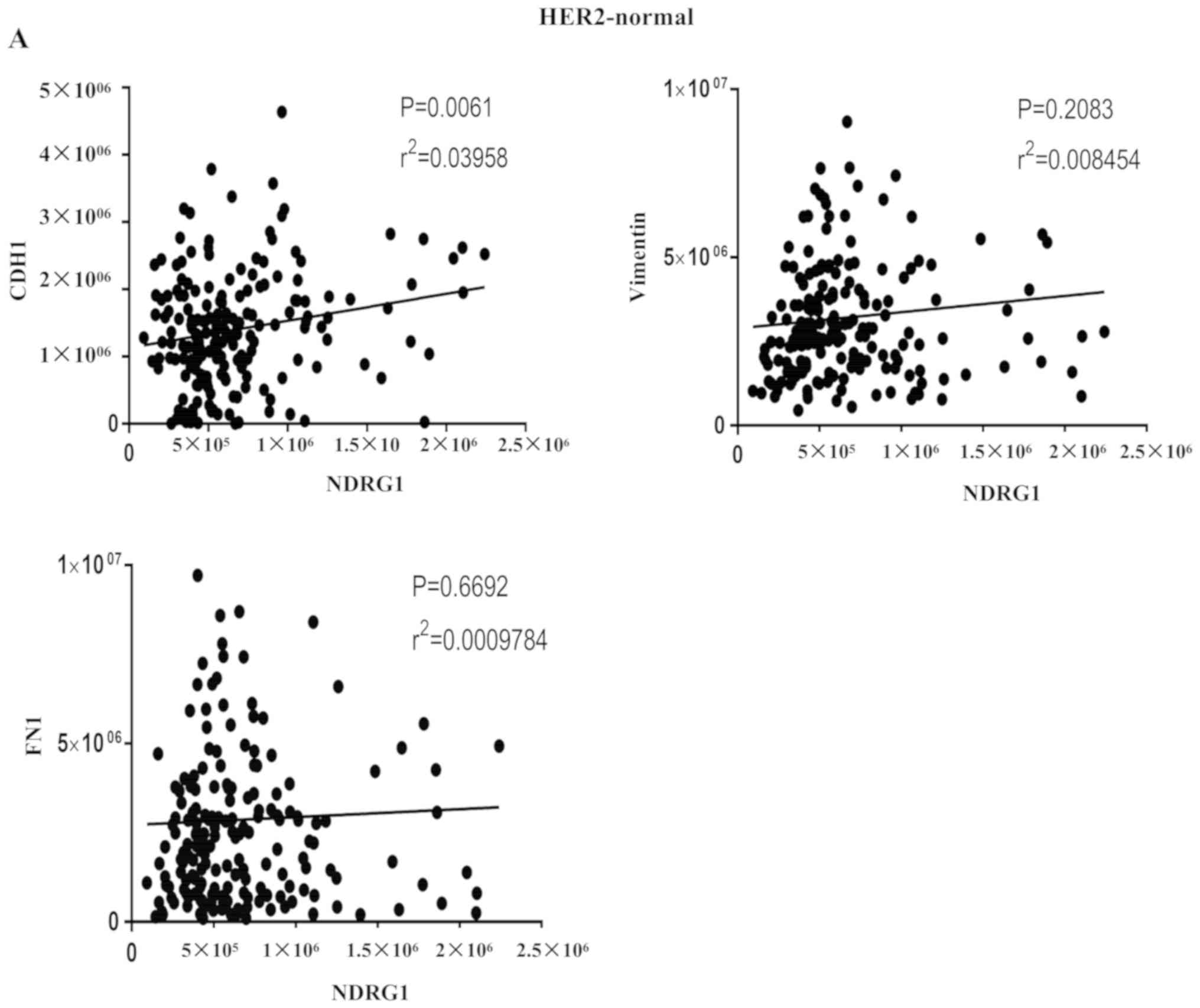
In addition, correlations between NDRG1 and
EMT-associated factors were investigated from two aspects. On the
one hand, in cases of GC from TCGA database, the results indicated
that NDRG1 and CDH1 (E-cadherin) were positively correlated in
HER2-normal and HER2-amplified samples, and that NDRG1 and
vimentin, and NDRG1 and fibronectin 1 were negatively correlated in
HER2-amplified samples at the RNA level (Fig. 5A and B). On the other hand,
correlations among NDRG1 and EMT-associated markers were determined
using Pearson's correlation in patients with GC. The results
revealed that NDRG1 has a negative correlation with Twist1, Snail
and vimentin (Fig. 5C, D and E,
respectively) and a positive correlation with E-cadherin at the
protein level (Fig. 5F). These
results suggest that NDRG1 may hinder EMT.
The results from IHC staining of VE-cadherin and
EMT-associated factors (Twist1, Snail, E-cadherin and vimentin) in
GC samples are presented in Fig. 3A.
In the NDRG1-positive group, HER2 0/1+ expression was identified in
58.82% (110/187), HER2 2+ expression was identified in 34.62%
(9/26) and HER2 3+ expression was identified in 40.00% (6/15) of
cases. In the NDRG1-negative group, HER2 0/1+ expression was
identified in 41.18% (77/187), HER2 2+ expression was identified in
65.38% (17/26) and HER2 3+ expression was identified in 60% (9/15)
of cases. There were significant differences in expression among
the HER2 0/+, HER2 2+ and HER2 3+ groups (χ2=6.826;
P=0.032; Table II, Fig. 3B). NDRG1 and HER2 expression was
negatively correlated based on Spearman's correlation analysis
(r=−0.149; P=0.025). At the same time, the analytical data from
TCGA indicated that NDRG1 expression was higher in HER2-amplified
samples compared with HER2-normal samples at the RNA level. The
results of the present study suggest that NDRG1 may decrease the
expression of HER2 (P<0.05); the molecular mechanisms underlying
this process require validation in the future.
Overall, the results of the present study indicate
that NDRG1 may weaken VM formation through EMT- and HER2-mediated
mechanisms.
Discussion
GC is characterized by a high growth rate, high
malignancy, and high rates of recurrence and metastasis (21,22). In
the present study, the metastasis rate of 228 cases of GC was
46.05%. The 5-year survival rate of 228 patients with GC was
30.71%. It is critical to investigate the novel molecules and
molecular mechanisms that are involved in the development of GC. It
has been indicated that VM serves an important role in regulating
the development of GC (4). In the
present study, 28.5% of patients with GC were VM-positive. Patients
with VM had a significantly poorer prognosis than those patients
without VM.
NDRG1 has been revealed to serve a key role in
physiological conditions, such as cellular differentiation and the
cell cycle, and pathophysiological conditions, such as cancer
pathology. NDRG1 inhibits tumor progression in the majority of
cancer types; however, the inhibitory effect of NDRG1 is tissue
specific, and the effects between tissues may be completely
different. For example, NDRG1 has been identified as a metastasis
suppressor gene in colon and prostate cancer, but the opposite
results were demonstrated in hepatocellular carcinoma and breast
cancer (23,24). Results of the present study indicated
that NDRG1 possesses inhibitory effects within patients with GC,
which is consistent with the results of a previous study (2). Another study reported that NDRG1
promotes the metastasis of human scirrhous GC cells through EMT
(25). Therefore, the inhibitory
effects of NDRG1 in GC require verification in future studies.
Across the cohort of patients included in the
present study, 54.8% of the patients with GC exhibited high NDRG1
protein expression. NDRG1 expression had a significant negative
correlation with TNM stage, lymph node metastasis, distant
metastasis, and recurrence and metastasis in GC. Furthermore,
patients with high NDRG1 expression levels had a significantly
lower VM-positive rate and longer survival. VM-positivity was
present in 37.8% of GC cases negative for NDRG1 expression. The
expression of NDRG1 had a negative correlation with VM formation.
VE-cadherin has been identified as a key marker for indicating the
presence of VM, and tumor cells lacking VE-cadherin are incapable
of forming VM tubes (26). The
results from the present study revealed that the presence of NDRG1
resulted in decreased expression of VE-cadherin, and that NDRG1 was
negatively correlated with VE-cadherin. Therefore, NDRG1-mediated
suppression of tumor metastasis in GC may occur through decreased
VM formation.
To further clarify the suppressive effects of NDRG1
on VM formation, the levels of EMT-associated factors (Twist1,
Snail, E-cadherin and vimentin) and HER2 were assessed. EMT has
been proposed as a key process in cancer progression, whereby
epithelial cells acquire mesenchymal properties and exhibit
decreased cell-matrix adhesion. Notably, tumor cells may gain
dedifferentiated phenotypes to convert onto endothelial cells via
EMT, as revealed in our previous study (6). Twist1 and Snail are transcription
factors that serve an important role in the EMT process, a process
associated with cancer progression and VM formation (6,27). The
results of present study and the previous study (6), indicated that NDRG1 significantly
enhanced the expression of the epithelial marker E-cadherin and
decreased the expression of the mesenchymal marker vimentin in GC
specimens. In addition, the results of the present study that were
derived from Metascape and GSEA were consistent with the analysis
of patients with GC. Furthermore, NDRG1 may decrease the expression
of the transcription factors Twist1 and Snail in GC specimens. A
similar conclusion was made by Lee et al (27); thus, NDRG1 may suppress VM formation
and metastasis in GC by inhibiting EMT.
The HER2 protein is a member of the epidermal growth
factor receptor family. It has been demonstrated that HER2
overexpression is associated with a poor prognosis in patients with
GC (28). In the present study,
patients with GC with HER2 3+ expression had a worse prognosis
compared with those with HER2 2+ and HER2 0/1+ expression, which is
consistent with the results of a previous study (29). Currently, HER2 is regarded as an
important and promising target in the treatment of HER2-positive
patients with GC (30,31). NDRG1 may downregulate the ErbB family
of receptors to inhibit oncogenic signaling pathways in other types
of tumor in humans (11).
Preliminary research has indicated that HER2 overexpression induces
VM formation through upregulation of VE-cadherin in invasive breast
carcinoma (32). The results from
the present study revealed that patients with GC with HER2 3+
expression had a higher VM formation rate compared with those with
HER2 2+ and HER2 0/1+ expression. Furthermore, samples with higher
expression of HER2 presented with a higher VM count, and NDRG1
decreased the HER2 expression levels. These results demonstrate
that NDRG1 may suppress VM formation and metastasis in GC by
decreasing HER2 expression.
It is reasonable to assume that NDRG1 may act as a
protective agent against GC progression. The results of the present
study indicate that NDRG1 may be a potentially important predictive
biomarker of recurrence, metastasis and poor outcome for patients
with GC, and may provide a basis for the development of targeted
treatments for patients with GC. However, the results of the
present study were limited in human specimens and, as such, further
studies are required in order to confirm these conclusions using
mechanistic experiments in vitro.
Acknowledgements
Not applicable.
Funding
The present study was supported by The National
Natural Science Foundation of China (grant no. 81572872), Key
project of the National Natural Science Foundation of China (grant
no. 81230050), and The Science & Technology Development Fund of
Tianjin Education Commission for Higher Education (grant no.
2018KJ074).
Availability of data and materials
The datasets used and/or analyzed during the present
study are available from the corresponding author upon reasonable
request.
Authors' contributions
XD and BS designed the study and wrote the
manuscript. YH, HS, CC and XZ performed the experiments and
analyzed data. All authors read and approved the final version of
the manuscript.
Ethics approval and consent to
participate
This study was approved by the Ethics Committee of
Tianjin Medical University. All patients provided written informed
consent. The privacy rights of human subjects were maintained.
Patient consent for publication
Not applicable.
Competing interests
The authors declare that they have no competing
interests.
References
|
1
|
Leung WK, Wu MS, Kakugawa Y, Kim JJ, Yeoh
KG, Goh KL, Wu KC, Wu DC, Sollano J, Kachintorn U, et al: Screening
for gastric cancer in Asia: Current evidence and practice. Lancet
Oncol. 9:279–287. 2008. View Article : Google Scholar : PubMed/NCBI
|
|
2
|
Chang X, Xu X, Ma J, Xue X, Li Z, Deng P,
Zhang S, Zhi Y, Chen J and Dai D: NDRG1 expression is related to
the progression and prognosis of gastric cancer patients through
modulating proliferation, invasion and cell cycle of gastric cancer
cells. Mol Biol Rep. 41:6215–6223. 2014. View Article : Google Scholar : PubMed/NCBI
|
|
3
|
McLemore MR: The role of the data safety
monitoring board: Why was the Avastin phase III clinical trial
stopped? Clin J Oncol Nurs. 10:153–154. 2006. View Article : Google Scholar : PubMed/NCBI
|
|
4
|
Qu B, Guo L, Ma JL and Lv Y:
Antiangiogenesis therapy might have the unintended effect of
promoting tumor metastasis by increasing an alternative circulatory
system. Med Hypotheses. 74:360–361. 2010. View Article : Google Scholar : PubMed/NCBI
|
|
5
|
Maniotis AJ, Folberg R, Hess A, Seftor EA,
Gardner LM, Pe'er J, Trent JM, Maniotis AJ, Folberg R, Hess A, et
al: Vascular channel formation by human melanoma cells in vivo and
in vitro: Vasculogenic mimicry. Am J Pathol. 155:739–752. 1999.
View Article : Google Scholar : PubMed/NCBI
|
|
6
|
Sun T, Zhao N, Zhao XL, Gu Q, Zhang SW,
Che N, Wang XH, Du J, Liu YX and Sun BC: Expression and functional
significance of Twist1 in hepatocellular carcinoma: Its role in
vasculogenic mimicry. Hepatology. 51:545–556. 2010. View Article : Google Scholar : PubMed/NCBI
|
|
7
|
Liu TJ, Sun BC, Zhao XL, Zhao XM, Sun T,
Gu Q, Yao Z, Dong XY, Zhao N and Liu N: CD133+ cells with cancer
stem cell characteristics associates with vasculogenic mimicry in
triple-negative breast cancer. Oncogene. 32:544–553. 2013.
View Article : Google Scholar : PubMed/NCBI
|
|
8
|
Du J, Sun B, Zhao X, Gu Q, Dong X, Mo J,
Sun T, Wang J, Sun R and Liu Y: Hypoxia promotes vasculogenic
mimicry formation by inducing epithelial-mesenchymal transition in
ovarian carcinoma. Gynecol Oncol. 133:575–583. 2014. View Article : Google Scholar : PubMed/NCBI
|
|
9
|
Sun J, Sun B, Zhu D, Zhao X, Zhang Y, Dong
X, Che N, Li J, Liu F, Zhao N, et al: HMGA2 regulates CD44
expression to promote gastric cancer cell motility and sphere
formation. Am J Cancer Res. 7:260–274. 2017.PubMed/NCBI
|
|
10
|
Richardson A, Kovacevic Z and Richardson
DR: Iron chelation: Inhibition of key signaling pathways in the
induction of the epithelial mesenchymal transition in pancreatic
cancer and other tumors. Crit Rev Oncog. 18:409–434. 2013.
View Article : Google Scholar : PubMed/NCBI
|
|
11
|
Kovacevic Z, Menezes SV, Sahni S,
Kalinowski DS, Bae DH, Lane DJ and Richardson DR: The metastasis
suppressor, N-MYC downstream-regulated gene-1 (NDRG1),
down-regulates the ErbB family of receptors to inhibit downstream
oncogenic signaling pathways. J Biol Chem. 291:1029–1052. 2016.
View Article : Google Scholar : PubMed/NCBI
|
|
12
|
Sun T, Sun BC, Zhao XL, Zhao N, Dong XY,
Che N, Yao Z, Ma YM, Gu Q, Zong WK and Liu ZY: Promotion of tumor
cell metastasis and vasculogenic mimicry by way of transcription
coactivation by Bcl-2 and Twist1: A study of hepatocellular
carcinoma. Hepatology. 54:1690–1706. 2011. View Article : Google Scholar : PubMed/NCBI
|
|
13
|
Broggini T, Wustner M, Harms C, Stange L,
Blaes J, Thome C, Harms U, Mueller S, Weiler M, Wick W, et al:
NDRG1 overexpressing gliomas are characterized by reduced tumor
vascularization and resistance to antiangiogenic treatment. Cancer
Lett. 380:568–576. 2016. View Article : Google Scholar : PubMed/NCBI
|
|
14
|
Bittner M, Meltzer P, Chen Y, Jiang Y,
Seftor E, Hendrix M, Radmacher M, Simon R, Yakhini Z, Ben-Dor A, et
al: Molecular classification of cutaneous malignant melanoma by
gene expression profiling. Nature. 406:536–540. 2000. View Article : Google Scholar : PubMed/NCBI
|
|
15
|
Rüschoff J, Dietel M, Baretton G, Arbogast
S, Walch A, Monges G, Chenard MP, Penault-Llorca F, Nagelmeier I,
Schlake W, et al: HER2 diagnostics in gastric cancer-guideline
validation and development of standardized immunohistochemical
testing. Virchows Arch. 457:299–307. 2010. View Article : Google Scholar : PubMed/NCBI
|
|
16
|
Huang P, Li S, Aronow WS, Wang Z, Nair CK,
Xue N, Shen X, Chen C and Cosgrove D: Double contrast-enhanced
ultrasonography evaluation of preoperative Lauren classification of
advanced gastric carcinoma. Arch Med Sci. 7:287–293. 2011.
View Article : Google Scholar : PubMed/NCBI
|
|
17
|
Sun B, Zhang S, Zhang D, Yin X, Wang S, Gu
Y and Wang Y: Doxycycline influences microcirculation patterns in
B16 melanoma. Exp Biol Med (Maywood). 232:1300–1307. 2007.
View Article : Google Scholar : PubMed/NCBI
|
|
18
|
Langfelder P and Horvath S: WGCNA: An R
package for weighted correlation network analysis. BMC
Bioinformatics. 9:5592008. View Article : Google Scholar : PubMed/NCBI
|
|
19
|
Subramanian A, Tamayo P, Mootha VK,
Mukherjee S, Ebert BL, Gillette MA, Paulovich A, Pomeroy SL, Golub
TR, Lander ES and Mesirov JP: Gene set enrichment analysis: A
knowledge-based approach for interpreting genome-wide expression
profiles. Proc Natl Acad Sci USA. 102:15545–15550. 2005. View Article : Google Scholar : PubMed/NCBI
|
|
20
|
Bosman FT, Carneiro F, Hruban RH and
Theise ND: WHO classification of tumours of the digestive system.
Fourth edition. IARC; France: 2010
|
|
21
|
Parkin DM, Bray F, Ferlay J and Pisani P:
Global cancer statistics, 2002. CA Cancer J Clin. 55:74–108. 2005.
View Article : Google Scholar : PubMed/NCBI
|
|
22
|
Wöhrer SS, Raderer M and Hejna M:
Palliative chemotherapy for advanced gastric cancer. Ann Oncol.
15:1585–1595. 2004. View Article : Google Scholar : PubMed/NCBI
|
|
23
|
Cheng J, Xie HY, Xu X, Wu J, Wei XY, Su R,
Zhang W, Lv Z, Zheng S and Zhou L: NDRG1 as a biomarker for
metastasis, recurrence and of poor prognosis in hepatocellular
carcinoma. Cancer Lett. 310:35–45. 2011. View Article : Google Scholar : PubMed/NCBI
|
|
24
|
Nagai MA, Gerhard R, Fregnani JH, Nonogaki
S, Rierger RB, Netto MM and Soares FA: Prognostic value of NDRG1
and SPARC protein expression in breast cancer patients. Breast
Cancer Res Treat. 126:1–14. 2011. View Article : Google Scholar : PubMed/NCBI
|
|
25
|
Ureshino H, Murakami Y, Watari K, Izumi H,
Kawahara A, Kage M, Arao T, Nishio K, Yanagihara K, Kinoshita H, et
al: N-myc downstream regulated gene 1 (NDRG1) promotes metastasis
of human scirrhous gastric cancer cells through epithelial
mesenchymal transition. PLoS One. 7:e413122012. View Article : Google Scholar : PubMed/NCBI
|
|
26
|
Hendrix MJ, Seftor EA, Meltzer PS, Gardner
LM, Hess AR, Kirschmann DA, Schatteman GC and Seftor RE: Expression
and functional significance of VE-cadherin in aggressive human
melanoma cells: Role in vasculogenic mimicry. Proc Natl Acad Sci
USA. 98:8018–8023. 2001. View Article : Google Scholar : PubMed/NCBI
|
|
27
|
Lee JC, Chung LC, Chen YJ, Feng TH and
Juang HH: N-myc downstream-regulated gene 1 downregulates cell
proliferation, invasiveness, and tumorigenesis in human oral
squamous cell carcinoma. Cancer Lett. 355:242–252. 2014. View Article : Google Scholar : PubMed/NCBI
|
|
28
|
Iqbal N and Iqbal N: Human epidermal
growth factor receptor 2 (HER2) in cancers: Overexpression and
therapeutic implications. Mol Biol Int. 2014:8527482014. View Article : Google Scholar : PubMed/NCBI
|
|
29
|
Yang Y, Liu Y, Guo R, Fu Y, Zhang Z, Zhang
P, Zhou P, Wang T, Huang T, Li X and Li C: The novel
dithiocarbamate, DpdtC suppresses HER2-overexpressed cancer cells
by up-regulating NDRG1 via inactivation of HER2-ERK 1/2 signaling.
Sci Rep. 8:33982018. View Article : Google Scholar : PubMed/NCBI
|
|
30
|
Menezes SV, Sahni S, Kovacevic Z and
Richardson DR: Interplay of the iron-regulated metastasis
suppressor NDRG1 with epidermal growth factor receptor (EGFR) and
oncogenic signaling. J Biol Chem. 292:12772–12782. 2017. View Article : Google Scholar : PubMed/NCBI
|
|
31
|
Lei YY, Huang JY, Zhao QR, Jiang N, Xu HM,
Wang ZN, Li HQ, Zhang SB and Sun Z: The clinicopathological
parameters and prognostic significance of HER2 expression in
gastric cancer patients: A meta-analysis of literature. World J
Surg Oncol. 15:682017. View Article : Google Scholar : PubMed/NCBI
|
|
32
|
Liu T, Sun B, Zhao X, Gu Q, Dong X, Yao Z,
Zhao N, Chi J, Liu N, Sun R and Ma Y: HER2/neu expression
correlates with vasculogenic mimicry in invasive breast carcinoma.
J Cell Mol Med. 17:116–122. 2013. View Article : Google Scholar : PubMed/NCBI
|