Introduction
Liver cancer had the seventh highest age-adjusted
incidence rate of all types of cancer in the United States of
America in 2014, often with high mortality (1–3). Liver
cancer requires the detection of small tumors that are often
present in asymptomatic individuals (4). Liver cancer is diagnosed by imaging
modalities, including computerized tomography and magnetic
resonance imaging scans, followed by confirmation via liver biopsy,
an invasive procedure (5–7). Although the diagnosis of liver cancer
has improved rapidly in recent years, <5% of patients with liver
cancer survive >5 years following diagnosis (8,9).
Previous studies suggest that high invasiveness and metastasis are
the main causes of poor prognosis (10,11).
Early liver cancer diagnosis is of paramount importance to therapy
and more effective biomarkers to predict the clinical outcome of
liver cancer are required.
The mannose receptor (CD206), also known as C-type
lectin, is expressed on the surface of macrophages and some subsets
of immature dendritic cells (12).
CD206 participates in antigen presentation, macrophage endocytosis
and is considered a hallmark of tumor-associated macrophages
(13). CD206 increased the growth
and migration of microglia by promoting their activation (14). Additionally, CD206 expressed on
lymphatic endothelium participates in the attachment of various
cancer cells to lymphatic endothelium to promote lymphatic
metastasis (15–17). Serum CD206 is elevated in patients
with multiple myeloma and is a prognostic marker for overall
survival (18,19). Furthermore, CD206 has been reported
as a novel biomarker for the diagnosis of patients with colorectal
and gastric cancer (20,21). Therefore, the investigation of the
potential role of CD206 in liver cancer may be beneficial to
patients.
The present study evaluated the clinical
significance of CD206 in the progression and prognosis of liver
cancer. A suspension culture was used to enrich liver cancer stem
cell (CSC)-like cells, which acquire the properties of liver CSCs
in term of self-renewal, differentiation, quiescence,
chemo-resistance and tumorigenicity (22,23). The
results obtained indicated that CD206 may act as a biomarker in
CSC-like cells to predict liver cancer occurrence. CD206 promoted
the motility and invasiveness of liver cancer cell lines in
vitro. Furthermore, it was revealed that the upregulation of
CD206 in liver cancer contributes to poor patient prognosis. Thus,
novel therapeutic agents targeting CD206 may be beneficial for
patients with liver cancer.
Materials and methods
Liver cancer samples and cell
lines
The tissue microarrays (TMAs) used in the current
study were purchased from Shanghai Biochip Co. Ltd. Written
informed consent was obtained from all the patients and the
protocol was approved by the Medical Ethics Review Committee of the
Zhejiang Provincial People's Hospital. The experiment began in
August 2013 and ended in August 2018. The inclusion criteria were
as follows: i) diagnosis of hepatocellular carcinoma, ii) patients
who signed informed consent and iii) patients with adequate
hepatocellular carcinoma tissue to make tissue microarrays
(cylindrical liver cancer tissue at least 1 mm in diameter and 1 mm
in length). In the tissue microarrays, all the patients had
complete clinical data. A total of 327 patients with liver cancer
were divided into the following groups according to their
respective characteristics: i) age (<55 or ≥55 years) (24), ii) gender (male or female), iii)
tumor size (diameter <5 or ≥5 cm) (25), iv) tumor number (single or multiple)
(26), v) Edmondson Grade (I+II or
III) (27), vi) metastasis (M0 or
M1) (28), vii) micro-vascular
invasion (absent or present) (29,30),
viii) Hepatitis B virus antigen (negative or positive) (31), ix) cirrhosis (negative or positive)
(32) and x) α-fetoprotein (AFP;
<20 or ≥20 µg/l) (33). The
patients included 266 males and 61 females (age range, 31–83 years;
median, 57.5 years). All the patients had follow-up records for 60
months. Survival time was calculated from the date of surgery to
the deadline of 60 months. The liver cancer cell lines HepG2 and
PLC/PRF/5 (Chinese Academy of Sciences) were cultured in medium
DMEM (HyClone; GE Healthcare Life Sciences), containing 10% fetal
bovine serum (Gibco; Thermo Fisher Scientific, Inc.), 1%
penicillin-streptomycin and 0.5% glutamine (2 mM) at 37°C in a
humidified atmosphere (95% air and 5% CO2).
Immunohistochemical staining
The hepatocellular carcinoma tissue samples were
fixed in 10% formalin for 24 h at 26°C and paraffin embedded.
Histological sections were cut into 4 µm and stored at 4°C. The
sections were dried for 2 h in an oven preheated to 65°C and
dewaxed using xylene and standard procedures using graded alcohol
at 37°C for 10 min each time for three times. Antigen retrieval was
performed by heating the sections in citrate buffer (pH=6.0; 0.01
M) at 180°C for 3 min. Endogenous peroxidase activity was inhibited
by 3% hydrogen peroxide solution at 37°C for 15 min. Sections were
blocked using goat serum (cat. no. 31430; 1:500; Thermo Fisher
Scientifc, Inc.) to prevent non-specific binding for 15 min at
37°C. Rabbit anti-CD206 primary monoclonal antibody (cat. no.
ab64693; 1:400; Abcam) was used to incubate the sections at 4°C
overnight. Following three washes with PBS, the biotin-labeled
rabbit antibody (cat. no. 31402; 1:500; Thermo Fisher Scientific,
Inc.) was used as a secondary antibody and incubated with sections.
Horseradish enzyme labeled streptomycin albumen (Thermo Fisher
Scientific, Inc.) was subsequently incubated for 0.5 h at 37°C.
TMAs were stained with 3,3′-diaminobenzidine (Fuzhou Maixin Biotech
Co., Ltd.) for 15 min at 37°C and counterstained with hematoxylin
for 15 sec at 37°C. The sections were dehydrated at 37°C as
follows: 75% alcohol immersion for 3 min, 85% alcohol immersion for
3 min, 95% alcohol immersion for 3 min, absolute alcohol immersion
for 5 min and two rounds of xylene immersion for 5 min. The
sections were dried, covered with neutral gum and mounted. The
sections were observed using a fluorescence microscope
(magnification, ×400).
Staining patterns and evaluation
The degree of CD206 immunostaining was evaluated
blindly by three pathologists. CD206 expression evaluation was
based on the intensity of stained tumor cells. A total of five
fields of view were randomly selected in each microarray. Scores
were used to represent the intensity of the staining in the
cytoplasm or membrane in the microarrays. ‘0’ was non-stained, ‘1’
was weakly stained as light yellow, ‘2’ was moderately stained as
brown and ‘3’ was heavily stained as dark brown. In total, five
fields of view were observed and scored according to the
aforementioned rules. CD206 scores were calculated with scores of
0–5 and 6–12 representing the low and high expression groups,
respectively.
Sphere culture and sphere passage
PLC/PRF/5 cells were resuspended to a cell density
of 5000 cells/ml, washed to remove serum and suspended in
serum-free DMEM/F12 (Gibco; Thermo Fisher Scientific, Inc.)
supplemented with 20 ng/ml human recombinant basic fibroblast
growth factor (Merck KGaA), 20 ng/ml human recombinant epidermal
growth factor (Merck KGaA), 2% B27 supplement without vitamin A
(Gibco; Thermo Fisher Scientific, Inc.), 100 IU/ml penicillin and
100 µg/ml of streptomycin in ultra-low attachment 6-well plates
(Corning Inc.). Fresh DMEM/F12 with 20 ng/ml human recombinant
basic fibroblast growth factor, 20 ng/ml human recombinant
epidermal growth factor, 2% B27 was added to the ultra-low
attachment plates every two days. Four days later, the tumor
spheres were collected by gentle centrifugation with 100 × g for 4
min at 37°C and digested by Accutase (Sigma-Aldrich; Merck KGaA)
for 5 min at 37°C to form a single cell suspension for subsequent
experiments. Tumor spheres were centrifuged with 100 × g for 3 min
at 37°C to remove the enzyme and resuspended with 20 ng/ml human
recombinant basic fibroblast growth factor, 20 ng/ml human
recombinant epidermal growth factor and 2% B27 in ultra-low
attachment 6-well plates (Corning Inc.) and allowed to reform
spheres. The spheres were passaged every 4 days.
Reverse-transcription quantitative
polymerase chain reaction (RT-qPCR)
HepG2 and PLC/PRF/5 cells transfected with shCD206
and shNC were washed three times with PBS, and the total RNA was
extracted using Trizol (Invitrogen; Thermo Fisher Scientific,
Inc.). The extacted RNA was reverse-transcribed into cDNA using
PrimeScript™ 1st Strand cDNA Synthesis kit (Takara Biotechnology
Co., Ltd.). qPCR was performed using a KAPA SYBR Green qPCR kit
(Roche Diagnostics). The following primer pairs were used: CD206
forward, 5′-GCAGAAGGAGTAACCCACCC-3′ and reverse,
5′-TGGCAAATGAAGGCGTTTGG-3′; Nanog homeobox (Nanog) forward,
5′-AAGGCCTCAGCACCTACCTA-3′ and reverse, 5′-ACATTAAGGCCTTCCCCAGC-3′;
POU class 5 homeobox 1 (Oct4) forward, 5′-GCCCGAAAGAGAAAGCGAAC-3′
and reverse, 5′-AACCACACTCGGACCACTCG-3′; SRY-box 2 (Sox2) forward,
5′-TTTGTCGGAGACGGAGAAGC-3′ and reverse, 5′-TAACTGTCCATGCGCTGGTT-3′;
MYC-binding protein (c-Myc) forward, 5′-GCATACATCCTGTCCGTCCA-3′ and
reverse, 5′-CGTCGTTTCCGCAACAAGTC-3′; CD44 molecule (CD44) forward,
5′-AGCAACTGAGACAGCAACCA-3′ and reverse, 5′-CGTACCAGCCATTTGTGTTGT-3′
and GAPDH forward, 5′-GCTCCCTCTTTCTTTGCAGC-3′ and reverse,
5′-GTTGTCATGGATGACCTTGGC-3′. The following thermocycling conditions
were used: Pre-denaturing at 95°C for 5 min, 35 cycles of 95°C for
10 sec, 60°C for 30 sec and, 72°C for 30 sec and extension at 72°C
for 10 min. mRNA levels were quantified using the 2−ΔΔCq
method (20) and normalized to
GAPDH. RT-qPCR was performed in triplicate.
Transfection
The short hairpin (sh) CD206
(5′-GCAGAAGGAGTAACCCACCC-3′) and sh negative control (NC)
(5′-GATCCGACTTCATAAGGCTTC-3′) were purchased from GeneCopoeia, Inc.
The QIAGEN Plasmid Mini kit (Qiagen GmbH), EndoFectin-Lenti™
(GeneCopoeia, Inc.) and TiterBoost™ reagents (GeneCopoeia, Inc.)
were used to generate plasmids delivering shNC and shCD206. The
plasmids were co-transfected into 293Ta cells (Chinese Academy of
Sciences) with Lenti-Pac™ HIV packaging mix (cat. no. HPK-LvTR-20,
GeneCopoeia, Inc.). The lentiviral particles were purified by
centrifugation at 3,500 × g and 4°C for 25 min. Lentiviral
particles were stored at −80°C until use. HepG2 and PLC/PRF/5 cells
seeded in 24-well plate (1×10⁵ cells/well). The
lenti-shCD206 and lenti-shNC were used to transfect HepG2 and
PLC/PRF/5 cell at a multiplicity of infection of 30. DMEM was
replaced 24 h following transfection. Puromycin at 1–10 µg/ml was
used to screen the HepG2 and PLC/PRF/5 cells stably expressing shNC
and shCD206.
Western blot analysis
HepG2 and PLC/PRF/5 cells stably expressing shCD206
or shNC were lysed using lysis buffer (Beyotime Institute of
Biotechnology), 1% complete mini-protease inhibitor cocktail (Roche
Diagnostics) and 5 mM sodium fluoride. The total protein was
quantified using a bicinchoninic acid assay (Thermo Fisher
Scientific, Inc.) and heated for 10 min at 100°C. A total of 30 µg
protein/lane was separated via SDS-PAGE on a 12% gel and
transferred to a nitrocellulose membrane (Merck KGaA). The membrane
was blocked for 1 h at 37°C and immunoblotted with primary
antibodies against CD206 (cat. no. ab8918; 1:1,000; Abcam), Nanog
(cat. no. ab109250; 1:1,000; Abcam), Oct4 (cat. no. ab18976;
1:1,000; Abcam), Sox2 (cat. no. ab79351; 1:1,000; Abcam), c-Myc
(cat. no. ab39688; 1:1,000; Abcam), CD44 (cat. no. ab189524;
1:1,000; Abcam) and GAPDH (cat. no. ab9485; 1:5,000; Abcam)
overnight at 4°C. Membranes were subsequently washed with TBST and
incubated with horseradish peroxidase-conjugated goat anti-rabbit
(cat no. HA1001; 1:50,000; HuaBio) or anti-mouse antibodies (cat.
no. HA1006; 1:5,000; HuaBio) for 1 h at room temperature. Protein
bands were detected using a ChemiDoc™ MP Imaging system (Bio-Rad
Laboratories, Inc.) with a super enhanced chemiluminescence
detection kit (Applygen Technologies, Inc.).
Wound healing assay
HepG2 and PLC/PRF/5 cells transfected with
lenti-shNC and lenti-shCD206 were seeded on separate 6-well plates
at a concentration of 1×10⁶ cells/ml. When the liver
cancer cells reached ~90% confluence, a marker pen was used to draw
a horizontal line on the bottom of the 6-well plates. A cross was
drawn every 0.5 cm and 5 lines were drawn across each well. A 20 µl
pipette tip was used to scratch the cell layer perpendicular to the
horizontal lines. PBS was used to wash the cells three times and
serum-free DMEM was added into the plates. The cells were cultured
at 37°C with 5% CO2 for 24 h. Images at 5 fields of view
were taken and migration as calculated as follows: (Width of the
scratch at 0 h-width of the scratch at 24 h)/width of the scratch
at 0 h ×100%.
Cell invasion assay
HepG2 and PLC/PRF/5 cells transfected with shCD206
and shNC were serum starved for 24 h prior to the invasion assay.
Transwell inserts were placed into 24-well plates, 24-well
transwell plate (8.0 µm pore size; Corning Inc.) coated with 20 µg
Matrigel and incubated for 120 min at 37°C to promote uniform gel
formation. A total of 100 µl cell suspension at a concentration of
3×10⁴ cells/ml was added to the upper chamber, and 500
µl 10% FBS-DMEM medium containing cells transfected with shCD206 or
shNC were added to the lower chamber. In the control group, 500 µl
10% FCS-DMEM medium was added to lower chamber. Following
incubation for 48 h at 37°C with 5% CO2, the cells were
fixed with 4% formaldehyde at 37°C for 15 min. Cells were stained
with 0.5% crystal violet dye at 37°C for 10 min. Cells on the
Matrigel and microporous membrane layer were removed using a cotton
swab and cells which invaded the lower microporous membrane were
retained. The number of cells in five randomly selected fields of
view was counted using an inverted light microscope (magnification,
×100).
Statistical analysis
The Statistical Package for the Social Sciences
(version 13.0; SPSS Inc.) was used for statistical analysis. Data
are presented as the mean ± standard deviation. For comparison
between 2 groups, significant differences were determined using the
Student's t-test or Wilcoxon rank test. The χ2 or Fisher
exact tests were used to assess CD206 expression in patients with
HCC. The Kaplan-Meier method and the log-rank test were used to
analyze survival curves. The multivariate Cox proportional hazards
regression model was used to analyze the univariate factors with
prognostic significance in HCC. P<0.05 was considered to
indicate a statistically significant difference.
Results
CD206 expression in liver cancer
samples and healthy tissue
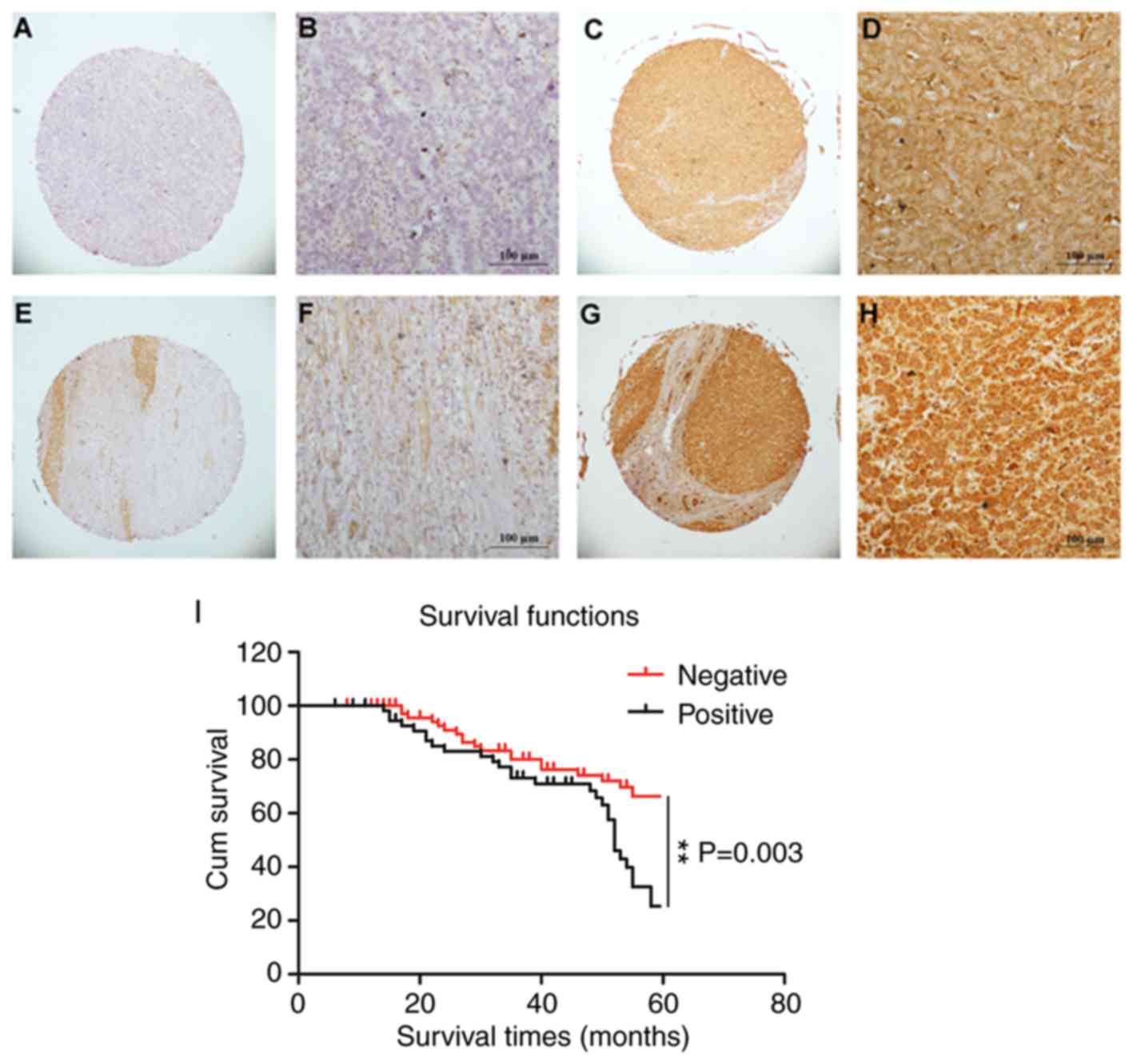
Immunohistochemical methods were used to assess the
expression of CD206 in liver cancer tissue. The staining for CD206
was observed predominantly in the membrane and cytoplasm of liver
cancer cells. Three pathologists independently evaluated the liver
cancer tissue microarrays under ×40 (Fig. 1A, C, E and G) and ×200 magnification
(Fig. 1B, D, F and H). CD206 showed
positive expression in the cell membrane and cytoplasm of liver
cancer cells. High levels of CD206 expression were detected in
204/327 (62.4%) of the patients with liver cancer. Positive
expression was observed in adjacent healthy liver tissue (5 mm from
the liver tumor and histopathologically confirmed) in 140/327
(42.8%) patients. Upon dividing samples into high expression (>6
points) and low expression groups (0–5 points), the expression
value of CD206 in liver cancer tissues was 7.69±3.11 points, and
the expression value of CD206 in adjacent healthy liver tissue was
3.60±2.17 points (P<0.05). Taken together, these data
demonstrated elevated CD206 expression in liver cancer tumors.
Patient characteristics and the
association between CD206 expression, liver cancer
clinicopathological features and prognosis
The present study investigated whether CD206
expression is associated with the progression of liver cancer.
CD206 immunopositivity was not associated with gender, age, tumor
number, Edmondson grade, microvascular invasion, hepatitis B virus
antigen, cirrhosis and AFP (Table
I). The survival time for patients with CD206-negative liver
cancer was 51.517±1.781 months and was significantly increased
compared with patients with CD206-postitive liver cancer
(46.067±2.183 months). The Kaplan-Meier survival curves indicated
that CD206 expression was significantly associated with overall
survival in patients with liver cancer (P=0.003; Fig. 1I). Additionally, prognosis factors in
liver cancer were analyzed by Cox-regression analysis. CD206
positivity was significantly associated with tumor size (P=0.039),
metastasis (P=0.022) and AFP value (P=0.002). There was no
statistically significant association between CD206 expression and
gender (P>0.05), age (P>0.05), cirrhosis (P>0.05),
metastasis (P>0.05), AFP (P>0.05) and tumor number
(P>0.05) as demonstrated by multivariate analysis (Table II). However, CD206 was significantly
associated with the Edmondson grade (P=0.009).
 | Table I.Expression of CD206 in liver cancer
tissue. |
Table I.
Expression of CD206 in liver cancer
tissue.
|
|
| CD206
expression |
|
|
|---|
|
|
|
|
|
|
|---|
| Clinical
parameter | Number | Low | Ηigh | χ2 | P-value |
|---|
| Age (years) |
|
|
| 1.601 | 0.206 |
|
<55 | 126 | 42 | 84 |
|
|
|
≥55 | 201 | 81 | 120 |
|
|
| Gender |
|
|
| 0.745 | 0.388 |
|
Male | 266 | 103 | 163 |
|
|
|
Female | 61 | 20 | 41 |
|
|
| Sizea |
|
|
| 6.913 | 0.009 |
|
<5 | 191 | 83 | 108 |
|
|
| ≥5 | 128 | 37 | 81 |
|
|
| Tumour number |
|
|
| 0.003 | 0.956 |
|
Single | 269 | 101 | 168 |
|
|
|
Multiple | 58 | 22 | 36 |
|
|
| Edmondson
gradea |
|
|
| 3.771 | 0.052 |
|
I+II | 202 | 68 | 134 |
|
|
|
III | 119 | 53 | 66 |
|
|
|
Metastasisa |
|
|
| 4.159 | 0.041 |
| M0 | 294 | 105 | 189 |
|
|
| M1 | 27 | 15 | 12 |
|
|
| Microvascular
invasiona |
|
|
| 0.118 | 0.732 |
|
Absence | 122 | 49 | 73 |
|
|
|
Presence | 121 | 46 | 75 |
|
|
| Hepatitis B virus
antigena |
|
|
| 3.101 | 0.078 |
|
Negtive | 62 | 29 | 3 |
|
|
|
Positive | 259 | 90 | 169 |
|
|
| Cirrhosis |
|
|
| 0.008 | 0.928 |
|
Negtive | 110 | 41 | 69 |
|
|
|
Positive | 217 | 82 | 135 |
|
|
|
α-fetoproteina |
|
|
| 0.090 | 0.764 |
|
<20 | 143 | 49 | 94 |
|
|
|
≥20 | 123 | 40 | 83 |
|
|
| Table II.Univariate and multivariate Cox
regression analysis of the clinicopathological parameters in
patients with liver cancer. |
Table II.
Univariate and multivariate Cox
regression analysis of the clinicopathological parameters in
patients with liver cancer.
|
|
| Univariate
analysis | Multivariate
analysis |
|---|
|
|
|
|
|
|---|
| Parameter | Number | Regression
coefficient | HR | 95% CI | P-value | Regression
coefficient | HR | 95.0% CI | P-value |
|---|
| Sex
(male/female) | 266/61 | −0.124 | 0.883 | 0.556–1.403 | 0.598 | −0.186 | 0.830 | 0.371–1.857 | 0.650 |
| Age (<55/≥55
years) | 126/201 | −0.027 | 0.973 | 0.571–1.658 | 0.920 | −0.493 | 0.611 | 0.289–1.293 | 0.198 |
| Tumor size
(<50/≥50 mm)a | 191/128 | 0.489 | 1.630 | 1.025–2.592 | 0.039 | 0.254 | 1.289 | 0.557–2.982 | 0.553 |
| Tumor number
(single/multiple)a | 268/58 | 0.059 | 1.060 | 0.582–1.933 | 0.848 | 1.008 | 2.740 | 1.051–7.143 | 0.039 |
| Edmondson grade
(I+II/III)a | 202/119 | 0.455 | 1.577 | 0.989–2.515 | 0.056 | 1.021 | 2.775 | 1.291–5.965 | 0.009 |
| Metastasis
(M0/M1)a | 294/27 | 0.160 | 1.173 | 0.629–2.188 | 0.022 | 1.293 | 3.644 | 1.314–10.104 | 0.013 |
| Microvascular
invasion (−/+)a | 122/121 | 0.606 | 1.834 | 1.089–3.087 | 0.615 | 0.310 | 1.364 | 0.578–3.219 | 0.479 |
| Hepatitis B virus
(−/+)a | 62/259 | 0.002 | 1.002 | 0.558–1.800 | 0.994 | −0.526 | 0.591 | 0.180–1.937 | 0.385 |
| Cirrhosis
(−/+) | 110/217 | −0.283 | 0.754 | 0.456–1.247 | 0.271 | 0.901 | 2.463 | 0.912–6.655 | 0.076 |
| α-fetoprotein
(<20/≥20 µg/l)a | 143/123 | 0.920 | 2.510 | 1.395–4.517 | 0.002 | 0.647 | 1.910 | 0.855–4.264 | 0.114 |
CD206 as a biomarker in cancer stem
cells may be used to predict liver cancer
The suspension culture in growth factor-defined
serum-free medium may be used to enrich cells associated with the
traits of CSCs (34,35). To validate whether liver cancer cell
lines acquire these traits when passed through the suspension
culture, the liver cancer cell line PLC/PRF/5 was subjected to
specific serum-free medium. In ultra-low attachment plates,
PLC/PRF/5 gradually formed non-adherent spheroid bodies, termed
sphere cells, following culture for 4–6 days (Fig. 2A). RT-qPCR was used to assess the
expression of recognized biomarker genes (Nanog, Oct4, Sox2, c-Myc
and CD44) and CD206 in the CSC spheres and the parent cells from
which they were derived (36–38). The
results revealed that, although the mRNA levels of CD206 did not
increase to the same levels as Nanog, CD44 and Sox2, CD206
expression was comparable to Oct4, and was significantly increased
compared with c-Myc (Fig. 2B). These
results were validated at the protein level through western blot
analysis, where increased expression of CD206 was observed in the
sphere cells compared with the parent cells (Fig. 2C). Taken together, these data suggest
that CD206, similar to the biomarkers Nanog, CD44, Sox2, Oct4 and
c-Myc, has the potential to predict cancer occurrence and
progression in CSC-like cells.
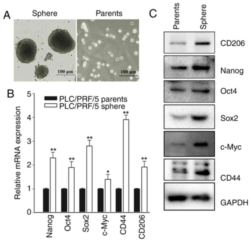 | Figure 2.CD206 is a biomarker in cancer stem
cells to predict the progression of liver cancer. (A) The liver
cancer cell line PLC/PRF/5 formed sphere bodies in suspension
culture/ (B) mRNA levels of Nanog, Oct4, Sox2, c-Myc, CD44 and
CD206. *P<0.05, **P<0.01 vs. PLC/PRF/5 parents. (C) The
expression level of CD206 increased in a similar manner to the
expression of the internationally recognized biomarker proteins
Nanog, Oct4, Sox2, c-Myc and CD44. CD206, mannose receptor; Nanog,
Nanog homeobox; Oct4, POU class 5 homeobox 1; Sox2, SRY-box 2;
c-Myc, MYC binding protein; CD44, CD44 molecule. |
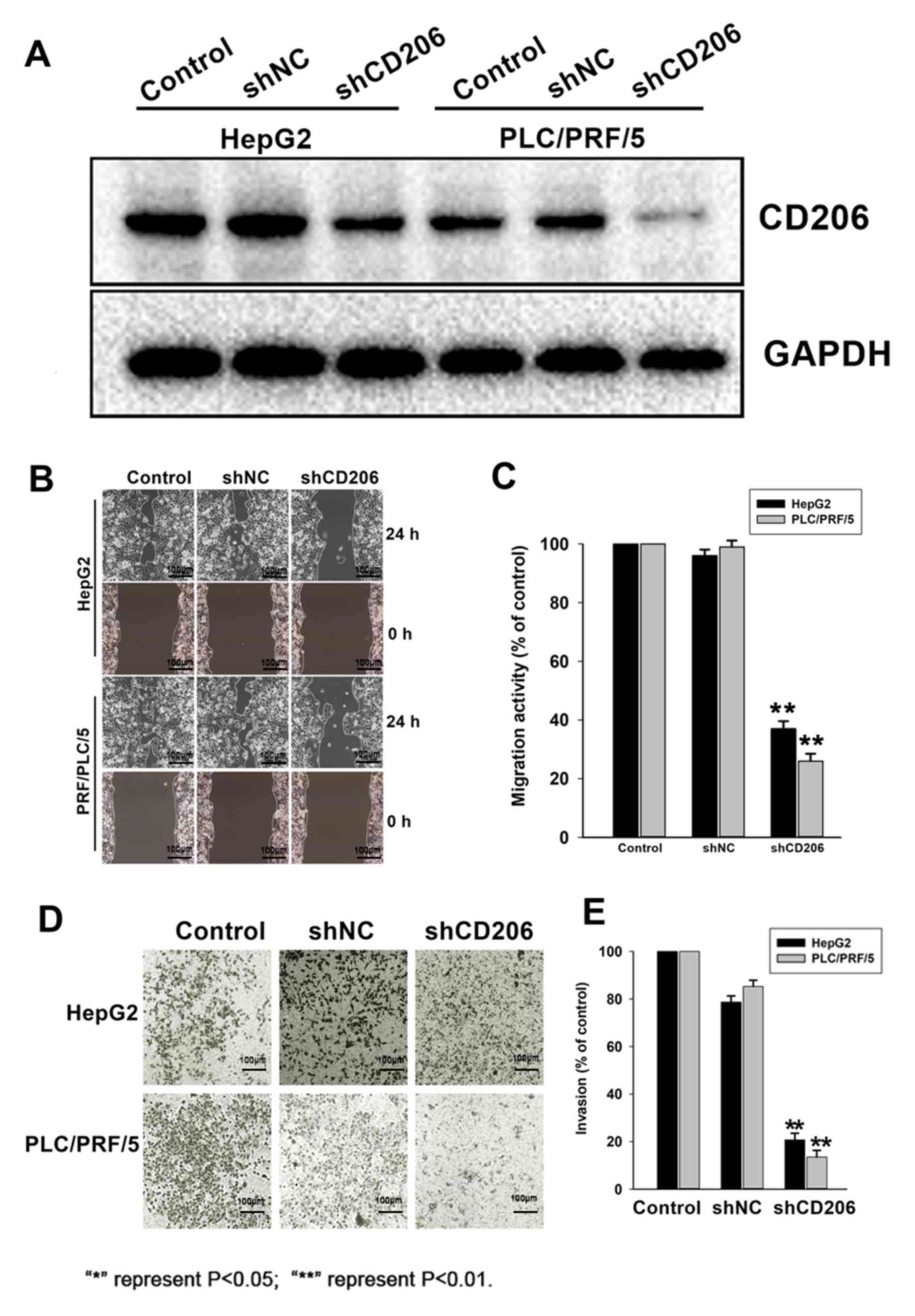
CD206 silencing decreases liver cancer
cell motility and invasion
To assess the role of CD206 in liver cancer cell
migration and invasion, HepG2 and PLC/PRF/5 cells were infected
with lentiviruses expressing CD206 shRNA (shCD206) or a scrambled
sequence (shNC) to obtain stable expression cell lines. CD206
silencing following shCD206 lentiviral transfection was confirmed
by Western Blot analysis (Fig. 3A).
Migration (Fig. 3B and C) and
invasion assays (Fig. 3D and E)
revealed that CD206 silencing significantly decreased HepG2 and
PLC/PRF/5 cell migration and invasion compared with the shNC group
(P<0.05). These observations indicated that CD206 promotes the
motility and invasiveness of liver cancer cell lines.
Discussion
Liver cancer is a multigene disease characterized by
a high degree of malignancy, rapid development, low survival rates
and late detection (9,39). Surgical interventions are often
ineffective due to late diagnosis (40). Novel liver cancer diagnostic and
therapeutic targets are therefore required.
CD206 is a pattern recognition receptor that
identifies the extracellular domains of specific carbohydrate
molecules and is highly expressed on the surface of macrophages and
immature dendritic cells (41). In
the present study, CD206 was expressed in the cytoplasm and on the
plasma membrane of liver cancer cells. Additionally, CD206
upregulation was observed in liver cancer tissue compared with
healthy adjacent tissue obtained from patients with liver cancer.
CD206 expression was not significantly associated with gender, age,
tumor number, Edmondson grade, microvascular invasion, hepatitis B
virus antigen and cirrhosis, but had a positive association with
tumor size, metastasis and the AFP value. The present study
therefore demonstrated a preliminary association between CD206 and
the occurrence and development of liver cancer. Previous studies
investigating liver cancer-associated proteins have demonstrated an
association between marker expression and cancer staging. The
present study revealed that CD206 expression was associated with
tumor size, metastasis and the AFP value, indicating that its
expression is associated with poor liver cancer prognosis.
Furthermore, the association between CD206 expression and survival
time revealed that the levels of CD206 were positively associated
with poor prognosis. Taken together, these data suggested that high
CD206 expression promotes the rapid growth and metastasis of liver
cancer.
Uncontrolled self-renewal directly contributes to
the progression of liver cancer and other types of carcinomas. The
same molecular pathways that regulate self-renewal in normal stem
cells also control CSCs (22,38,42).
The present study revealed that CD206 is an important biomarker for
liver cancer progression in CSCs, similar to other known markers
including Nanog, Oct4, Sox2, c-Myc and CD44 (15,43–46). The
expression of these markers in CSCs are recognized as evidence of
stem cell carcinogenesis (37,47,48). The
present study detected the expression levels of CD206 in CSCs and
demonstrated high expression at both the mRNA and protein levels,
which were comparable with the levels of Oct4 and c-Myc. The
results obtained in the current study suggested that CD206 may be
used as a biomarker in CSCs to predict liver cancer, highlighting
its diagnostic value.
The present study investigated the direct effects of
CD206 knockdown on liver cancer cells lines. The migration and
invasion abilities of liver cancer cells decreased when CD206 was
silenced, suggesting that high CD206 expression levels promote
tumor metastasis and poor prognosis in patients.
Hepatitis C virus (HCV) infection is a main cause of
liver cancer (49,50). However, the present study did not
investigate the important role of HCV infection in liver cancer and
this is a limitation of the study. Future studies investigating the
association between HCV infection and CD206 expression are
required.
In summary, the present study revealed that CD206
may be used as a diagnostic tool to screen for liver cancer, and
its upregulation in liver tumors represents a therapeutic target to
reduce liver cancer cell metastasis. Future investigation of the
efficacy of CD206 inhibitors on liver cancer cells as well as an
evaluation of the potential toxic effects on normal hepatocytes is
required. Such studies may lay the theoretical foundations for
future clinical liver cancer diagnosis and treatment.
Acknowledgements
Not applicable.
Funding
The present study was supported by the National
Science Foundation of China (grant nos. 81602706 and 81570198),
Zhejiang Medical Technology Plan Project (grant no. WKJ-ZJ-1709)
and the State Administration of Traditional Chinese Medicine of
Zhejiang (grant nos. 2016ZZ007 and 2017ZB006).
Availability of data and materials
The datasets used during the present study are
available from the corresponding author upon reasonable
request.
Authors' contributions
SW, LZ and XT conceived and designed the study. WF,
XY and FH performed the experiments. SW wrote the paper. SW, LZ and
XM reviewed and edited the manuscript. All authors read and
approved the manuscript.
Ethical approval and consent to
participate
The study was approved by the Ethics Committee of
Zhejiang Provincial People's Hospital and all patients provided
written informed consent.
Patient consent for publication
Not applicable.
Competing interests
The authors declare that they have no competing
interests.
References
|
1
|
Ghouri YA, Mian I and Rowe JH: Review of
hepatocellular carcinoma: Epidemiology, etiology, and
carcinogenesis. J Carcinog. 16:12017. View Article : Google Scholar : PubMed/NCBI
|
|
2
|
Altekruse SF, Henley SJ, Cucinelli JE and
McGlynn KA: Changing hepatocellular carcinoma incidence and liver
cancer mortality rates in the United States. Am J Gastroenterol.
109:542–553. 2014. View Article : Google Scholar : PubMed/NCBI
|
|
3
|
Ferlay J, Soerjomataram I, Dikshit R, Eser
S, Mathers C, Rebelo M, Parkin DM, Forman D and Bray F: Cancer
incidence and mortality worldwide: Sources, methods and major
patterns in GLOBOCAN 2012. Int J Cancer. 136:E359–E386. 2015.
View Article : Google Scholar : PubMed/NCBI
|
|
4
|
Tsai WC, Kung PT, Wang YH, Kuo WY and Li
YH: Influence of the time interval from diagnosis to treatment on
survival for early-stage liver cancer. PLoS One. 13:e01995322018.
View Article : Google Scholar : PubMed/NCBI
|
|
5
|
Ronot M, Clift AK, Vilgrain V and Frilling
A: Functional imaging in liver tumours. J Hepatol. 65:1017–1030.
2016. View Article : Google Scholar : PubMed/NCBI
|
|
6
|
Marrero JA, Ahn J and Rajender Reddy K;
Americal College of Gastroenterology, : ACG clinical guideline: The
diagnosis and management of focal liver lesions. Am J
Gastroenterol. 109:1328–1348. 2014. View Article : Google Scholar : PubMed/NCBI
|
|
7
|
Elsayes KM, Hooker JC, Agrons MM, Kielar
AZ, Tang A, Fowler KJ, Chernyak V, Bashir MR, Kono Y, Do RK, et al:
2017 version of LI-RADS for CT and MR Imaging: An update.
Radiographics. 37:1994–2017. 2017. View Article : Google Scholar : PubMed/NCBI
|
|
8
|
Ryerson AB, Eheman CR, Altekruse SF, Ward
JW, Jemal A, Sherman RL, Henley SJ, Holtzman D, Lake A, Noone AM,
et al: Annual report to the nation on the status of cancer,
1975–2012, featuring the increasing incidence of liver cancer.
Cancer. 122:1312–1337. 2016. View Article : Google Scholar : PubMed/NCBI
|
|
9
|
Sia D, Villanueva A, Friedman SL and
Llovet JM: Liver cancer cell of origin, molecular class, and
effects on patient prognosis. Gastroenterology. 152:745–761. 2017.
View Article : Google Scholar : PubMed/NCBI
|
|
10
|
Bhoori S and Mazzaferro V: Current
challenges in liver transplantation for hepatocellular carcinoma.
Best Pract Res Clin Gastroenterol. 28:867–879. 2014. View Article : Google Scholar : PubMed/NCBI
|
|
11
|
Chen W, Fan W, Ru G, Huang F, Lu X, Zhang
X, Mou X and Wang S: Gemcitabine combined with an engineered
oncolytic vaccinia virus exhibits a synergistic suppressive effect
on the tumor growth of pancreatic cancer. Oncol Rep. 41:67–76.
2019.PubMed/NCBI
|
|
12
|
Staines K, Hunt LG, Young JR and Butter C:
Evolution of an expanded mannose receptor gene family. PLoS One.
9:e1103302014. View Article : Google Scholar : PubMed/NCBI
|
|
13
|
Lee SH, Charmoy M, Romano A, Paun A,
Chaves MM, Cope FO, Ralph DA and Sacks DL: Mannose receptor high,
M2 dermal macrophages mediate nonhealing Leishmania major infection
in a Th1 immune environment. J Exp Med. 215:357–375. 2018.
View Article : Google Scholar : PubMed/NCBI
|
|
14
|
Peng H, Geil Nickell CR, Chen KY, McClain
JA and Nixon K: Increased expression of M1 and M2 phenotypic
markers in isolated microglia after four-day binge alcohol exposure
in male rats. Alcohol. 62:29–40. 2017. View Article : Google Scholar : PubMed/NCBI
|
|
15
|
Salmi M, Karikoski M, Elima K, Rantakari P
and Jalkanen S: CD44 binds to macrophage mannose receptor on
lymphatic endothelium and supports lymphocyte migration via
afferent lymphatics. Circ Res. 112:1577–1582. 2013. View Article : Google Scholar : PubMed/NCBI
|
|
16
|
van Lessen M, Shibata-Germanos S, van
Impel A, Hawkins TA, Rihel J and Schulte-Merker S: Intracellular
uptake of macromolecules by brain lymphatic endothelial cells
during zebrafish embryonic development. Elife. 6(pii): e259322017.
View Article : Google Scholar : PubMed/NCBI
|
|
17
|
Azad AK, Rajaram MV, Metz WL, Cope FO,
Blue MS, Vera DR and Schlesinger LS: γ-Tilmanocept, a new
radiopharmaceutical tracer for cancer sentinel lymph nodes, binds
to the mannose receptor (CD206). J Immunol. 195:2019–2029. 2015.
View Article : Google Scholar : PubMed/NCBI
|
|
18
|
Andersen MN, Andersen NF, Rodgaard-Hansen
S, Hokland M, Abildgaard N and Moller HJ: The novel biomarker of
alternative macrophage activation, soluble mannose receptor
(sMR/sCD206): Implications in multiple myeloma. Leuk Res.
39:971–975. 2015. View Article : Google Scholar : PubMed/NCBI
|
|
19
|
Bianchi G and Munshi NC: Pathogenesis
beyond the cancer clone(s) in multiple myeloma. Blood.
125:3049–3058. 2015. View Article : Google Scholar : PubMed/NCBI
|
|
20
|
Kume H, Muraoka S, Kuga T, Adachi J,
Narumi R, Watanabe S, Kuwano M, Kodera Y, Matsushita K, Fukuoka J,
et al: Discovery of colorectal cancer biomarker candidates by
membrane proteomic analysis and subsequent verification using
selected reaction monitoring (SRM) and tissue microarray (TMA)
analysis. Mol Cell Proteomics. 13:1471–1484. 2014. View Article : Google Scholar : PubMed/NCBI
|
|
21
|
Rodgaard-Hansen S, Rafique A, Christensen
PA, Maniecki MB, Sandahl TD, Nexo E and Moller HJ: A soluble form
of the macrophage-related mannose receptor (MR/CD206) is present in
human serum and elevated in critical illness. Clin Chem Lab Med.
52:453–461. 2014. View Article : Google Scholar : PubMed/NCBI
|
|
22
|
Sun C, Shui B, Zhao W, Liu H, Li W, Lee
JC, Doran R, Lee FK, Sun T, Shen QS, et al: Central role of
IP3R2-mediated Ca2+ oscillation in self-renewal of liver
cancer stem cells elucidated by high-signal ER sensor. Cell Death
Dis. 10:3962019. View Article : Google Scholar : PubMed/NCBI
|
|
23
|
Cao J, Zhao M, Liu J, Zhang X, Pei Y, Wang
J, Yang X, Shen B and Zhang J: RACK1 promotes self-renewal and
chemoresistance of cancer stem cells in human hepatocellular
carcinoma through stabilizing nanog. Theranostics. 9:811–828. 2019.
View Article : Google Scholar : PubMed/NCBI
|
|
24
|
Yang X, Li S, Wang H, Chen W, Mou X and
Wang S: Expression of coxsackie and adenovirus receptor is
correlated with inferior prognosis in liver cancer patients. Oncol
Lett. 17:2485–2490. 2019.PubMed/NCBI
|
|
25
|
Petrizzo A and Buonaguro L: Application of
the Immunoscore as prognostic tool for hepatocellular carcinoma. J
Immunother Cancer. 4:712016. View Article : Google Scholar : PubMed/NCBI
|
|
26
|
Luo Y, Pandey A, Ghasabeh MA, Pandey P,
Varzaneh FN, Zarghampour M, Khoshpouri P, Ameli S, Li Z, Hu D and
Kamel IR: Prognostic value of baseline volumetric multiparametric
MR imaging in neuroendocrine liver metastases treated with
transarterial chemoembolization. Eur Radiol. Mar 15–2019.doi:
10.1007/s00330-019-06100-3 (Epub ahead of print). View Article : Google Scholar
|
|
27
|
Colecchia A, Scaioli E, Montrone L,
Vestito A, Di Biase AR, Pieri M, D'Errico-Grigioni A,
Bacchi-Reggiani ML, Ravaioli M, Grazi GL and Festi D: Pre-operative
liver biopsy in cirrhotic patients with early hepatocellular
carcinoma represents a safe and accurate diagnostic tool for tumour
grading assessment. J Hepatol. 54:300–305. 2011. View Article : Google Scholar : PubMed/NCBI
|
|
28
|
Kong M and Hong SE: Optimal follow-up
duration for evaluating objective response to radiotherapy in
patients with hepatocellular carcinoma: A retrospective study. Chin
J Cancer. 34:79–85. 2015. View Article : Google Scholar : PubMed/NCBI
|
|
29
|
Gan W, Zhang MX, Wang JX, Fu YP, Huang JL,
Yi Y, Jing CY, Fan J, Zhou J and Qiu SJ: Prognostic impact of
lactic dehydrogenase to albumin ratio in hepatocellular carcinoma
patients with Child-Pugh I who underwent curative resection: A
prognostic nomogram study. Cancer Manag Res. 10:5383–5394. 2018.
View Article : Google Scholar : PubMed/NCBI
|
|
30
|
Couri T and Pillai A: Goals and targets
for personalized therapy for HCC. Hepatol Int. 13:125–137. 2019.
View Article : Google Scholar : PubMed/NCBI
|
|
31
|
Zhu R, Huang H, Zhang H, Wang Z, Hu X,
Zhai W, Lin Y, Wang J and Zhu H: Prognostic analysis in chronic
hepatitis B patients: A retrospective study of 216 cases about
Scheuer scores, in situ expression of viral antigens and tissue
hepatitis B virus DNA levels. Liver Int. 26:82–89. 2006. View Article : Google Scholar : PubMed/NCBI
|
|
32
|
Zhu AX, Kang YK, Yen CJ, Finn RS, Galle
PR, Llovet JM, Assenat E, Brandi G, Pracht M, Lim HY, et al:
Ramucirumab after sorafenib in patients with advanced
hepatocellular carcinoma and increased alpha-fetoprotein
concentrations (REACH-2): A randomised, double-blind,
placebo-controlled, phase 3 trial. Lancet Oncol. 20:282–296. 2019.
View Article : Google Scholar : PubMed/NCBI
|
|
33
|
Shim JH, Yoon DL, Han S, Lee YJ, Lee SG,
Kim KM, Lim YS, Lee HC, Chung YH and Lee YS: Is serum
alpha-fetoprotein useful for predicting recurrence and mortality
specific to hepatocellular carcinoma after hepatectomy? A test
based on propensity scores and competing risks analysis. Ann Surg
Oncol. 19:3687–3696. 2012. View Article : Google Scholar : PubMed/NCBI
|
|
34
|
Zhong M, Zhong C, Cui W, Wang G, Zheng G,
Li L, Zhang J, Ren R, Gao H, Wang T, et al: Induction of
tolerogenic dendritic cells by activated TGF-beta/Akt/Smad2
signaling in RIG-I-deficient stemness-high human liver cancer
cells. BMC Cancer. 19:4392019. View Article : Google Scholar : PubMed/NCBI
|
|
35
|
Wei X, You X, Zhang J and Zhou C:
MicroRNA-1305 inhibits the stemness of LCSCs and Tumorigenesis by
repressing the UBE2T-dependent Akt-signaling pathway. Mol Ther
Nucleic Acids. 16:721–732. 2019. View Article : Google Scholar : PubMed/NCBI
|
|
36
|
Bamodu OA, Kuo KT, Yuan LP, Cheng WH, Lee
WH, Ho YS, Chao TY and Yeh CT: HDAC inhibitor suppresses
proliferation and tumorigenicity of drug-resistant chronic myeloid
leukemia stem cells through regulation of hsa-miR-196a targeting
BCR/ABL1. Exp Cell Res. 370:519–530. 2018. View Article : Google Scholar : PubMed/NCBI
|
|
37
|
Choi HS, Kim JH, Kim SL, Deng HY, Lee D,
Kim CS, Yun BS and Lee DS: Catechol derived from aronia juice
through lactic acid bacteria fermentation inhibits breast cancer
stem cell formation via modulation Stat3/IL-6 signaling pathway.
Mol Carcinog. 57:1467–1479. 2018. View
Article : Google Scholar : PubMed/NCBI
|
|
38
|
Mani SK, Zhang H, Diab A, Pascuzzi PE,
Lefrancois L, Fares N, Bancel B, Merle P and Andrisani O:
EpCAM-regulated intramembrane proteolysis induces a cancer stem
cell-like gene signature in hepatitis B virus-infected hepatocytes.
J Hepatol. 65:888–898. 2016. View Article : Google Scholar : PubMed/NCBI
|
|
39
|
Liu M, Jiang L and Guan XY: The genetic
and epigenetic alterations in human hepatocellular carcinoma: A
recent update. Protein Cell. 5:673–691. 2014. View Article : Google Scholar : PubMed/NCBI
|
|
40
|
Orcutt ST and Anaya DA: Liver resection
and surgical strategies for management of primary liver cancer.
Cancer Control. 25:10732748177446212018. View Article : Google Scholar : PubMed/NCBI
|
|
41
|
Scodeller P, Simon-Gracia L, Kopanchuk S,
Tobi A, Kilk K, Saalik P, Kurm K, Squadrito ML, Kotamraju VR,
Rinken A, et al: Precision targeting of tumor macrophages with a
CD206 binding peptide. Sci Rep. 7:146552017. View Article : Google Scholar : PubMed/NCBI
|
|
42
|
Maehara O, Ohnishi S, Asano A, Suda G,
Natsuizaka M, Nakagawa K, Kobayashi M, Sakamoto N and Takeda H:
Metformin regulates the expression of CD133 through the AMPK-CEBPβ
pathway in hepatocellular carcinoma cell lines. Neoplasia.
21:545–556. 2019. View Article : Google Scholar : PubMed/NCBI
|
|
43
|
Palla AR, Piazzolla D, Abad M, Li H,
Dominguez O, Schonthaler HB, Wagner EF and Serrano M: Reprogramming
activity of NANOGP8, a NANOG family member widely expressed in
cancer. Oncogene. 33:2513–2519. 2014. View Article : Google Scholar : PubMed/NCBI
|
|
44
|
Jerabek S, Merino F, Scholer HR and
Cojocaru V: OCT4: Dynamic DNA binding pioneers stem cell
pluripotency. Biochim Biophys Acta. 1839:138–154. 2014. View Article : Google Scholar : PubMed/NCBI
|
|
45
|
Guvench O: Revealing the mechanisms of
protein disorder and N-glycosylation in CD44-hyaluronan binding
using molecular simulation. Front Immunol. 6:3052015. View Article : Google Scholar : PubMed/NCBI
|
|
46
|
Yoshida GJ: Emerging roles of Myc in stem
cell biology and novel tumor therapies. J Exp Clin Cancer Res.
37:1732018. View Article : Google Scholar : PubMed/NCBI
|
|
47
|
Suzuki Y, Haraguchi N, Takahashi H, Uemura
M, Nishimura J, Hata T, Takemasa I, Mizushima T, Ishii H, Doki Y,
et al: SSEA-3 as a novel amplifying cancer cell surface marker in
colorectal cancers. Int J Oncol. 42:161–167. 2013.PubMed/NCBI
|
|
48
|
Botchkina GI, Zuniga ES, Das M, Wang Y,
Wang H, Zhu S, Savitt AG, Rowehl RA, Leyfman Y, Ju J, et al:
New-generation taxoid SB-T-1214 inhibits stem cell-related gene
expression in 3D cancer spheroids induced by purified colon
tumor-initiating cells. Mol Cancer. 9:1922010. View Article : Google Scholar : PubMed/NCBI
|
|
49
|
Kanda T, Goto T, Hirotsu Y, Moriyama M and
Omata M: Molecular mechanisms driving progression of liver
cirrhosis towards hepatocellular carcinoma in chronic Hepatitis B
and C infections: A review. Int J Mol Sci. 20(pii): E13582019.
View Article : Google Scholar : PubMed/NCBI
|
|
50
|
Zahra M, Azzazy H and Moustafa A:
Transcriptional regulatory networks in Hepatitis C virus-induced
hepatocellular carcinoma. Sci Rep. 8:142342018. View Article : Google Scholar : PubMed/NCBI
|