Introduction
Second primary tumors (SPTs) are a common occurrence
in daily cancer practice and are a major cause of mortality
(1). In patients with head and neck
(H&N) squamous cell carcinoma (SCC), SPTs are frequently
detected in the H&N region, lungs, or esophagus years after
treatment (2). Therefore, long-term
follow-up is critical to detect SPTs as well as recurrence and
metastasis (3,4). However, there are no broadly accepted
guidelines for lifelong follow-up using computed tomography (CT)
(5). Here, we report the case of a
patient with metachronous SCC of the tongue, SCC of the lip, and
type A thymoma. Thymoma was incidentally detected during follow-up
using contrast-enhanced CT 9 years after the second tumor
resection. Following the detection, we performed further
examinations to clinically diagnose the mass and made preparations
to treat the mass; however, the patient initially refused the
treatment for the thymoma.
Case report
In October 2005, a 66-year-old male patient was
admitted to our hospital with a 6-month history of pain and ulcer
involving the left side of the lower lip. The disease was initially
diagnosed from biopsy as SCC and treated with cryotherapy (liquid
nitrogen) and chemotherapy (local injection of oil bleomycin) at
another clinic 2.5 years before the presentation (April 2003).
Although the lip mass temporarily disappeared following cryotherapy
and chemotherapy, it recurred and pain gradually worsened. Physical
examination revealed a hard, elastic 1.3×1.0-cm mass with an ulcer
of the left lower lip. No other lesions were observed on the lip or
oral cavity, and no palpable neck lymphadenopathy was detected. The
patient was a former moderate smoker and former moderate drinker,
and his medical history included tongue SCC resection on the right
edge of the tongue followed by radical neck dissection on the right
side and adjuvant radiotherapy of the neck at another hospital at
the age of 33 years (in February 1973). Moreover, the patient had
an adenomatous goiter on presentation; however, he has remained
stable till date. Regarding family history, his mother had colon
cancer and younger brother had esophageal cancer. Contrast-enhanced
CTs of the H&N and chest revealed the enhanced mass in the left
lower lip, but no lesions of the tongue, cervical lymph nodes,
lungs, mediastinum, or bone were revealed. Similarly, ultrasound
and magnetic resonance imaging revealed no neck lesions, and upper
gastrointestinal examination revealed no tumorous lesions. The
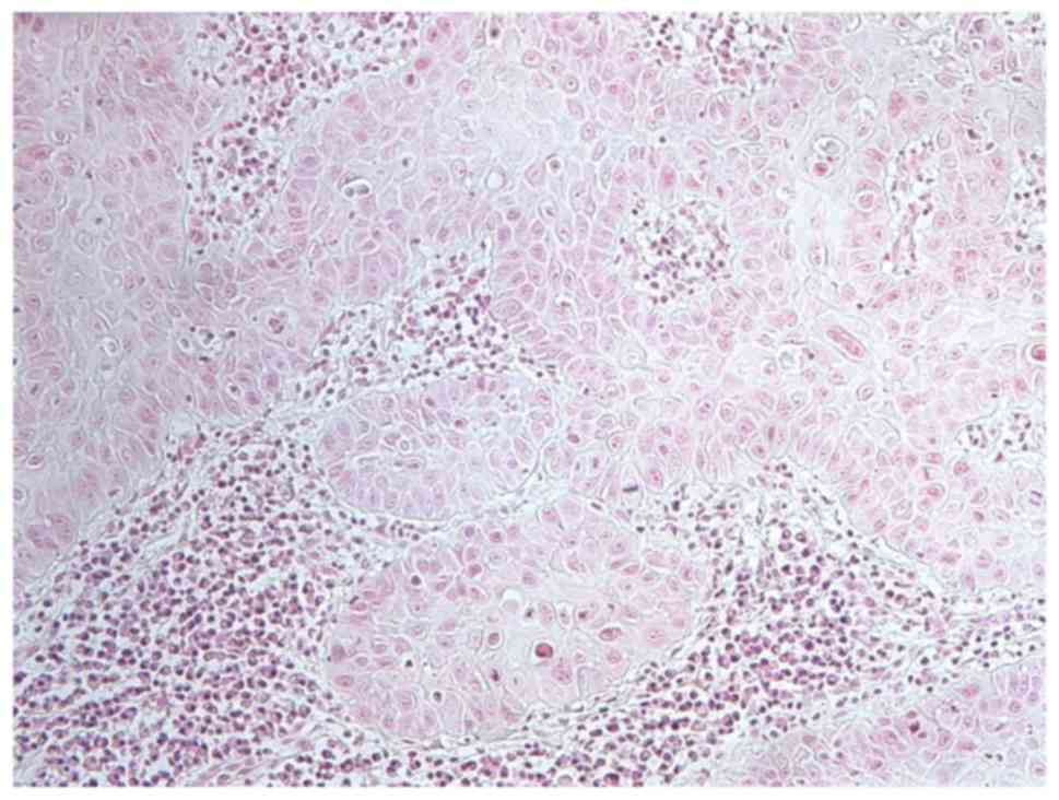
biopsy of the lip confirmed SCC recurrence (Fig. 1). Based on radiographic and clinical
assessments, SCC of the lip was graded as RT1N0M0, stage I,
according to the Union for International Cancer Control TNM
classification (6). The patient
underwent local excision of the lip tumor after receiving
neoadjuvant chemotherapy with bleomycin (a total dose of 105 mg for
approximately 1 month) plus 450 mg uracil/tegafur per day
(approximately 1 month), as previously described (7,8).
Histopathological examination of the resected tumor further
confirmed SCC. No adjuvant therapy was performed.
The patient remained disease-free for 9 years
following the treatment for SCC of the lip. During this period, he
visited our outpatient clinic regularly for clinical examinations.
However, CT had not been performed for 7 years (from 2 years after
lip resection) because his clinical lesions were stable. We
recommended the patient to perform the CT to detect hidden SPTs or
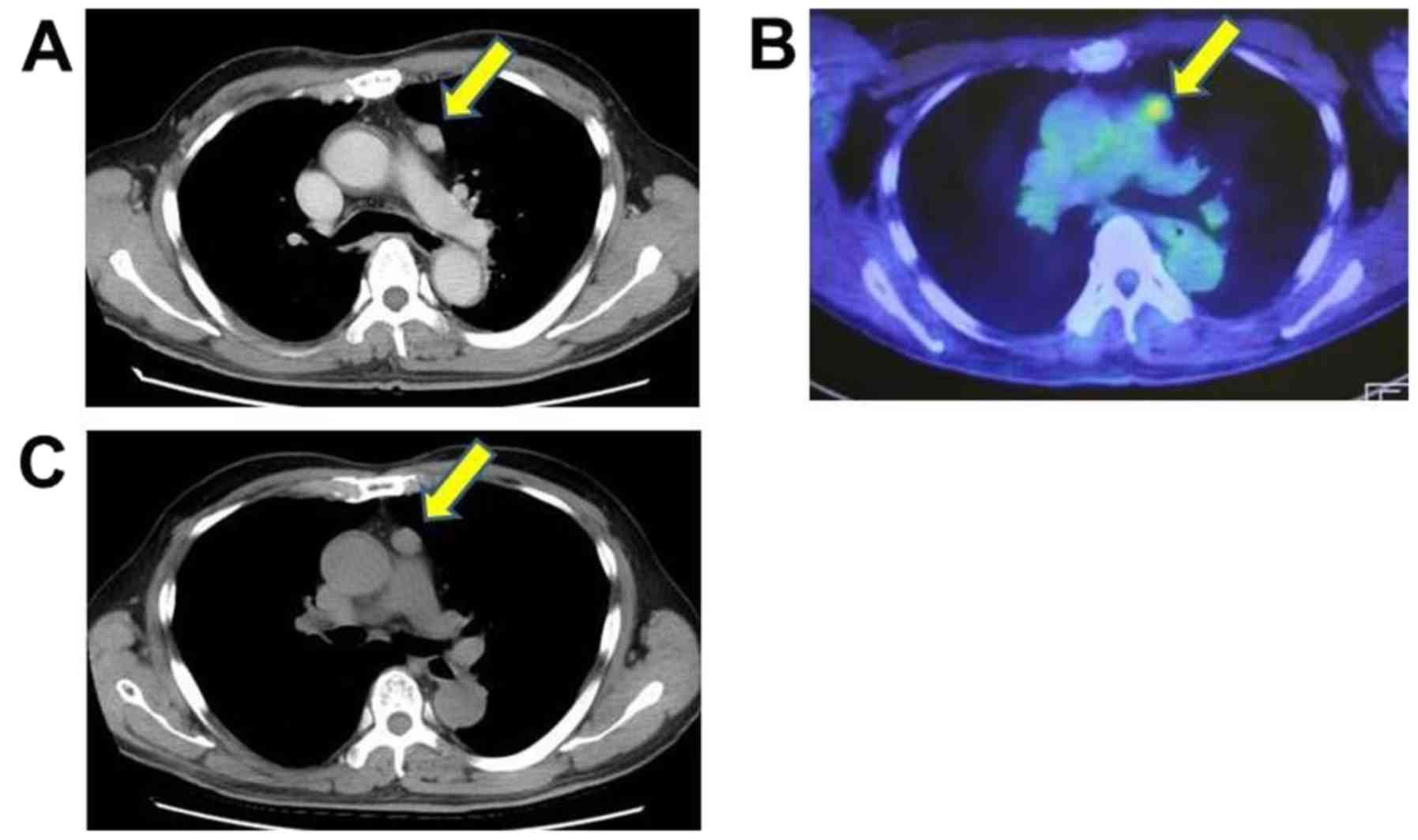
SCC metastasis. Contrast-enhanced CTs of the H&N and chest (in
August 2015) revealed no H&N lesion but a mass lesion in the
mediastinum (Fig. 2A). At the time,
no clinical symptoms were found. Subsequent
2-[18F]-fluoro-2-deoxy-D- glucose positron emission
tomography (FDG-PET)/CT revealed high FDG uptake by the mediastinal
mass (standardized uptake value max=4.45) (Fig. 2B). Thereafter, gastroendoscopy was
performed to examine the gastroesophageal lesion related to the
mediastinum mass; however, no lesion was found. Preliminary
clinical and radiological diagnosis was mediastinal lymph node
metastasis of lip or tongue cancer or metachronous SPT. However,
contrary to our advice, the patient initially rejected any further
examination or treatment of the mass owing to stress experienced by
him because of numerous examinations conducted. However, after
6-month observation period, follow-up using CT revealed mass growth
(Fig. 2C). Following an explanation
by the thoracic surgeon, the patient finally agreed to treatment
(in February 2016). To definitively diagnose the mediastinal
lesion, subsequent excision biopsy with video-assisted thoracic
surgery was performed, and histological examination revealed
thymoma [World Health Organisation (WHO) classification: Type A;
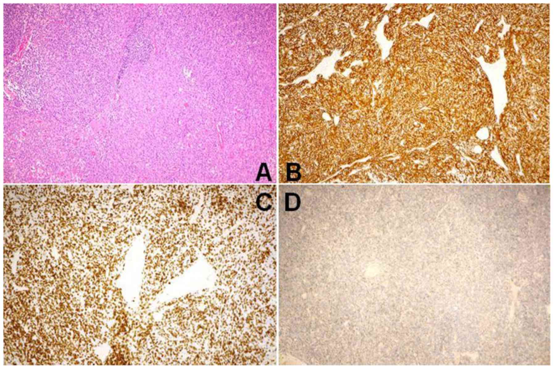
Masaoka stage I] (Fig. 3). In the
resected mass, the adhesive proliferation of spindle cells that
interspersed with lymphocytes was found in hematoxylin and eosin
staining (Fig. 3A).
Immunohistochemically, the spindle cells were positive for CK5/6
(Fig. 3B), whereas they were
negative for AE1/AE3, CD5, and p16 (data not shown). The
lymphocytes was mostly positive for CD3 (Fig. 3C) and CD1a (data not shown) and
partly positive for TdT (Fig. 3D).
Approximately 10–20% of the tumor cells were positive for MIB-1 on
the lymphocytes (which were also positive for MIB-1) (data not
shown). No oral cancer metastasis was histologically found. The
study patient was subsequently diagnosed with metachronous triple
primary tumors (9,10). After a follow-up period of 2.5 years,
the patient is alive and well with no evidence of tumor recurrence,
metastatic disease, or any more SPTs.
Written informed consent was obtained from the
patient for publication of this case report and the accompanying
images. The Ethics Committee of the University of the Ryukyus
waived the requirement for review per institutional protocol, as
the study does not contain content that requires ethical approval,
and approved the submission and publication of this case
report.
Discussion
There are three notable aspects of this case. First,
to the best of our knowledge, the specific combination of tumors
(SCC of the tongue, SCC of the lip, and type A thymoma) has not
been reported previously. Second, we incidentally detected the
asymptomatic thymus tumor using CT after an unusually long (9 year
and 9 months) asymptomatic period following the treatment for the
lip SCC. Third, the patient initially refused any further
examination or treatment of the new SPT, we believe that patient
background is an important prognostic factor for SPT treatment.
Through this case, in addition to presenting the case report, we
decided to perform an adequate literature review to identify novel
cases based on the three aspects of the present case.
To identify previous cases of thymoma with SPT, we
performed a literature search for cases with the combined
occurrence of H&N cancer and thymoma (including WHO type C,
i.e., thymic carcinoma) reported in English using PubMed
(https://www.ncbi.nlm.nih.gov/pubmed/)
and Google Scholar (https://scholar.google.co.jp/) between 1832 (11) (when thymus was initially reported)
and 2018; we excluded non-English literature or conference
proceedings, with no other exclusion criteria (Table I) (12–41).
Although we identified several cases of thymoma with SPT, we found
no case matching the current case of SCC of the tongue, SCC of the
lip, and thymoma in sequence.
 | Table I.Cases of combined thymoma and other
extrathymic cancers. |
Table I.
Cases of combined thymoma and other
extrathymic cancers.
| Year |
Age/sexa | World Health
Organisation classification of thymoma | Masaoka stage of
thymoma | First cancer | Second cancer | Third cancer | Fourth cancer | Fifth cancer | Time interval
between thymoma and H&N malignancy | Autoimmune
disease | Thymoma
detection | Outcome | (Refs.) |
|---|
| 1962 | NA/NA | NA | NA | Thymoma | Tongue cancer | (−) | (−) | (−) | NA | NA | NA | NA | (14) |
| 1962 | NA/NA | NA | NA | Thymoma | Thyroid cancer and
rectal cancer | (−) | (−) | (−) | NA | NA | NA | NA | (14) |
| 1968 | NA/NA (3
cases) | NA | NA | Thymoma | Thyroid cancer | (−) | (−) | (−) | NA | NA | NA | NA | (13) |
| 1977 | NA/male (2
cases) | NA | NA | Papillary carcinoma
of the thyroid gland | Thymoma | (−) | (−) | (−) | NA | NA | NA | NA | (15) |
| 1983 | 58/female | NA | NA | Endometrial
adenocarcinoma | Thymoma (7 years
after the first cancer treatment) | Papillary thyroid
carcinoma | (−) | (−) | Synchronous
(thymoma and thyroid cancer) | MG | CT for initial
clinical symptoms | NA | (16) |
| 1987 | 62/male | NA | NA | Hodgkin's disease
of the neck | Thymoma | (−) | (−) | (−) | 7 years | NA | Autopsy | Died from severe
dyspnea 7 years after the first malignancy diagnosis | (17) |
| 1989 | Around
67/female | NA | NA | Thymoma | Breast cancer | SCC of the upper
lip | (−) | (−) | Approximately 8
years (from thymoma to lip cancer) |
Hypogamma-globulinemia | NA | Died 2 years after
thediagnosis of SCC of the lip from congestive heart failure with
malabsorption syndrome | (18) |
| 1990 | NA/NA | NA | NA | Maxillary
sarcoma | Thymoma | (−) | (−) | (−) | 2 years | NA | NA | NA | (19) |
| 1990 | NA/NA | NA | NA | Laryngeal
cancer | Thyroid cancer
(follicular) | Thymoma | (−) | (−) | Within 1 year (from
first and second cancer to thymoma) | NA | NA | NA | (19) |
| 1992 | 48/female | NA | NA | Thymoma | Thyroid papillary
carcinoma and bilateral jugular lymph node metastasis | (−) | (−) | (−) | 10 days | MG | CT for MG
lesions | Alive without tumor
recurrence | (20) |
| 1995 | NA/NA | NA | NA | Thymoma | Thyroid
carcinoma | (−) | (−) | (−) | NA | NA | NA | NA | (21) |
| 1997 | NA/NA | NA | NA | H&N SCC | Thymoma | (−) | (−) | (−) | NA | NA | CT and chest
radiography within 60 days of initial H&N SCC diagnosis | NA | (22) |
| 1997 | NA/NA | NA | NA | H&N SCC | Thymoma | (−) | (−) | (−) | NA | NA | CT and chest
radiography within 60 days of initial H&N SCC diagnosis | NA | (22) |
| 1998 | 68 or
69/female | NA | NA | Thymoma | Kaposi's sarcoma of
the lower limbs (7 months after thymoma) | Kaposi's sarcoma of
the tongue | (−) | (−) | 13 months (thymoma
to tongue malignancy) | None | NA | Died 34 months
after thymoma treatment from myocardiopathy secondary to
hemosiderosis caused bymultiple transfusions | (23) |
| 1999 | NA/NA (5
cases) | NA | NA | Thymoma | Thyroid cancer | (−) | (−) | (−) | NA | NA | NA | NA | (24) |
| 2001 | 67/male | AB | II | Thymoma | SCC of the
larynx | (−) | (−) | (−) | 0 month | NA | NA | Alive and well 121
months after thymoma diagnosis | (25) |
| 2002 | NA/NA (2
cases) | NA | NA | Thymoma | Thyroid cancer | (−) | (−) | (−) | Synchronous | NA | NA | NA | (26) |
| 2002 | NA/NA (1 case) | NA | NA | Thymoma | Thyroid cancer | Breast cancer
(synchronous with the other 2 tumors ?) | (−) | (−) | Synchronous | NA | NA | NA | (26) |
| 2003 | 85/female | NA | NA | Gliosarcoma of the
brain | Poorly
differentiated adenocarcinoma of the cecum | Follicular variant
of papillary thyroid carcinoma | Meningioma | Malignant
(invasive) thymoma | NA | None | Autopsy | Death from first
cancer without treatment | (27) |
| 2003 | NA/NA (3
cases) | NA | NA | Thymoma | Oral cavity or
pharyngeal cancer (1 case), laryngeal cancer (2 cases) | (−) | (−) | (−) | NA | NA | NA | NA | (28) |
| 2004 | NA/NA | NA | NA | Thymoma | Thyroid papillary
cancer | Breast cancer | (−) | (−) | Tumor order
unknown | None of MG | NA | NA | (29) |
| 2004 | NA/NA | NA | NA | Thymoma | Thyroid papillary
cancer | (−) | (−) | (−) | Tumor order
unknown | MG | NA | NA | (29) |
| 2008 | 42/male | NA | NA | Malignant
thymoma | Papillary thyroid
cancer | (−) | (−) | (−) | Within 12
months | NA | NA | Death from thyroid
cancer | (30) |
| 2011 | 45/female | B2 | I | Thymoma | Nasopharyngeal
carcinoma | (−) | (−) | (−) | 50 months | MG | NA | Death from
nasopharyngeal cancer 60 months after treatment | (31) |
| 2011 | 47/female | C | IVb | Cerebellar
paraganglioma | Thymic
carcinoma | (−) | (−) | (−) | 11 months | None | NA | Death from thymic
tumor 32 months after treatment | (31) |
| 2012 | 42/female | C | IVb | Papillary thyroid
carcinoma | Thymic
carcinoma | (−) | (−) | (−) | Synchronous | None | Cervical node for
thyroid cancer treatment pathologically diagnosed as thymic
carcinoma | Death from cancer 7
months after treatment | (32) |
| 2012 | 59/female | NA | I | Breast cancer | Thymoma (3 years
after breast cancer) | Papillary thyroid
carcinoma (synchronous with the second tumor) | (−) | (−) | Synchronous
(thymoma to thyroid cancer) | Graves' disease and
MG | CT for initial
clinical symptoms | NA | (33) |
| 2012 | 38/female | NA | NA | Follicular
carcinoma of thyroid | Thymoma | (−) | (−) | (−) | Synchronous | MG | CT for initial
clinical symptoms | NA | (34) |
| 2013 | 67/male | AB | IV | Thymoma | Colon carcinoma (3
years after thymoma) | Rectal carcinoma (6
years after thymoma) | SCC of the
scalp | (−) | 7 years after
thymoma | None | NA | NA | (35) |
| 2013 | NA/NA (3
cases) | NA | NA | Thymoma | Thyroid cancer | (−) | (−) | (−) | NA | NA | NA | NA | (36) |
| 2013 | NA/NA (1 case) | NA | NA | Thymoma | Thyroid cancer | (−) | (−) | (−) | Synchronous | NA | NA | NA | (36) |
| 2013 | NA/NA (2
cases) | NA | NA | Thyroid cancer | Thymoma | (−) | (−) | (−) | NA | NA | NA | NA | (36) |
| 2013 | NA/NA (1 case) | NA | NA | H&N cancer | Thymoma | (−) | (−) | (−) | NA | NA | NA | NA | (36) |
| 2013 | NA/NA (1 case) | NA | NA | Thymoma | H&N cancer | (−) | (−) | (−) | NA | NA | NA | NA | (36) |
| 2014 | NA/NA (5
cases) | NA | NA | Thyroid cancer (2
cases), H&N cancer (3 cases) | Thymoma | (−) | (−) | (−) | NA | NA | NA | NA | (37) |
| 2014 | NA/NA (4
cases) | NA | NA | Thymoma | Thyroid cancer (3
cases), H&N cancer (1 case) | (−) | (−) | (−) | NA | NA | NA | NA | (37) |
| 2015 | 49/female | B2 | NA | Neck
paraganglioma | Thymoma | (−) | (−) | (−) | 5 years | None | Incidentally
detected during follow-up MRI | No symptoms 1 year
after thymoma treatment | (38) |
| 2016 | 63/female | AB | NA | Thyroid papillary
carcinoma | Thymoma | Undifferentiated
thymic carcinoma | (−) | (−) | Synchronous in the
same mass (left anterior mediastinum) | None | CT for thyroid
tumor | Under going
treatment for pulmonary metastases | (39) |
| 2016 | 53/female | B2 | NA | Thymoma | Oligodendriglioma
of the brain WHO grade III | (−) | (−) | (−) | Synchronous | None | CT for initial
clinical symptoms | No symptoms 1 year
after treatment | (40) |
| 2017 | NA/NA (2
cases) | NA | NA | Thymoma | H&N cancer | (−) | (−) | (−) | NA | NA | NA | NA | (12) |
| 2017 | NA/NA (2
cases) | NA | NA | H&N cancer | Thymoma | (−) | (−) | (−) | NA | NA | NA | NA | (12) |
| 2018 | NA/NA | NA | NA | Thyroid cancer | Thymoma | (−) | (−) | (−) | NA | NA | NA | NA | (41) |
| 2018 | NA/NA | NA | NA | Thyroid cancer | Thymic
carcinoma | (−) | (−) | (−) | NA | NA | NA | NA | (41) |
| 2018 | 49/female | B1 | II | Thymoma | Thyroid cancer | (−) | (−) | (−) | 43 months | NA | NA | Alive | (41) |
| 2018 | 58/male | B2 | II | Gastric and
esophageal cancer | Thymoma | Gingival
cancer | (−) | (−) | 16 months (thymoma
to gingival cancer) | NA | NA | Death from gingival
cancer | (41) |
| 2019 | 36/male | A | I | SCC of the
tongue | SCC of the lower
lip | Thymoma | (−) | (−) | First: 42 years 6
months; second 9 years 9 months | None | Incidentally
detected during follow-up CT | No symptoms 2.5
years after treatment | The current
case |
The present case of thymoma was detected at the
small size stage, which may have contributed to successful outcome.
There are three major advantages of early thymoma detection. i)
Prognosis is worsened by size and invasion (42–45);
moreover, progression increases the risk of SPT (36). Unfortunately, thymoma is a relatively
slow growing tumor and therefore, it tends to be asymptomatic for
long periods. Hence, its initial detection often occurs
incidentally by imaging. ii) Thymic carcinoma can occur within the
thymoma (40,46–48),
which also worsens prognosis (49).
For instance, Kuo and Chan (49)
have reported that four of five thymoma cases progressing to thymic
carcinoma died within 15 months. Karino et al (39) have performed the clonality analysis
of coexisting thymoma and thymic carcinoma and suggested
transformation from a preexisting thymoma to a malignant tumor.
iii) Thymoma is associated with several potentially fatal diseases,
particularly autoimmune diseases such as myasthenia gravis (MG)
arising as a paraneoplastic syndrome (50–53).
Therefore, early thymoma detection is crucial for clinicians.
Thymoma is a rare tumor, with reported incidence of
only 0.13 per 100,000 person-years in the United States according
to the Surveillance Epidemiology End Results (SEER) program
(54). Actually, the incidence of
thymoma is low and varies between countries (54,55).
However, through the case, we recommend the lifelong follow-up
using CT in patients with H&N cancer for three reasons. One,
the person-years of all individuals in the countries was low; on
the other hand, in patients with H&N cancer, there was a
significant occurrence of thymoma (56). Two, as described above, thymoma
should be detected and treated as soon as possible. Three, for
patients with H&N cancer, SPT (any type of tumor) tends to
occur particularly in the ‘H&N, lung, and esophagus’ and to
develop for long periods (such as ≥10 years) after the treatment
(2,57). However, no guidelines for lifelong
follow-up using CT exist to date (5). Therefore, for patients with H&N
cancer, CT facilitates the detection of all other SPTs as well as
thymomas.
Patients with thymoma, however, frequently develop a
subsequent (synchronous and metachronous) SPT, i.e., ‘SPT following
thymoma,’ which has been well reported to date (12,13,28,36,37,42,56,58–62).
Those studies were conducted to manage patients with thymoma.
Alternatively, there are relatively fewer reports of ‘SPT before
thymoma’ (similar to the present case of H&N cancer), and those
reports did not regard ‘SPT before thymoma’ as important (12,36,37,42,56,59,61).
However, ‘SPT before thymoma’ has significantly occurred in
patients with H&N cancer (56),
indicating that thymoma has significantly occurred as SPT in
patients with H&N cancer. Therefore, to determine the new
management protocol for patients with H&N cancer, we postulated
that there was greater number of hidden ‘SPTs before thymoma’ cases
than we have noticed to date.
Despite being a rare disease, thymoma tends to occur
as an SPT in patients with H&N cancer, as described above. In
the current case, thymoma was incidentally detected using CT during
follow-up 9 years after SCC of the lip. During post-treatment
follow-up of patients with H&N cancer, CT can be used to detect
SPTs as well as cancer recurrence and metastasis (63). However, the National Comprehensive
Cancer Network guidelines for long-term radiological follow-up of
H&N cancer are ambiguous (5). In
most cases in Table I, the time
interval between thymoma and H&N malignancy was synchronized;
however, some previous cases of thymoma as well as the present
case, were diagnosed as SPT >5 years after the preceding cancer
(Table I) (17,38). The
current patient exhibited three risk factors for SPT. First, SPT
can be induced by radiotherapy and/or chemotherapy (64). The patient had received the
postoperative radiotherapy for SCC of the tongue and neck
metastasis. The radiotherapy might have induced the second lip
cancer (65). Second, he was a
former heavy smoker and former frequent drinker (64,66).
Finally, the patient had a family history of cancer (67,68).
However, thymoma as SPT is not applicable to these theories because
the underlying cause of the occurrence of thymoma remains unknown
owing to its rarity (51,54,69). On
the other hand, thymoma is related to autoimmune diseases such as
MG (50,56). However, the present patient did not
have any autoimmune disease, including MG. Furthermore, thymoma is
associated with lichen planus (70);
however, the patient exhibited no clinical or pathological lesions
indicative of lichen planus. Through our literature review, we
attempted to identify possible reasons for thymoma following other
extrathymic cancers, but previous cases show no common
characteristics (see also Table IV of Engels) (54).
Further, the SPT of thymoma has been well discussed
in recent papers (71,72). Theories on the causes of extrathymic
tumor before or after thymoma have also been debated till date.
Thymoma itself is associated with SPTs (25,41,56,73), and
several patients with thymoma succumb to subsequent SPTs as well as
to thymoma recurrence and metastasis or related autoimmune diseases
(74). Therefore, ‘SPTs following
thymoma’ is widely recognized as a critical issue. In contrast,
‘SPT before thymoma’ is rare and there has been relatively little
investigation of possible causes, even in studies documenting such
cases (12,36,37,41,42,56,60,61,75,76).
Evidently, ‘SPT before thymoma’ is unrelated to thymoma therapy and
not directly caused by thymoma itself (35,54,73).
Travis et al (59) have
reported a non-significant odds ratio for ‘SPTs before thymoma’
based on SEER program data from 1973 to 2000 (O/E=1.33; 95%
CI=1.0–1.73; O=56). On the other hand, several studies have
described ‘SPT before thymoma’ as well as ‘following thymoma’
(56,61,77). It
has been suggested that ‘SPT before thymoma’ arises from the
dysfunction of cortical thymic epithelial cells in nascent thymoma
without clinical symptoms (61).
This theory is based on observations that the time interval from
SPT to thymoma diagnosis is significantly shorter than that for
other sequential cancers (61).
Alternatively, Filosso et al (36) have suggested that the autoimmunity
disorder associated with thymoma may cause prior SPTs. In the
present patient, however, thymoma occurred 9 years 9 months after
SCC of the lip (also, 42 years and 6 months after SCC of the
tongue); therefore, these explanations are unlikely. Another study
has reported that SPT was diagnosed at 6.8±5.9 years (median, 5
years; range 1–29 years) prior to thymoma diagnosis (56). Further, the occurrence of ‘SPTs
before thymoma’ has significantly been reported in patients with
H&N cancer (56). However, the
authors did not describe the reason for such frequent occurrence.
Thymoma itself is a rarely occurring lesion; therefore, the
clinical information of ‘SPTs before thymoma’ cases is required to
determine the hint of the occurence. For example, H&N cancer is
sometimes treated with radiotherapy such as in our case. We
hypothesized that the radiotherapy of H&N or chest lesion may
have affected the thymus. To understand the reason underlying the
occurrence of subsequent thymoma in patients with H&N cancer
and to detect the risk factor, we summarized the ‘H&N cancer
followed by thymoma’ cases in Table
II. The current case was the first on ‘thymoma following
H&N cancer’ wherein the patient has a history of radiotherapy
or chemotherapy, smoking, drinking, and family members with cancer.
On the other hand, we were unable to confirm the clinical
information of other reported patients (16,27).
Therefore, we could not perform statistical analysis considering
the limited clinical information. Additional cases with
well-described clinical information are required to detect the
etiology of ‘thymoma following H&N cancer.’ According to the
summary in Table I, the time
interval between ‘H&N cancer before thymoma’ and thymoma
diagnosis ranges from 0 to 7 years (17,19,31,38,41)
including several cases occurring 5 to 7 years before thymoma.
Therefore, although the etiology remains uncertain, long-term
follow-up appears vital for patients with H&N cancer to detect
subsequent thymoma.
| Table II.The cases of H&N cancer followed
by thymoma. |
Table II.
The cases of H&N cancer followed
by thymoma.
| Year |
Age/sexa | First cancer | Second cancer | Third cancer | Fourth cancer | Fifth cancer |
Radiotherapy/chemotherapy before thymoma
occurence | Smoker or alcoholic
before thymoma occurence | Family malignancy
history | Autoimmune
disease | (Refs.) |
|---|
| 1977 | NA/male (2
cases) | Papillary carcinoma
of the thyroid gland | Thymoma | (−) | (−) | (−) | NA | NA | NA | NA | (15) |
| 1987 | 62/male | Hodgkins disease of
the neck | Thymoma | (−) | (−) | (−) | NA | NA | NA | NA | (17) |
| 1990 | NA/NA | Maxillary
sarcoma | Thymoma | (−) | (−) | (−) | NA | NA | NA | NA | (19) |
| 1990 | NA/NA | Laryngeal
cancer | Thyroid cancer
(follicular) | Thymoma | (−) | (−) | NA | NA | NA | NA | (19) |
| 1997 | NA/NA | H&N SCC | Thymoma | (−) | (−) | (−) | NA | NA | NA | NA | (22) |
| 1997 | NA/NA | H&N SCC | Thymoma | (−) | (−) | (−) | NA | NA | NA | NA | (22) |
| 2003 | 85/female | Gliosarcoma of the
brain | Poorly
differentiated adenocarcinoma of the cecum | Follicular variant
of papillary thyroid carcinoma | Meningioma | Malignant
(invasive) thymoma | (−) | No history of
smoking or alcohol abuse | (−) | None | (27) |
| 2011 | 47/female | Cerebellar
paraganglioma | Thymic
carcinoma | (−) | (−) | (−) | NA | NA | NA | None | (31) |
| 2013 | NA/NA (2
cases) | Thyroid cancer | Thymoma | (−) | (−) | (−) | NA | NA | NA | NA | (36) |
| 2013 | NA/NA (1 case) | H&N cancer | Thymoma | (−) | (−) | (−) | NA | NA | NA | NA | (36) |
| 2014 | NA/NA (5
cases) | Thyroid cancer (2
cases), H&N cancer (3 cases) | Thymoma | (−) | (−) | (−) | NA | NA | NA | NA | (37) |
| 2015 | 49/female | Neck
paraganglioma | Thymoma | (−) | (−) | (−) | NA | NA | NA | None | (38) |
| 2017 | NA/NA (2
cases) | H&N cancer | Thymoma | (−) | (−) | (−) | NA | NA | NA | NA | (12) |
| 2018 | NA/NA | Thyroid cancer | Thymoma | (−) | (−) | (−) | NA | NA | NA | NA | (41) |
| 2018 | NA/NA | Thyroid cancer | Thymic
carcinoma | (−) | (−) | (−) | NA | NA | NA | NA | (41) |
| 2018 | 58/male | Gastric and
esophageal cancer | Thymoma | Gingival
cancer | (−) | (−) | NA | NA | NA | NA | (41) |
| 2019 | 36/male | SCC of the
tongue | SCC of the lower
lip | Thymoma | (−) | (−) | Radiotherapy of the
neck/intraveneous chemotherapy | (+) | (+) | None | The current
case |
The patient initially refused to treat the thymoma
because experienced stress owing to the numerous examinations, such
as CT, FDG-PET, and gastroendoscopy. For patients with H&N
cancer, the risk of SPT significantly increased (2). Further, despite the medical advances
for controlling the index H&N cancer, SPT currently poses a
high mortality risk for the patients (2,78). To
date, in numerous SPT cases, risk factors, such as treatment
(radiotherapy, chemotherapy) for index cancer, environmental factor
(e.g., smoking, alcohol consumption), or HPV infection, have been
reported (64,79). The mechanism (in terms of genomics
and proteomics) of SPTs in patients with H&N cancer is crucial
and has been well analyzed and reported. For example, Bunbanjerdsuk
et al have analyzed the oncoproteomics and gene expression
and reported ITPR3, KMT2D, and EMILIN1 as prognostic
factors in SPT for patients with H&N cancer (80). da Silva et al have reported
that epithelial-mesenchymal transition markers such as
E-cadherin and beta-catenin, exhibit a significant
prognostic impact in multiple primary oral SCC cases (81). Sun et al have suggested that
Fas and FasL polymorphisms may modify SPT risk in
oropharyngeal or other types of H&N SCC (82). Those studies were conducted to
achieve better patient outcomes. Moreover, we strongly suggest that
patient background is an important prognostic factor. To date, the
cases with ≥3 SPTs, including H&N cancer (similar to our case),
have also been well reported, and one of the reasons for the poor
outcome was ‘patient's refusal for examination or treatment’
(83). Guy et al have
reported that lost productivity costs was higher for cancer
survivors than for individuals without cancer history and that such
economic burden may affect the management of SPTs (84). Moreover, there are several types of
stress for cancer survivors (84,85). In
the current case, the tongue or lip could be directly examined;
however the other sites of the H&N, lung, and esophagus must be
examined using approaches such as CT, PET, and gastroendoscopy. The
patient might experience stress from those examinations as
described above.
Limitations of our study include the retrospective
design and the absence of additional case patients. In addition,
our literature review was based on only two search services, PubMed
and Google Scholar; therefore, additional cases may yet emerge.
Nonetheless, this case report may enhance the clinical awareness of
possible thymoma years after H&N cancer and provide beneficial
information for thymoma detection.
In conclusion, we report a case with a previously
undocumented combination of tumors. To ensure that the presence of
thymomas in patients with H&N cancer is not overlooked, we
suggest regular lifelong follow-up using contrast-enhanced CT. Our
literature review revealed that thymomas significantly occur in
patients with H&N cancer, and similar to other SPTs, thymomas
should be detected as soon as possible to increase the chances of
successful treatment outcome.
Acknowledgements
Not applicable.
Funding
No funding was received.
Availability of data and materials
All data generated or analyzed during this study are
included in this published article.
Authors' contributions
NM and TM acquired the data, performed the
literature review and edited the manuscript. TM and AA
substantially contributed to the concept and design of the study.
TS, TN, TT, AMata, JF, YK and KN acquired the data and provided
clinical advice. AA revised the manuscript. AMats, KK and NY
evaluated the specimens and provided histopathological advice. TM
played a major role in preparation of the manuscript. All authors
read and approved the final manuscript.
Ethics approval and consent to
participate
The report was submitted for ethical review to the
Ethics Committee of the University of the Ryukyus (Okinawa, Japan),
who waived the requirement for review per institutional protocol
owing to the study not containing content that requires ethical
approval. The Ethics Committee approved the submission and
publication of the manuscript.
Patient consent for publication
Written informed consent was obtained from the
patient for the publication of this case report and the
accompanying images.
Competing interests
The authors declare that they have no competing
interests.
Glossary
Abbreviations
Abbreviations:
|
CT
|
computed tomography
|
|
FDG-PET
|
2-[18F]-
fluoro-2-deoxy-D-glucose positron emission tomography
|
|
H&N
|
head and neck
|
|
MG
|
myasthenia gravis
|
|
SCC
|
squamous cell carcinoma
|
|
SEER
|
the Surveillance Epidemiology End
Results
|
|
SPT
|
second primary tumor
|
References
|
1
|
Ko HH, Cheng SL, Lee JJ, Chen HM, Wang CW,
Cheng SJ and Kok SH: Factors influencing the incidence and
prognosis of second primary tumors in patients with oral squamous
cell carcinoma. Head Neck. 38:1459–1466. 2016. View Article : Google Scholar : PubMed/NCBI
|
|
2
|
Morris LG, Sikora AG, Hayes RB, Patel SG
and Ganly I: Anatomic sites at elevated risk of second primary
cancer after an index head and neck cancer. Cancer Causes Control.
22:671–679. 2011. View Article : Google Scholar : PubMed/NCBI
|
|
3
|
Brands MT, Brennan PA, Verbeek ALM, Merkx
MAW and Geurts SME: Follow-up after curative treatment for oral
squamous cell carcinoma. A critical appraisal of the guidelines and
a review of the literature. Eur J Surg Oncol. 44:559–565. 2018.
View Article : Google Scholar : PubMed/NCBI
|
|
4
|
Mehra R, Seiwert TY, Gupta S, Weiss J,
Gluck I, Eder JP, Burtness B, Tahara M, Keam B, Kang H, et al:
Efficacy and safety of pembrolizumab in recurrent/metastatic head
and neck squamous cell carcinoma: Pooled analyses after long-term
follow-up in KEYNOTE-012. Br J Cancer. 119:153–159. 2018.
View Article : Google Scholar : PubMed/NCBI
|
|
5
|
National Comprehensive Cancer Network:
Head and Neck Cancers Version 2, 2018. https://www.nccn.org/professionals/physician_gls/pdf/head-and-neck.pdfOctober
22–2018
|
|
6
|
Sobin LH and Wittekind C: International
Union against cancer: TNM Classification of Malignant Tumours. 6th.
Wiley-Liss; New York, NY: 2002
|
|
7
|
Licitra L, Grandi C, Guzzo M, Mariani L,
Lo Vullo S, Valvo F, Quattrone P, Valagussa P, Bonadonna G,
Molinari R and Cantù G: Primary chemotherapy in resectable oral
cavity squamous cell cancer: A randomized controlled trial. J Clin
Oncol. 21:327–333. 2003. View Article : Google Scholar : PubMed/NCBI
|
|
8
|
Yamamoto E, Kohama G, Sunakawa H, Iwai M
and Hiratsuka H: Mode of invasion, bleomycin sensitivity, and
clinical course in squamous cell carcinoma of the oral cavity.
Cancer. 51:2175–2180. 1983. View Article : Google Scholar : PubMed/NCBI
|
|
9
|
Moertel CG, Dockerty MB and Baggenstoss
AH: Multiple primary malignant neoplasms. I. Introduction and
presentation of data. Cancer. 14:221–230. 1961. View Article : Google Scholar : PubMed/NCBI
|
|
10
|
Warren S and Gates O: Multiple primary
malignant tumors: A survey of the literature and a statistical
study. Am J Cancer. 16:13581932.
|
|
11
|
Cooper A: The Anatomy of the Thymus Gland.
Longman; London, Rees, Orme, Green, and Brown, London: 1832
|
|
12
|
Kamata T, Yoshida S, Wada H, Fujiwara T,
Suzuki H, Nakajima T, Iwata T, Nakatani Y and Yoshino I:
Extrathymic malignancies associated with thymoma: A forty-year
experience at a single institution. Interact Cardiovasc Thorac
Surg. 24:576–581. 2017.PubMed/NCBI
|
|
13
|
Souadjian JV, Silverstein MN and Titus JL:
Thymoma and cancer. Cancer. 22:1221–1225. 1968. View Article : Google Scholar : PubMed/NCBI
|
|
14
|
Lattes R: Thymoma and other tumors of the
thymus. An analysis of 107 cases. Cancer. 15:1224–1260. 1962.
View Article : Google Scholar
|
|
15
|
LeGolvan DP and Abell MR: Thymomas.
Cancer. 39:2142–2157. 1977. View Article : Google Scholar : PubMed/NCBI
|
|
16
|
Donaldson JO, Grunnet ML and Thompson HG:
Concurrence of myasthenia gravis, thymoma, and thyroid carcinoma.
Arch Neurol. 40:122–124. 1983. View Article : Google Scholar : PubMed/NCBI
|
|
17
|
Nemoto K, Ishikawa H, Ohnishi Y, Nakamura
T and Ohsaki N: Hodgkin's disease accompanied with thymoma. Acta
Pathol Jpn. 37:1505–1512. 1987.PubMed/NCBI
|
|
18
|
Rothberg MS, Eisenbud L and Griboff S:
Chronic mucocutaneous candidiasis-thymoma syndrome. A case report.
Oral Surg Oral Med Oral Pathol. 68:411–413. 1989. View Article : Google Scholar : PubMed/NCBI
|
|
19
|
Couture MM and Mountain CF: Thymoma. Semin
Surg Oncol. 6:110–114. 1990. View Article : Google Scholar : PubMed/NCBI
|
|
20
|
Senga O, Hikita H, Kinoshita T, Hara K and
Miyakawa M: Myasthenia gravis with thymoma associated with occult
thyroid carcinoma. Surg Today. 22:66–68. 1992. View Article : Google Scholar : PubMed/NCBI
|
|
21
|
Blumberg D, Port JL, Weksler B, Delgado R,
Rosai J, Bains MS, Ginsberg RJ, Martini N, McCormack PM, Rusch V,
et al: Thymoma: A multivariate analysis of factors predicting
survival. Ann Thorac Surg. 60:908–913. 1995. View Article : Google Scholar : PubMed/NCBI
|
|
22
|
Reiner B, Siegel E, Sawyer R, Brocato RM,
Maroney M and Hooper F: The impact of routine CT of the chest on
the diagnosis and management of newly diagnosed squamous cell
carcinoma of the head and neck. AJR Am J Roentgenol. 169:667–671.
1997. View Article : Google Scholar : PubMed/NCBI
|
|
23
|
Perez E, Barnadas MA, Garcia-Patos V,
Pedro C, Curell R, Sander CA, Kind P, de Moragas JM and Alomar A:
Kaposi's sarcoma in a patient with erythroblastopenia and thymoma:
Reactivation after topical corticosteroids. Dermatology.
197:264–267. 1998. View Article : Google Scholar : PubMed/NCBI
|
|
24
|
Wilkins KB, Sheikh E, Green R, Patel M,
George S, Takano M, Diener-West M, Welsh J, Howard S, Askin F and
Bulkley GB: Clinical and pathologic predictors of survival in
patients with thymoma. Ann Surg. 230:562–572. 1999. View Article : Google Scholar : PubMed/NCBI
|
|
25
|
Pan CC, Chen PC, Wang LS, Chi KH and
Chiang H: Thymoma is associated with an increased risk of second
malignancy. Cancer. 92:2406–2411. 2001. View Article : Google Scholar : PubMed/NCBI
|
|
26
|
Evoli A, Minisci C, Di Schino C, Marsili
F, Punzi C, Batocchi AP, Tonali PA, Doglietto GB, Granone P,
Trodella L, et al: Thymoma in patients with MG-Characteristics and
long-term outcome. Neurology. 59:1844–1850. 2002. View Article : Google Scholar : PubMed/NCBI
|
|
27
|
Welsh JS, Thurman SA and Howard SP:
Thymoma and multiple malignancies: A case of five synchronous
neoplasms and literature review. Clin Med Res. 1:227–232. 2003.
View Article : Google Scholar : PubMed/NCBI
|
|
28
|
Engels EA and Pfeiffer RM: Malignant
thymoma in the United States: Demographic patterns in incidence and
associations with subsequent malignancies. Int J Cancer.
105:546–551. 2003. View Article : Google Scholar : PubMed/NCBI
|
|
29
|
Evoli A, Punzi C, Marsili F, Di Schino C,
Cesario A, Galetta D, Margaritora S and Granone P: Extrathymic
malignancies in patients with thymoma. Ann Oncol. 15:692–693. 2004.
View Article : Google Scholar : PubMed/NCBI
|
|
30
|
Omu O, Ozcan Z, Yazici B, Akgun A, Oral A
and Ozkilic H: Multiple primary tumors in differentiated thyroid
carcinoma and relationship to thyroid cancer outcome. Endocr J.
55:365–372. 2008. View Article : Google Scholar : PubMed/NCBI
|
|
31
|
Yen YT, Lai WW, Wu MH, Lin MY, Chang JM,
Hsu IL and Tseng YL: Thymic neuroendocrine carcinoma and thymoma
are both associated with increased risk of extrathymic malignancy:
A 20-year review of a single institution. Ann Thorac Surg.
91:219–225. 2011. View Article : Google Scholar : PubMed/NCBI
|
|
32
|
Doumasa S, Ioannis A, Voutsadakisbd,
Fourkasc N, Papagiannic M and Papandreou CN: Concomitant thymic
carcinoma with cervical and supraclavicular node metastases and
thyroid carcinoma: Report of a case and literature review. J Med
Cases. 3:181–184. 2012.
|
|
33
|
Anzai T, Yokoyama J, Ohba S, Ito S,
Fujimaki M, Kojima M and Ikeda K: An important initial diagnosis of
a patient with Graves' disease associated with myasthenia gravis,
thyroid carcinoma, and thymoma. Head Neck Oncol. 4:562012.
|
|
34
|
Ni H and Htet A: Follicular thyroid
carcinoma in a patient with myasthenia gravis and thymoma: A rare
association. Ecancermedicalscience. 6:2742012.PubMed/NCBI
|
|
35
|
Thongprayoon C, Tantrachoti P,
Phatharacharukul P, Buranapraditkun S and Klaewsongkram J:
Associated immunological disorders and cellular immune dysfunction
in thymoma: A study of 87 cases from Thailand. Arch Immunol Ther
Exp (Warsz). 61:85–93. 2013. View Article : Google Scholar : PubMed/NCBI
|
|
36
|
Filosso PL, Galassi C, Ruffini E,
Margaritora S, Bertolaccini L, Casadio C, Anile M and Venuta F:
Thymoma and the increased risk of developing extrathymic
malignancies: A multicentre study. Eur J Cardiothorac Surg.
44:219–224. 2013. View Article : Google Scholar : PubMed/NCBI
|
|
37
|
Filosso PL, Venuta F, Oliaro A, Ruffini E,
Rendina EA, Margaritora S, Casadio C, Terzi A, Rena O, Lococo F and
Guerrera F: Thymoma and inter-relationships between clinical
variables: A multicentre study in 537 patients. Eur J Cardiothorac
Surg. 45:1020–1027. 2014. View Article : Google Scholar : PubMed/NCBI
|
|
38
|
Bano G, Sennik D, Kenchaiah M, Kyaw Y,
Snape K, Tripathi V, Wilson P, Vlahos I, Hunt I and Hodgson S: A
case of co-existing paraganglioma and thymoma. Springerplus.
4:6322015. View Article : Google Scholar : PubMed/NCBI
|
|
39
|
Karino F, Yokose T, Matsukuma S, Miyagi Y,
Murakami S, Ito H, Nakayama H and Yamada K: Clonality analysis
performed using human androgen receptor assay in a rare case of
undifferentiated thymic carcinoma coexisting with type AB thymoma.
Pathol Int. 66:398–403. 2016. View Article : Google Scholar : PubMed/NCBI
|
|
40
|
Vaziri M and Rad K: Synchronous thymoma
and oligodendroglioma: A rare association. Int J Surg Case Rep.
21:95–98. 2016. View Article : Google Scholar : PubMed/NCBI
|
|
41
|
Hamaji M, Kawaguchi A, Omasa M, Nakagawa
T, Sumitomo R, Huang CL, Fujinaga T, Ikeda M, Shoji T, Katakura H,
et al: Low incidence of and mortality from a second malignancy
after resection of thymic carcinoma. Interact Cardiovasc Thorac
Surg. 28:375–379. 2018. View Article : Google Scholar
|
|
42
|
Mariano C, Ionescu DN, Cheung WY, Ali RH,
Laskin J, Evans K, Carolan H and Murray N: Thymoma: A
population-based study of the management and outcomes for the
province of British Columbia. J Thorac Oncol. 8:109–117. 2013.
View Article : Google Scholar : PubMed/NCBI
|
|
43
|
Johnson SB, Eng TY, Giaccone G and Thomas
CR Jr: Thymoma: Update for the new millenium. The Oncologist.
6:239–246. 2001. View Article : Google Scholar : PubMed/NCBI
|
|
44
|
Okumura M, Ohta M, Tateyama H, Nakagawa K,
Matsumura A, Maeda H, Tada H, Eimoto T, Matsuda H and Masaoka A:
The World Health Organization histologic classification system
reflects the oncologic behavior of thymoma: A clinical study of 273
patients. Cancer. 94:624–632. 2002. View Article : Google Scholar : PubMed/NCBI
|
|
45
|
Okumura M, Shiono H, Minami M, Inoue M,
Utsumi T, Kadota Y and Sawa Y: Clinical and pathological aspects of
thymic epithelial tumors. Gen Thorac Cardiovasc Surg. 56:10–16.
2008. View Article : Google Scholar : PubMed/NCBI
|
|
46
|
Hosaka Y, Tsuchida M, Umezu H, Eimoto T,
Hashimoto T, Shinohara H and Hayashi J: Primary thymic
adenocarcinoma coexisting with type AB thymoma: A rare case with
long-term survival. Gen Thorac Cardiovasc Surg. 58:488–491. 2010.
View Article : Google Scholar : PubMed/NCBI
|
|
47
|
Hsu NY, Lin JW, Hsieh MJ, Lai YF, Kao CL
and Chang JP: Thymic lymphoepithelioma-like carcinoma associated
with thymoma in a patient with ocular myasthenia. Scand Cardiovasc
J. 32:105–107. 1998. View Article : Google Scholar : PubMed/NCBI
|
|
48
|
Suster S and Moran CA: Primary thymic
epithelial neoplasms showing combined features of thymoma and
thymic carcinoma. A clinicopathologic study of 22 cases. Am J Surg
Pathol. 20:1469–1480. 1996. View Article : Google Scholar : PubMed/NCBI
|
|
49
|
Kuo TT and Chan JK: Thymic carcinoma
arising in thymoma is associated with alterations in
immunohistochemical profile. Am J Surg Pathol. 22:1474–1481. 1998.
View Article : Google Scholar : PubMed/NCBI
|
|
50
|
Evoli A, Minicuci GM, Vitaliani R,
Battaglia A, Della Marca G, Lauriola L and Fattorossi A:
Paraneoplastic diseases associated with thymoma. J Neurol.
254:756–762. 2007. View Article : Google Scholar : PubMed/NCBI
|
|
51
|
Venuta F, Rendina EA, Anile M, de Giacomo
T, Vitolo D and Coloni GF: Thymoma and thymic carcinoma. Gen Thorac
Cardiovasc Surg. 60:1–12. 2012. View Article : Google Scholar : PubMed/NCBI
|
|
52
|
Gong L, Zhang P, Liu XY and Fang M: A rare
thymoma case with seven paraneoplastic syndromes. Int J Clin Exp
Med. 8:19517–19523. 2015.PubMed/NCBI
|
|
53
|
Qiao J, Zhou G, Ding Y, Zhu D and Fang H:
Multiple paraneoplastic syndromes: Myasthenia gravis, vitiligo,
alopecia areata, and oral lichen planus associated with thymoma. J
Neurol Sci. 308:177–179. 2011. View Article : Google Scholar : PubMed/NCBI
|
|
54
|
Engels EA: Epidemiology of thymoma and
associated malignancies. J Thorac Oncol. 5 (Suppl 4):S260–S265.
2010. View Article : Google Scholar : PubMed/NCBI
|
|
55
|
Jeong DY, Lee KS, Chung MJ, Zo JI, Shim YM
and Moon JW: JOURNAL CLUB: Doubling time of thymic epithelial
tumors correlates with world health organization histopathologic
classification. AJR Am J Roentgenol. 209:W202–W210. 2017.
View Article : Google Scholar : PubMed/NCBI
|
|
56
|
Weksler B, Nason KS, Mackey D, Gallagher A
and Pennathur A: Thymomas and extrathymic cancers. Ann Thorac Surg.
93:884–888. 2012. View Article : Google Scholar : PubMed/NCBI
|
|
57
|
Curtis RE, Freedman DM, Ron E, Ries LAG,
Hacker DG, Edwards BK, Tucker MA and Fraumeni JF Jr: New
malignancies among cancer survivors: SEER Cancer Registries,
1973–2000. National Cancer Institute. NIH Publ. No 05-5302;
Bethesda, MD: 2006, https://seer.cancer.gov/archive/publications/mpmono/MPMonograph_complete.pdfMarch
17–2019
|
|
58
|
Gadalla SM, Rajan A, Pfeiffer R,
Kristinsson SY, Bjorkholm M, Landgren O and Giaccone G: A
population-based assessment of mortality and morbidity patterns
among patients with thymoma. Int J Cancer. 128:2688–2694. 2011.
View Article : Google Scholar : PubMed/NCBI
|
|
59
|
Travis LB, Boice JD Jr and Travis WD:
Second primary cancers after thymoma. Int J Cancer. 107:868–870.
2003. View Article : Google Scholar : PubMed/NCBI
|
|
60
|
Welsh JS, Wilkins KB, Green R, Bulkley G,
Askin F, Diener-West M and Howard SP: Association between thymoma
and second neoplasms. JAMA. 283:1142–1143. 2000. View Article : Google Scholar : PubMed/NCBI
|
|
61
|
Granato F, Ambrosio MR, Spina D, Lazzi S,
Rocca BJ, Voltolini L, Bongiolatti S, Luzzi L, Gotti G, Leoncini L
and Tosi P: Patients with thymomas have an increased risk of
developing additional malignancies: Lack of immunological
surveillance? Histopathology. 60:437–442. 2012. View Article : Google Scholar : PubMed/NCBI
|
|
62
|
Basta I, Pekmezovic T, Peric S, Nikolic A,
Rakocevic-Stojanovic V, Stevic Z, Marjanovic I and Lavrnic D:
Extrathymic malignancies in a defined cohort of patients with
myasthenia gravis. J Neurol Sci. 346:80–84. 2014. View Article : Google Scholar : PubMed/NCBI
|
|
63
|
Maruyama T, Nakasone T, Maruyama N,
Matayoshi A and Arasaki A: Synchronous quadruple multiple primary
cancers of the tongue, bilateral breasts, and kidney in a female
patient with a disease-free survival time of more than 5 years: A
case report. World J Surg Oncol. 13:2632015. View Article : Google Scholar : PubMed/NCBI
|
|
64
|
Babacan NA, Aksoy S, Cetin B, Ozdemir NY,
Benekli M, Uyeturk U, Ali Kaplan M, Kos T, Karaca H, Oksuzoglu B,
et al: Multiple primary malignant neoplasms: Multi-center results
from Turkey. J BUON. 17:770–775. 2012.PubMed/NCBI
|
|
65
|
Martin OA, Yin X, Forrester HB, Sprung CN
and Martin RF: Potential strategies to ameliorate risk of
radiotherapy-induced second malignant neoplasms. Semin Cancer Biol.
37-38:65–76. 2016. View Article : Google Scholar : PubMed/NCBI
|
|
66
|
Kotnis A, Namkung J, Kannan S,
Jayakrupakar N, Park T, Sarin R and Mulherkar R: Multiple
pathway-based genetic variations associated with tobacco related
multiple primary neoplasms. PLoS One. 7:e300132012. View Article : Google Scholar : PubMed/NCBI
|
|
67
|
Hung CY, Ueng SH, Lin YC and Chou WC:
Metastatic carcinoma of the urinary bladder in a 67-year-old female
with underlying triple primary cancers. Journal of Cancer Research
and Practice. 3:49–53. 2016. View Article : Google Scholar
|
|
68
|
Forrest J, Slaney G, Crocker J, Hallam J
and Taylor AM: Multiple malignancy with a familial tendency. Clin
Oncol. 7:357–364. 1981.PubMed/NCBI
|
|
69
|
Travi WD, Brambilla E, Burke AP, Marx A
and Nicholson AG: WHO classification of tumours of the lung,
pleura, thymus and heart. IARC Press; Lyon, France: 7. 4th.
2015
|
|
70
|
Motegi S, Uchiyama A, Yamada K, Toki S,
Amano H and Ishikawa O: Lichen planus complicated with thymoma:
Report of three Japanese cases and review of the published work. J
Dermatol. 42:1072–1077. 2015. View Article : Google Scholar : PubMed/NCBI
|
|
71
|
Mou H, Liao Q, Hou X, Chen T and Zhu Y:
Clinical characteristics, risk factors, and outcomes after adjuvant
radiotherapy for patients with thymoma in the United States:
Analysis of the surveillance, epidemiology, and end results (SEER)
registry (1988–2013). Int J Radiat Biol. 94:495–502. 2018.
View Article : Google Scholar : PubMed/NCBI
|
|
72
|
Hamaji M, Sozu T, Machida R, Omasa M,
Menju T, Aoyama A, Sato T, Chen-Yoshikawa TF, Sonobe M and Date H:
Second malignancy versus recurrence after complete resection of
thymoma. Asian Cardiovasc Thorac Ann. 26:290–295. 2018. View Article : Google Scholar : PubMed/NCBI
|
|
73
|
Scorsetti M, Leo F, Trama A, D'Angelillo
R, Serpico D, Macerelli M, Zucali P, Gatta G and Garassino MC:
Thymoma and thymic carcinomas. Crit Rev Oncol Hematol. 99:332–350.
2016. View Article : Google Scholar : PubMed/NCBI
|
|
74
|
Huang J, Detterbeck FC, Wang Z and Loehrer
PJ Sr: Standard outcome measures for thymic malignancies. J Thorac
Oncol. 5:2017–2023. 2010. View Article : Google Scholar : PubMed/NCBI
|
|
75
|
Masaoka A, Yamakawa Y, Niwa H, Fukai I,
Saito Y, Tokudome S, Nakahara K and Fujii Y: Thymectomy and
malignancy. Eur J Cardiothorac Surg. 8:251–253. 1994. View Article : Google Scholar : PubMed/NCBI
|
|
76
|
Owe JF, Cvancarova M, Romi F and Gilhus
NE: Extrathymic malignancies in thymoma patients with and without
myasthenia gravis. J Neurol Sci. 290:66–69. 2010. View Article : Google Scholar : PubMed/NCBI
|
|
77
|
Szolkowska M, Langfort R, Winiarski S,
Zaremba J, Prochorec-Sobieszek M, Rymkiewicz G, Jaskiewicz P and
Kowalski DM: Two neoplasms rich in small lymphocytes, B1B2 thymoma
and small lymphocytic lymphoma, intermingled in one tumor mass. A
case report. Pol J Pathol. 68:75–81. 2017. View Article : Google Scholar : PubMed/NCBI
|
|
78
|
Rose BS, Jeong JH, Nath SK, Lu SM and Mell
LK: Population-based study of competing mortality in head and neck
cancer. J Clin Oncol. 29:3503–3509. 2011. View Article : Google Scholar : PubMed/NCBI
|
|
79
|
Yamashita T, Araki K, Tomifuji M, Tanaka
Y, Harada E, Suzuki T, Miyamoto S and Shiotani A: Clinical features
and treatment outcomes of Japanese head and neck cancer patients
with a second primary cancer. Asia Pac J Clin Oncol. 13:172–178.
2017. View Article : Google Scholar : PubMed/NCBI
|
|
80
|
Bunbanjerdsuk S, Vorasan N, Saethang T,
Pongrujikorn T, Pangpunyakulchai D, Mongkonsiri N, Arsa L, Thokanit
N, Pongsapich W, Anekpuritanang T, et al: Oncoproteomic and gene
expression analyses identify prognostic biomarkers for second
primary malignancy in patients with head and neck squamous cell
carcinoma. Mod Pathol. Feb 8–2019.(Epub ahead of print). doi:
10.1038/s41379-019-0211-2. View Article : Google Scholar : PubMed/NCBI
|
|
81
|
da Silva SD, Morand GB, Alobaid FA, Hier
MP, Mlynarek AM, Alaoui-Jamali MA and Kowalski LP:
Epithelial-mesenchymal transition (EMT) markers have prognostic
impact in multiple primary oral squamous cell carcinoma. Clin Exp
Metastasis. 32:55–63. 2015. View Article : Google Scholar : PubMed/NCBI
|
|
82
|
Sun Y, Yu W, Sturgis EM, Peng W, Lei D,
Wei Q, Song X and Li G: Site disparities in apoptotic variants as
predictors of risk for second primary malignancy in patients with
squamous cell carcinoma of the head and neck. BMC Cancer.
16:702016. View Article : Google Scholar : PubMed/NCBI
|
|
83
|
Zhou S, Lu Z, Wu H, Gu CY, Zhang DY, Sun
WL, Ma X and Liu HC: Synchronous multiple primary gallbladder and
gastric malignancies: Report of two cases and review of the
literature. Mol Clin Oncol. 7:869–873. 2017.PubMed/NCBI
|
|
84
|
Guy GP Jr, Yabroff KR, Ekwueme DU, Smith
AW, Dowling EC, Rechis R, Nutt S and Richardson LC: Estimating the
health and economic burden of cancer among those diagnosed as
adolescents and young adults. Health Aff (Millwood). 33:1024–1031.
2014. View Article : Google Scholar : PubMed/NCBI
|
|
85
|
Choi J, Lee M, Ki M, Lee JY, Song YJ, Kim
M, Lee S, Park S and Lim J: Risk factors for feelings of sadness
and suicide attempts among cancer survivors in South Korea:
Findings from nationwide cross-sectional study (KNHANES IV–VI). BMJ
Open. 7:e0161302017. View Article : Google Scholar : PubMed/NCBI
|