Introduction
Colorectal cancer (CRC) is one of the most common
types of cancer worldwide, and accounted for >1 million new
cases in 2014 (1). However, the
mechanisms underlying the development and progression of CRC remain
unclear as CRC results from co-occurrence and interaction of
multiple risk factors in the majority of cases (2). In addition, previous studies reported
that CRC is the fourth most common cause of cancer-associated
mortality worldwide, although the mortality rate of patients with
CRC has progressively declined in the last decades (1,3). Tumor
metastasis and anticancer drug resistance are the major causes of
the poor prognosis of CRC, which mainly result from the
dysregulation of cancer-associated genes (4,5).
Overall, the identification of CRC-associated genes may provide
novel therapeutic targets and biomarkers for predicting the
development, progression and prognosis of CRC.
In our previous study, 13 potential genes associated
with the metastasis of CRC were identified via microarray screening
(6). However, the effects of eight
of these genes, including magnesium transporter 1 (MAGT1), on the
development and progression of CRC remain unknown. MAGT1 protein is
a critical regulator of the intracellular free Mg2+
levels, which serves an important role in temporally coordinating
natural killer (NK) and CD8+ T cell activation (7). Notably, previous studies reported
aberrant expression of MAGT1, which is associated with therapeutic
effect and prognosis of cancer (8–10).
However, the underlying mechanism of action of MAGT1 remains
elusive.
The present study aimed to investigate the
association between MAGT1 and the progression of CRC by detecting
the expression levels of MAGT1 in clinical CRC samples and Gene
Expression Omnibus (GEO) datasets. The results suggested that MAGT1
may be a novel predictive biomarker and feasible therapeutic target
for CRC.
Materials and methods
Cell culture
CRC cell lines HT-15, HT-8, HCT116, LS174T, CACO2,
SW480, SW620, LOVO and the normal epithelial cell line FHC were
obtained from American Type Culture Collection. All cells were
maintained as previously described (6), authenticated by short tandem repeat
profiling prior to receipt and were propagated for <6 months
following resuscitation. The cells were grown in RPMI-1640 medium
(Thermo Fisher Scientific, Inc.) supplemented with 10% fetal bovine
serum (Invitrogen; Thermo Fisher Scientific, Inc.), streptomycin
(100 µg/ml; Sigma-Aldrich; Merck KGaA) and penicillin (100 U/ml;
Sigma-Aldrich; Merck KGaA), and placed at 37°C in a humidified
incubator containing 5% CO2.
Clinical population and public dataset
analysis
Clinical data were obtained for 51 patients (18
women and 33 men, aged between 50 and 60 years) who were diagnosed
with primary CRC between January 2015 and December 2017 at the
Department of Pathology of Zhuajiang Hospital of Southern Medical
University in Guangdong, China. The 51 pairs of CRC tissues with
matched normal mucosa (isolated 10 cm from the edge of the tumor),
which were obtained following surgical resection, were diagnosed by
the Department of Pathology of Zhuajiang Hospital, using the
Tumor-Node-Metastasis (TNM) pathological staging system (11). The analysis was restricted to
population-based cases, not selected on the basis of family
history. The present study was approved by the Ethics Committees of
Southern Medical University, and all aspects of the present study
complied with the criteria of the Declaration of Helsinki (12). MAGT1 expression profiling studies in
CRC samples including relevant clinical information were identified
by searching in GEO datasets GSE39852 (n=585) (13) and GSE87211 (n=363) (14).
RNA isolation and reverse
transcription-quantitative PCR (RT-qPCR)
Total RNA was extracted from tissues using
TRIzol® (Invitrogen; Thermo Fisher Scientific, Inc.). To
quantify the transcription of MAGT1, total RNA was subjected to
polyadenylation and RT using a ThermoScript RT-PCR system (85°C for
15 min followed by 37°C for 5 sec) (Invitrogen; Thermo Fisher
Scientific, Inc.). qPCR analysis was carried out using a SYBR Green
PCR master mix (Applied Biosystems; Thermo Fisher Scientific, Inc.)
on an ABI 7500HT system (Thermo Fisher Scientific, Inc.). The
thermocycling conditions were as follows: Stage1, 95°C for 30 sec;
stage2, 95°C for 5 sec and 60°C for 34 sec for 40 cycles; and stage
3, 95°C for 15 sec, 60°C for 1 min and 95°C for 15 sec. GAPDH was
used as an endogenous control. All samples were normalized to
internal controls, and fold changes were calculated by relative
quantification (2−ΔΔCq) (15). qPCR for target genes was performed as
previously described (16). The
primers used are shown in Table
SI.
Western blot analysis
Cells were washed twice with cold PBS and lysed in
RIPA buffer containing protease inhibitors (Sigma-Aldrich; Merck
KGaA). Protein quantification was performed with bicinchoninic acid
assay (Sigma-Aldrich; Merck KGaA). Proteins (40 µg) were separated
on 10% SDS-PAGE and transferred onto polyvinylidene fluoride
membranes (EMD Millipore). Membranes were blocked with 5% skimmed
milk dissolved in TBS, incubated with primary antibodies at 4°C
overnight and with horseradish peroxidase-conjugated secondary
antibodies at 37°C for 1 h. The rabbit primary antibody against
MAGT1 (cat. no. ab90478; 1:1,000) and the mouse primary antibody
against GAPDH (cat. no. ab8245; 1:1,000) were from Abcam. The
secondary antibodies goat anti-mouse (cat. no. ab205719; 1:10,000)
and goat anti-rabbit (cat. no. ab6721; 1:10,000) were from
Sigma-Aldrich Merck KGaA. Bands were detected using enhanced
chemiluminescence substrate (EMD Millipore).
Statistical analysis
Data were analyzed using SPSS version 19.0 software
(SPSS, Inc.). Unpaired Student's t-test and paired t-test were
carried out to evaluate statistical differences between groups.
Pearson's χ2 test, Kaplan-Meier survival analysis,
log-rank test and Cox regression analysis were performed using SPSS
software. All statistical tests were two-sided. Data are presented
as the means ± standard deviation. Each experiment was repeated
three times. P<0.05 was considered to indicate a statistically
significant difference.
Results
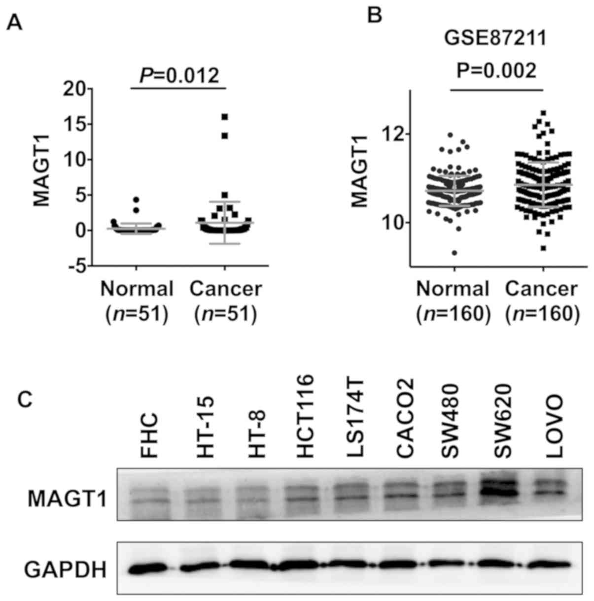
MAGT1 is upregulated in human CRC
tissues and cell lines
To investigate the role of MAGT1 in the development
of CRC, the expression level of MAGT1 in 51 primary tumor and
matched adjacent normal tissues was investigated using qPCR
(Table SII). The present study
revealed that the expression levels of MAGT1 were significantly
upregulated in primary CRC tissues (Fig.
1A). Similar results were found in samples from GSE87211
dataset (Fig. 1B). Additionally,
increased protein level of MAGT1 was observed in six out of eight
CRC cell lines, including HCT116, LS174T, CACO2, SW480, SW620 and
LOVO, compared with FHC cell line (Fig.
1C).
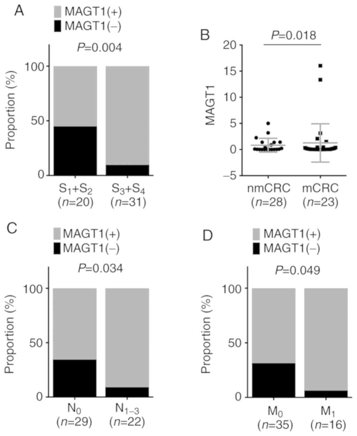
MAGT1 is associated with the
clinicopathological features of CRC
To further explore the roles of MAGT1 in CRC, the
association between the overexpression of MAGT1 and
clinicopathological features in 51 patients with CRC was analyzed.
The present study revealed that more tumors with advanced stages
were identified in the MAGT1-high expression group compared with in
the low expression group (Fig. 2A).
In addition, the expression levels of MAGT1 in metastatic CRC
tissues were identified to be higher than those in non-metastatic
CRC tissues (Fig. 2B). The
MAGT1-high expression group exhibited a slightly higher proportion
of patients with CRC with either lymphatic or distal metastasis
compared with the low expression group (Fig. 2C and D). To further confirm the
results of the present study, the GEO GSE39582 dataset was adopted
to analyze the expression of MAGT1 in 566 patients with CRC. These
patients with CRC were divided into MAGT1-high expression (n=283)
and MAGT1-low (n=283) expression groups according to the median
value of MAGT1. The present study demonstrated that there were more
patients with CRC with advanced tumor stages, lymphatic and distal
metastasis in the MAGT1-high expression group compared with the low
expression group (Table I). In
addition, the results of the present study indicated that
upregulation of MAGT1 was associated with higher incidence of CRC
in distal locations (Table I). These
findings suggested a positive association between MAGT1
overexpression and the progression of CRC.
 | Table I.Association of patient characteristics
and MAGT1 expression in 566 colorectal cancer samples from the
GSE39582 dataset. |
Table I.
Association of patient characteristics
and MAGT1 expression in 566 colorectal cancer samples from the
GSE39582 dataset.
|
|
| MAGT1 expression |
|
|---|
|
|
|
|
|
|---|
| Characteristics | Total, n | Low, n (%) | High, n (%) | P-valuea |
|---|
| Sex |
|
|
| 0.128 |
|
Female | 256 | 137 (53.5) | 119 (46.5) |
|
| Male | 310 | 146 (47.1) | 164 (52.9) |
|
| Age at diagnosis,
years |
|
|
| 0.332 |
|
<50 | 76 | 34 (44.7) | 42 (55.3) |
|
| ≥50 | 489 | 248 (50.7) | 241 (49.3) |
|
| Tumor location |
|
|
| 0.025a |
|
Proximal | 224 | 125 (55.8) | 99 (44.2) |
|
|
Distal | 342 | 158 (46.2) | 184 (53.8) |
|
| T classification |
|
|
| 0.648 |
|
Tis+T0+T1+T2 | 60 | 32 (53.3) | 28 (46.7) |
|
|
T3+T4 | 486 | 244 (50.2) | 242 (49.8) |
|
| N classification |
|
|
| 0.033a |
| N0 | 302 | 165 (54.6) | 137 (45.4) |
|
|
N1+N2+N3 | 238 | 108 (45.4) | 130 (54.6) |
|
| M
classification |
|
|
| 0.001a |
| M0 | 482 | 255 (52.9) | 227 (47.1) |
|
| M1 | 61 | 18 (29.5) | 43 (70.5) |
|
| Stage |
|
|
| 0.015a |
|
Stage1+Stage2 | 301 | 165 (54.8) | 136 (45.2) |
|
|
Stage3+Stage4 | 265 | 118 (44.5) | 147 (55.5) |
|
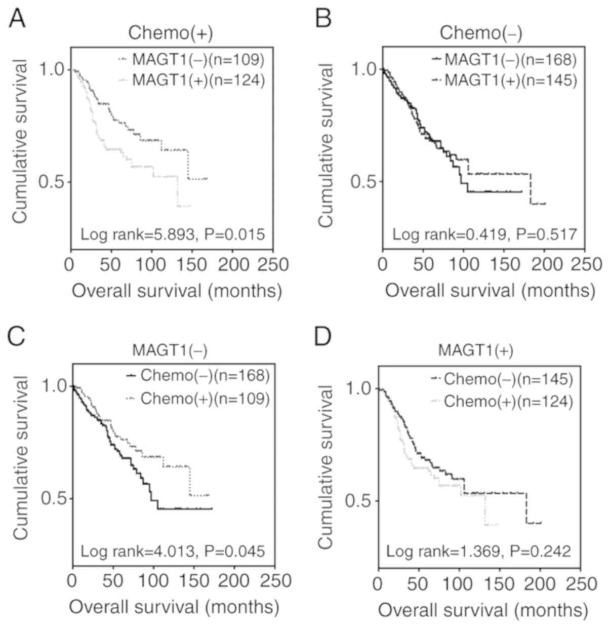
Upregulation of MAGT1 is associated
with the prognosis of patients with CRC treated with
chemotherapy
Since chemotherapy data from 19 healthy controls and
16 patients with CRC, and survival data from 4 patients with CRC
were missing in the GSE39582 dataset (n=585), Kaplan-Meier and Cox
regression analyses were applied to 546 patients with CRC to
further investigate the predictive role of MAGT1 in the prognosis
of CRC. Notably, for patients with postoperative chemotherapy, the
MAGT1-low expression group had a longer overall survival (OS) time
than the high expression group (Fig.
3A). However, no statistically significant difference in OS was
identified between the high and low expression groups in patients
who did not undergo chemotherapy (Fig.
3B). Therefore, it was hypothesized that MAGT1 may be
associated with anticancer drug resistance in patients with CRC. As
expected, patients treated with chemotherapy had a better prognosis
only in the MAGT1-low expression group (Fig. 3C and D), which supported the
aforementioned hypothesis. In addition, the 5-year overall survival
rates in the MAGT1-low expression group were 38.01% (without
chemotherapy) and 53.21% (with chemotherapy), while in the
MAGT1-high expression group survival rates were 43.45% (without
chemotherapy) and 36.29% (with chemotherapy). Therefore, univariate
and multivariate Cox regression analyses were performed only for
patients with CRC treated with chemotherapy. Univariate Cox
regression analysis demonstrated that MAGT1, tumor stage, and T and
M classification were indicators of poor prognosis, while no
statistically significant difference was identified for age, sex, N
classification and tumor location (Table II). However, multivariate Cox
regression analysis revealed that only T and M classification, but
not MAGT1, were independent factors for poor prognosis (Table III). Multivariate analysis was used
to analyze the effect of independent factors on patients' survival,
whereas tumor stage was determined by TNM classification. Stage was
therefore excluded while applying multivariate Cox regression
analysis. Collectively, MAGT1 was a valid but not an independent
prognostic factor for CRC.
| Table II.Univariate Cox regression analysis of
factors associated with overall survival in patients with
colorectal cancer. |
Table II.
Univariate Cox regression analysis of
factors associated with overall survival in patients with
colorectal cancer.
|
|
| 95% CI for
Exp(B) |
|
|---|
|
|
|
|
|
|---|
| Characteristic | Exp(B) | Lower | Upper | P-value |
|---|
| Age | 1.009 | 0.990 | 1.020 | 0.353 |
| Sex | 1.223 | 0.769 | 1.943 | 0.395 |
| T
classification | 2.743 | 1.735 | 4.338 | <0.001 |
| N
classification | 1.191 | 0.891 | 1.592 | 0.238 |
| M
classification | 8.584 | 4.803 | 15.343 | <0.001 |
| Location | 0.767 | 0.477 | 1.233 | 0.273 |
| Stage | 2.924 | 1.846 | 4.630 | <0.001 |
| MAGT1 | 1.768 | 1.107 | 2.822 | 0.017 |
| Table III.Multivariate Cox regression analysis
of factors associated with overall survival in patients with
colorectal cancer. |
Table III.
Multivariate Cox regression analysis
of factors associated with overall survival in patients with
colorectal cancer.
|
|
| 95% CI for
Exp(B) |
|
|---|
|
|
|
|
|
|---|
| Factor | Exp(B) | Lower | Upper | P-value |
|---|
| T
classification | 1.788 | 1.103 | 2.900 | 0.018 |
| M
classification | 5.946 | 3.183 | 11.111 | <0.001 |
| MAGT1 | 1.409 | 0.849 | 2.339 | 0.185 |
Discussion
A previous study revealed that CRC is the third most
common type of cancer (17), and the
fourth most common cause of cancer-associated mortality worldwide
(1,3). Furthermore, a recent study reported
that the 5-year survival rate of patients with primary CRC has
increased to 90%; however the 5-year survival rate of patients with
advanced CRC is only 13% (4). Since
tumor metastasis and drug resistance are the major reasons for the
poor prognosis of patients with CRC (18,19), it
is crucial to identify novel factors and mechanisms contributing to
the aforementioned processes. In addition, investigation of valid
biomarkers for predicting the development, progression and
prognosis of CRC is also necessary.
MAGT1 is a well-known chromosome X-linked gene that
encodes a highly selective Mg2+ transporter (20). An immunologic study demonstrated that
mutations in MAGT1 lead to T-cell deficiency by disturbing the
homeostasis of intracellular free Mg2+, which is an
important second messenger among multiple cellular activities
(7,21,22).
However, the pathophysiological significance of MAGT1 remains
elusive. The present study demonstrated that MAGT1 was upregulated
in CRC tissues compared with adjacent normal tissues, indicating a
positive association between the upregulation of MAGT1 and
incidence of CRC. In addition, patients with CRC with high
expression levels of MAGT1 had advanced tumor stage, and were more
likely to exhibit lymphatic or distal metastasis. Similar results
have previously been reported in hepatocellular carcinoma (8). Additionally, a previous study in breast
cancer revealed that decreased MAGT1 expression serves an important
role in reducing the viability of cancer cells (9). Collectively, MAGT1 may be a valid
biomarker for predicting the development and metastasis of CRC.
Further investigations are required to clarify the exact effects of
MAGT1 on the biological activities of CRC cells.
Notably, a growing body of epidemiological studies
determined that high magnesium intake is associated with a lower
incidence of CRC (23–25), which seems to contradict the
aforementioned association between MAGT1 and CRC. However, due to
the crucial role of MAGT1 in the regulation of NK and
CD8+ T-cells by mediating transient Mg2+
influx (7,21), this discrepancy may be explained by
the high magnesium intake that may reduce the incidence of CRC by
activating cytotoxic T-cells. In addition, the concentration of
intracellular Mg2+ in epithelial and CRC cells following
high magnesium intake is unknown. Therefore, the role of
MAGT1-mediated alteration of Mg2+ in regulating the
development and progression of CRC remains unclear.
To further investigate the association between MAGT1
and CRC, Kaplan-Meier survival and Cox regression analyses were
performed in the present study. Notably, the Kaplan-Meier survival
analysis revealed that MAGT1 expression was negatively associated
with OS time only in patients with CRC treated with chemotherapy.
Therefore, MAGT1 may be involved in regulating the mechanisms
underlying anticancer drug resistance in CRC. Furthermore,
univariate and multivariate Cox regression analyses performed in
patients with CRC treated with chemotherapy suggested that MAGT1
was a valid but not an independent prognostic factor for CRC.
Notably, patients with CRC with postoperative chemotherapy had an
improved OS only in the MAGT1-low expression group. This finding
suggested that MAGT1 could be a valid biomarker for predicting the
chemotherapeutic efficacy in CRC.
In conclusion, the present study identified an
association between CRC development and progression and MAGT1
expression level. Upregulation of MAGT1 may be associated with
tumor metastasis and anticancer drug resistance, and MAGT1 could be
a valid biomarker for predicting the development, progression and
prognosis of CRC.
Supplementary Material
Supporting Data
Acknowledgements
Not applicable.
Funding
This study was supported by the National Natural
Science Foundation of China (grant no. 81600444).
Availability of data and materials
The datasets used and/or analyzed during the current
study are available from the corresponding author on reasonable
request.
Authors' contributions
KZ and QY performed the experiments. LX and ZQ
performed the statistical analysis. YH, YL and LT assisted in
tissue sample collection. LT performed data analysis and
interpretation. CC designed the study and prepared the manuscript.
All authors read and approved the final manuscript.
Ethics approval and consent to
participate
The study was approved by the Ethics Committee of
Southern Medical University and all aspects of the study complied
with the Declaration of Helsinki.
Patients consent for publication
Not applicable.
Competing interests
The authors declare that they have no competing
interests.
Glossary
Abbreviations
Abbreviations:
|
CRC
|
colorectal cancer
|
|
GEO
|
Gene Expression Omnibus
|
|
MAGT1
|
magnesium transporter 1
|
|
OS
|
overall survival
|
References
|
1
|
Siegel R, Desantis C and Jemal A:
Colorectal cancer statistics, 2014. CA Cancer J Clin. 64:104–117.
2014. View Article : Google Scholar : PubMed/NCBI
|
|
2
|
Brenner H, Kloor M and Pox CP: Colorectal
cancer. Lancet. 383:1490–1502. 2014. View Article : Google Scholar : PubMed/NCBI
|
|
3
|
Welch HG and Robertson DJ: Colorectal
cancer on the decline-why screening can't explain it all. N Engl J
Med. 374:1605–1607. 2016. View Article : Google Scholar : PubMed/NCBI
|
|
4
|
Phipps AI, Robinson JR, Campbell PT, Win
AK, Figueiredo JC, Lindor NM and Newcomb PA: Prediagnostic alcohol
consumption and colorectal cancer survival: The colon cancer family
registry. Cancer. 123:1035–1043. 2017. View Article : Google Scholar : PubMed/NCBI
|
|
5
|
World Cancer Research Fund International
(WCRF), . Continuous Update Project. Alcohol and Cancer. http://www.wcrf.org/int/cancer-facts-figures/link-between-lifestyle-cancer-risk/alcohol-cancerDecember
18–2017
|
|
6
|
Zheng K, Zhou X, Yu J, Li Q, Wang H, Li M,
Shao Z, Zhang F, Luo Y, Shen Z, et al: Epigenetic silencing of
miR-490-3p promotes development of an aggressive colorectal cancer
phenotype through activation of the Wnt/β-catenin signaling
pathway. Cancer Lett. 376:178–187. 2016. View Article : Google Scholar : PubMed/NCBI
|
|
7
|
Chaigne-Delalande B, Li FY, O'Connor GM,
Lukacs MJ, Jiang P, Zheng L, Shatzer A, Biancalana M, Pittaluga S,
Matthews HF, et al: Mg2+ regulates cytotoxic functions of NK and
CD8 T cells in chronic EBV infection through NKG2D. Science.
341:186–191. 2013. View Article : Google Scholar : PubMed/NCBI
|
|
8
|
Molee P, Adisakwattana P, Reamtong O,
Petmitr S, Sricharunrat T, Suwandittakul N and Chaisri U:
Up-regulation of AKAP13 and MAGT1 on cytoplasmic membrane in
progressive hepatocellular carcinoma: A novel target for prognosis.
Int J Clin Exp Pathol. 8:9796–9811. 2015.PubMed/NCBI
|
|
9
|
Uddin MB, Balaravi Pillai B, Tha KK,
Ashaie M, Karim ME and Chowdhury EH: Carbonate apatite
nanoparticles-facilitated intracellular delivery of siRNA(s)
targeting calcium ion channels efficiently kills breast cancer
cells. Toxics. 6:E342018. View Article : Google Scholar : PubMed/NCBI
|
|
10
|
Willis S, Villalobos VM, Gevaert O,
Abramovitz M, Williams C, Sikic BI and Leyland-Jones B: Single gene
prognostic biomarkers in ovarian cancer: A meta-analysis. PLoS One.
11:e01491832016. View Article : Google Scholar : PubMed/NCBI
|
|
11
|
Rice TW, Gress DM, Patil DT, Hofstetter
WL, Kelsen DP and Blackstone EH: Cancer of the esophagus and
esophagogastric junction-major changes in the American joint
committee on cancer eighth edition cancer staging manual. CA Cancer
J Clin. 67:304–317. 2017. View Article : Google Scholar : PubMed/NCBI
|
|
12
|
Issue Information-Declaration of Helsinki.
J Bone Mineral Res. 32:BMi–BMii. 2017.
|
|
13
|
Marisa L, de Reyniès A, Duval A, Selves J,
Gaub MP, Vescovo L, Etienne-Grimaldi MC, Schiappa R, Guenot D,
Ayadi M, et al: Gene expression classification of colon cancer into
molecular subtypes: Characterization, validation, and prognostic
value. PLoS Med. 10:e10014532013. View Article : Google Scholar : PubMed/NCBI
|
|
14
|
Hu Y, Gaedcke J, Emons G, Beissbarth T,
Grade M, Jo P, Yeager M, Chanock SJ, Wolff H, Camps J, et al:
Colorectal cancer susceptibility loci as predictive markers of
rectal cancer prognosis after surgery. Genes Chromosomes Cancer.
57:140–149. 2018. View Article : Google Scholar : PubMed/NCBI
|
|
15
|
Livak KJ and Schmittgen TD: Analysis of
relative gene expression data using real-time quantitative PCR and
the 2(-Delta Delta C(T)) method. Methods. 25:402–408. 2001.
View Article : Google Scholar : PubMed/NCBI
|
|
16
|
Wang H, An H, Wang B, Liao Q, Li W, Jin X,
Cui S, Zhang Y, Ding Y and Zhao L: miR-133a represses tumour growth
and metastasis in colorectal cancer by targeting LIM and SH3
protein 1 and inhibiting the MAPK pathway. Eur J Cancer.
49:3924–3935. 2013. View Article : Google Scholar : PubMed/NCBI
|
|
17
|
Ferlay J, Shin HR, Bray F, Forman D,
Mathers C and Parkin DM: Estimates of worldwide burden of cancer in
2008: GLOBOCAN 2008. Int J Cancer. 127:2893–2917. 2010. View Article : Google Scholar : PubMed/NCBI
|
|
18
|
Zarour LR, Anand S, Billingsley KG, Bisson
WH, Cercek A, Clarke MF, Coussens LM, Gast CE, Geltzeiler CB,
Hansen L, et al: Colorectal cancer liver metastasis: Evolving
paradigms and future directions. Cell Mol Gastroenterol Hepatol.
3:163–173. 2017. View Article : Google Scholar : PubMed/NCBI
|
|
19
|
Siegel RL, Miller KD, Fedewa SA, Ahnen DJ,
Meester RGS, Barzi A and Jemal A: Colorectal cancer statistics,
2017. CA Cancer J Clin. 67:177–193. 2017. View Article : Google Scholar : PubMed/NCBI
|
|
20
|
de Baaij JH, Hoenderop JG and Bindels RJ:
Magnesium in man: Implications for health and disease. Physiol Rev.
95:1–46. 2015. View Article : Google Scholar : PubMed/NCBI
|
|
21
|
Li FY, Chaigne-Delalande B, Kanellopoulou
C, Davis JC, Matthews HF, Douek DC, Cohen JI, Uzel G, Su HC and
Lenardo MJ: Second messenger role for Mg2+ revealed by human T-cell
immunodeficiency. Nature. 475:471–476. 2011. View Article : Google Scholar : PubMed/NCBI
|
|
22
|
Takaya J, Higashino H and Kobayashi Y: Can
magnesium act as a second messenger? Current data on translocation
induced by various biologically active substances. Magnes Res.
13:139–146. 2000.PubMed/NCBI
|
|
23
|
Ko HJ, Youn CH, Kim HM, Cho YJ, Lee GH and
Lee WK: Dietary magnesium intake and risk of cancer: A
meta-analysis of epidemiologic studies. Nutr Cancer. 66:915–923.
2014. View Article : Google Scholar : PubMed/NCBI
|
|
24
|
Gorczyca AM, He K, Xun P, Margolis KL,
Wallace JP, Lane D, Thomson C, Ho GY, Shikany JM and Luo J:
Association between magnesium intake and risk of colorectal cancer
among postmenopausal women. Cancer Causes Control. 26:1761–1769.
2015. View Article : Google Scholar : PubMed/NCBI
|
|
25
|
Meng Y, Sun J, Yu J, Wang C and Su J:
Dietary intakes of calcium, iron, magnesium, and potassium elements
and the risk of colorectal cancer: A meta-analysis. Biol Trace Elem
Res. 189:325–335. 2019. View Article : Google Scholar : PubMed/NCBI
|