Introduction
Tongue squamous cell carcinoma (TSCC) is the most
common type of oral squamous cell carcinoma and exhibits aggressive
biological characteristics (1–3). Despite
advances in therapeutic techniques and management, TSCC has a poor
prognosis due to the frequent incidence of invasion and metastasis
in the early stages (4).
Radiotherapy, as an important adjuvant treatment modality, reduces
relapse rates and increases survival rates in patients with TSCC;
however, severe complications, including mucositis, hyposalivation,
taste loss, osteoradionecrosis, dysphagia and swallowing
deterioration, limit the success of clinical application (5–7).
Therefore, it is important to understand the mechanisms underlying
the growth, invasion and metastasis of these tumors in order to
develop novel therapeutic strategies against TSCC.
High linear energy transfer (LET) heavy ion
irradiation, including carbon ions, has several advantages over
conventional low-LET X-rays, as it delivers a high dose to the
tumor, while minimizing the dose delivered to surrounding healthy
tissues (8). Therefore, heavy ion
radiotherapy is increasingly being regarded as an effective
therapeutic strategy for suppressing the invasive and metastatic
radiation-resistant malignant tumors (9,10).
Clinical data have demonstrated that numerous types of tumor of
various pathological types, including squamous cell carcinoma, lung
cancer and non-squamous cell types, such as adenocarcinoma, adenoid
cystic carcinoma, bone and soft-tissue tumors, can be treated with
carbon ions (10). Carbon ion
radiation suppresses the angiogenesis potential of human
endothelial ECV304 cells via inhibiting matrix metalloproteinase
(MMP)-2 activity, and additionally suppresses the migration and
invasion of human lung adenocarcinoma A549 cells by downregulating
expression of the anillin gene (11,12).
However, heavy ion radiation has not received much attention in
oral squamous cell carcinoma, which is well known for its invasive
and metastatic characteristic (3,4).
Angiogenesis and degradation of the extracellular matrix (ECM)
serve an important role in tumor growth, invasion and metastasis of
TSCC (13). Vascular endothelial
growth factor (VEGF), epidermal growth factor and certain enzymes
secreted by tumor cells, including MMPs, promote migration and
metastasis (13). Substantial
evidence has demonstrated that the overexpression of VEGF, MMP-2
and MMP-9 is associated with infiltration and metastasis of TSCC
(14,15); therefore, it is conceivable that
their downregulation may inhibit invasion and metastasis in
TSCC.
The aim of the present study was to investigate the
expression of VEGF, MMP-2 and MMP-9 in TSCC Tca8113 cells following
exposure to high-LET carbon ions and low-LET X-rays, and to provide
some basic data for the efficacy of heavy ion radiation therapy
against TSCC.
Materials and methods
Cell culture
The human TSCC cell line Tca8113 was obtained from
the China Center for Type Culture Collection (Wuhan, China). Cells
were cultured in RPMI-1640 medium (Thermo Fisher Scientific, Inc.,
Waltham, MA, USA) supplemented with 10% heat-inactivated fetal
bovine serum (FBS; Minghai Biochem, Lanzhou, China), 100 U/ml
penicillin and 100 µg/ml streptomycin, 1% sodium pyruvate and 2 mM
glutamine (all obtained from Thermo Fisher Scientific, Inc.) at
37°C with 5% CO2 and 95% humidity. Cells were seeded in
35 mm glass Petri dishes (Nunc GmbH, Wiesbaden, Germany), and cell
density was adjusted to 5×10⁵ cells/ml at 24 h prior to
irradiation. The cultured cells were divided into two groups; one
group of cells were treated by X-ray, while the other were treated
by heavy ion beam.
Irradiation procedure
The two groups of Tca8113 cells were irradiated at
room temperature with carbon ions or X-rays. The irradiation with
carbon ions was conducted in the Heavy Ion Research Facility in
Lanzhou (HIRFL) at the Institute of Modern Physics, Chinese Academy
of Sciences (Lanzhou, China). Tca8113 cells were exposed to a
carbon ion beam with energy of 250 MeV/u supplied by HIRFL. The LET
value of the beam was adjusted to 75 keV/µm and the dose rate was
~1 Gy/min. For X-ray irradiation, cell samples were treated at room
temperature with an X-ray irradiation system (Faxitron RX-650;
Faxitron Bioptics LLC, Tuscon, AZ, USA) at 50 kVp with a dose rate
of 0.5 Gy/min. The doses applied in this experiment were 0, 1, 2
and 4 Gy for both irradiations. Following irradiation, the cells
were incubated under normal culture conditions for 12, 24 and 48 h.
The control cells were sham-irradiated.
Cell invasion assay
Cell invasion assay was performed using Transwell
inserts with 8-µm pores and coated with Matrigel (BD Biosciences,
Franklin Lakes, NJ, USA), which was used as a basement membrane
equivalent, and is composed of extracellular matrix designed to
mimic the typical matrices that tumor cells encounter during the
invasion process in vivo. Cells irradiated with carbon ions
or X-rays at doses of 0, 1 and 4 Gy were treated with trypsinase
and resuspended in serum-free RPMI-1640, after which 200 µl
(2×10⁵ cells/ml) cell suspension was added to the upper
chambers. The lower chambers were filled with 600 µl RPMI-1640
supplemented with 10% FBS as a chemo-attractant. Following
incubation under normal culture conditions for 24 h, the
non-invading cells on the upper surface of the filters were scraped
away using a cotton swab. The cells that had invaded to the lower
surface of the filters were fixed in ethanol and stained with
Giemsa at room temperature for 20 min. Subsequently, the cells were
counted in five random fields under a light microscope (Olympus,
Tokyo, Japan) at ×100 magnification.
Western blotting
Cells irradiated with carbon ions or X-rays at doses
of 0, 1 and 4 Gy were collected at 12, 24 and 48 h
post-irradiation. The cells were rinsed twice with ice-cold PBS
prior to being lysed in lysis buffer (Wuhan Boster Biological
Technology, Ltd., Wuhan, China). Subsequently, the cell lysates
were centrifuged at 16,100 × g for 10 min at 4°C. The supernatant
was collected, and the amount of protein was estimated using a
bicinchoninic acid protein assay kit (Bioworld Technology, Inc.,
St. Louis Park, MN, USA). Equal quantities of protein (30 µg) were
loaded onto an 8% SDS-PAGE gel and the separated proteins were
transferred onto polyvinylidene difluoride membranes (Roche
Diagnostics, Basel, Switzerland) for 1 h at 100 V. After transfer,
the membranes were blocked in 5% bovine serum albumin (BSA; Lanzhou
Minhai Bioengineering Co., Ltd.) at room temperature for 2 h and
subsequently incubated overnight at 4°C with anti-MMP-2 (cat. no.
4022), anti-MMP-9 (cat. no. 3852; Cell Signaling Technology, Inc.,
Danvers, MA, USA) or anti-β-actin (cat. no. TA-09; all used at
1:1,000; Beijing Zhongshan Golden Bridge Biotech Co., Ltd.,
Beijing, China). Blots were rinsed with TBS (pH 7.4) and 0.05%
Tween-20, and incubated with horseradish peroxidase-conjugated
anti-mouse monoclonal antibody (cat. no. ZB-2305) or anti-rabbit
polyclonal antibody (cat. no. ZB-2301; both 1:2,000; Beijing
Zhongshan Golden Bridge Biotech Co., Ltd.) for 2 h at 37°C.
Immunoreactive proteins were detected using the ECL western
blotting kit (Bioworld Technology, Inc.). Bands were scanned and
detected using a chemiluminescent imaging system (Tanon 4200 SF;
Tanon Science and Technology Co., Ltd., Shanghai, China) and the
density of bands were quantified using the Quantity One software
(v4.4, Bio-Rad Laboratories, Inc., Hercules, CA, USA).
Indirect immunofluorescence assay
For indirect immunofluorescence, Tca8113 cells
(1×10⁵ cells/ml) were grown on glass coverslips and
cultured at 37°C at 5% CO2 for 24 h until the cells were
fully attached to the glass coverslips prior to irradiation. In
order to confirm whether the effect of irradiation was
dose-dependent, cells were irradiated with carbon ions or X-rays at
doses of 0, 1, 2 and 4 Gy and collected at 12, 24 and 48 h
post-irradiation. Cells were fixed in 4% paraformaldehyde for 15
min at room temperature, then permeabilized with 0.1% Triton X-100
for 2 min, washed in PBS, and blocked in 5% BSA (Lanzhou Minhai
Bioengineering Co., Ltd.) at room temperature for 30 min.
Subsequently, the coverslips were washed and incubated with mouse
anti-human monoclonal antibody VEGF (cat. no. ab1316; 1:200; Abcam,
Cambridge, MA, USA) at 37°C for 2 h, washed three times in PBS, and
incubated with fluorescein-isothiocyanate-labeled donkey anti-mouse
antibody IgG (cat. no. ab7057; 1:800; Abcam, Cambridge, MA, USA),
at 37°C for 1 h. Nuclear DNA was stained with DAPI (Sangon Biotech
Co., Ltd., Shanghai, China) at room temperature for 10 min. The
coverslips were evaluated and images were captured using a
fluorescence microscope (Olympus) at ×1,000 magnification.
Statistical analysis
All experiments were repeated at least three times.
Data are presented as the mean ± standard deviation and analyzed
using SPSS version 13.0 (SPSS, Inc., Chicago, IL, USA). Groups were
compared using ANOVA with Bonferroni's post-hoc test. P<0.05 was
considered to indicate a statistically significant difference.
Results
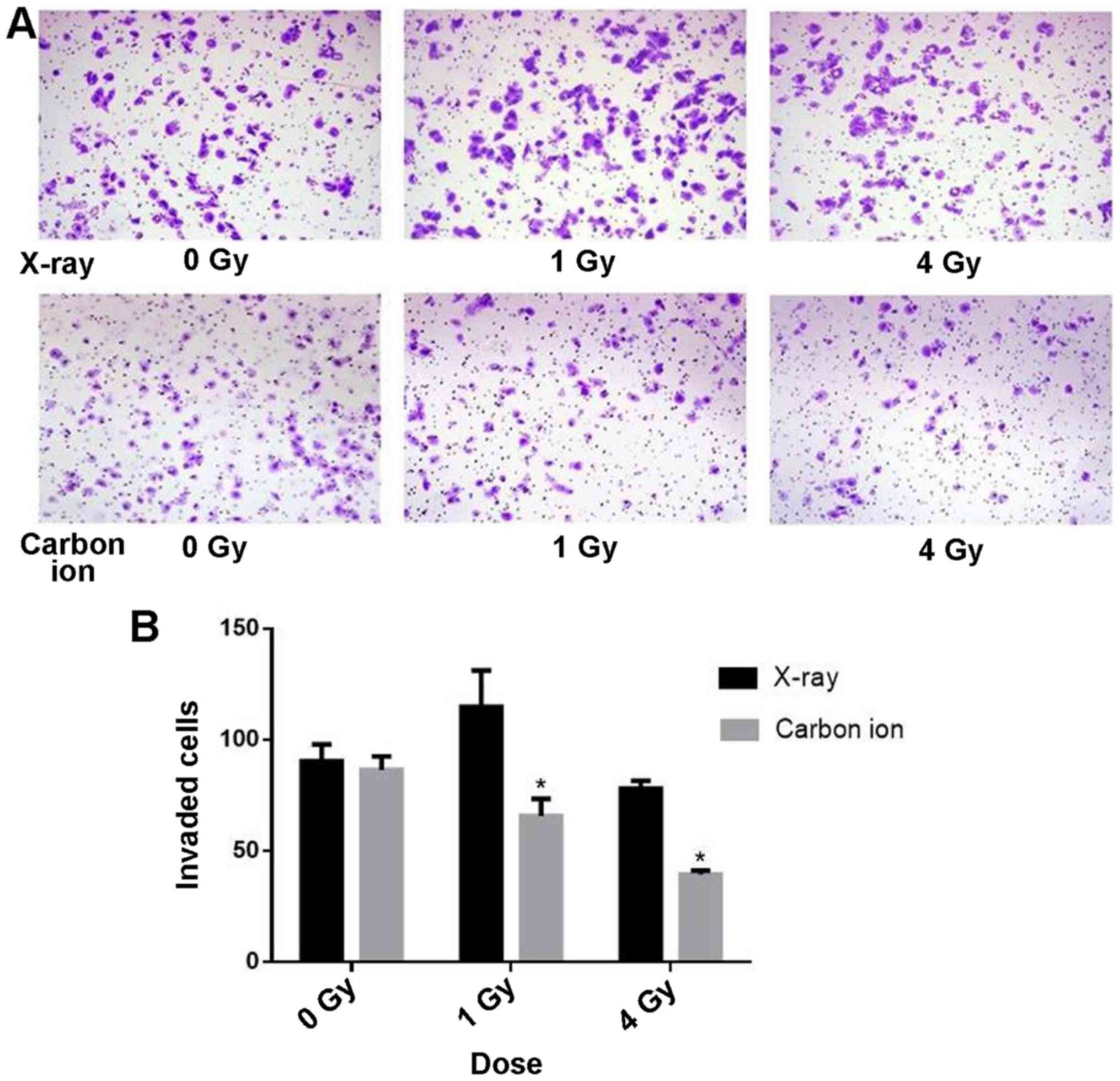
Effects of irradiation on cell
invasion
The cell invasion assay was used to investigate the
effects of irradiation with carbon ions and X-rays on the invasion
of Tca8113 cells. The rate of invasion in cells irradiated with
carbon ions was decreased with increasing doses of radiation, and
was significantly decreased compared with cells exposed to X-ray
irradiation at the same dose (P<0.05; Fig. 1).
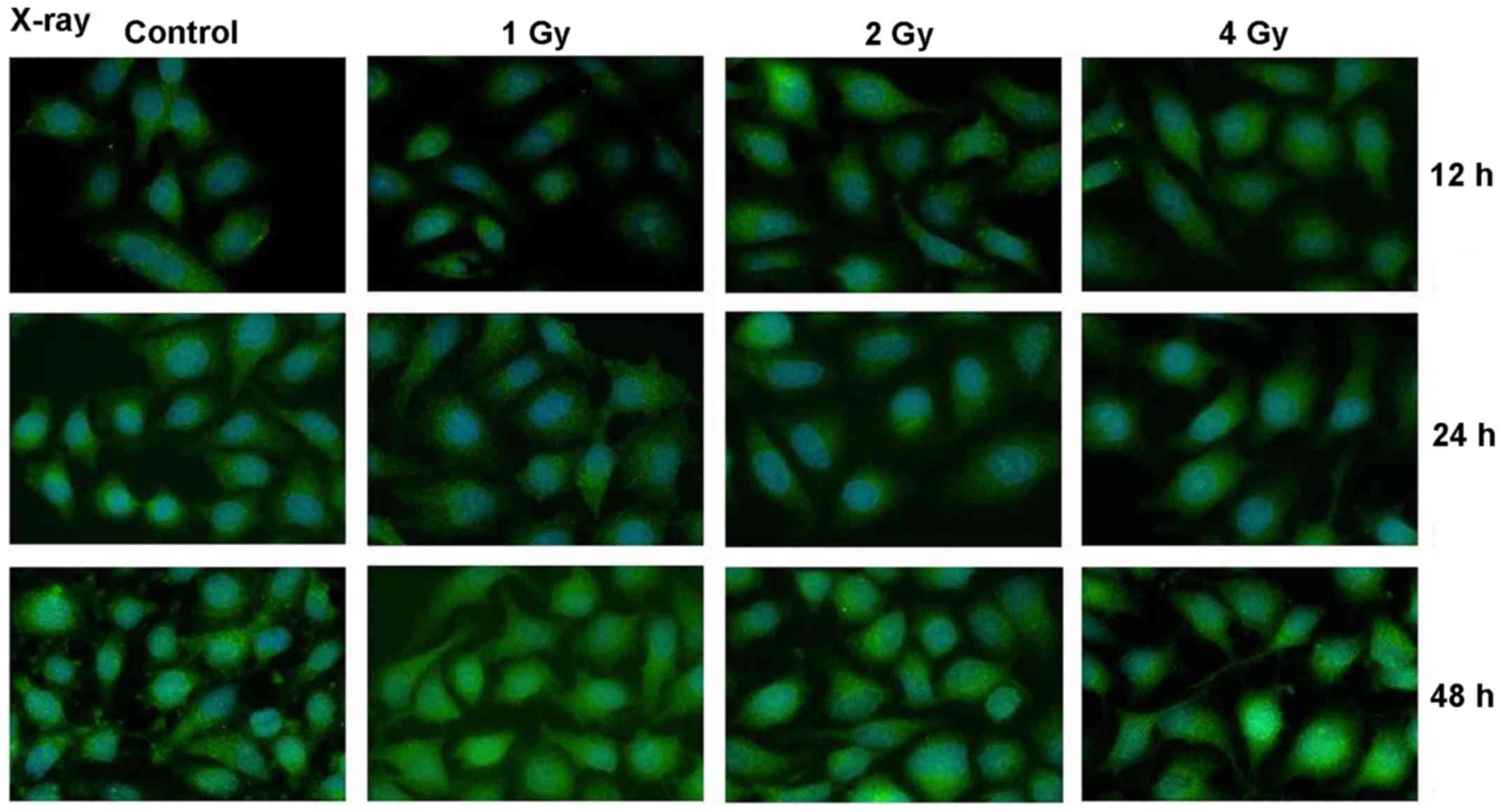
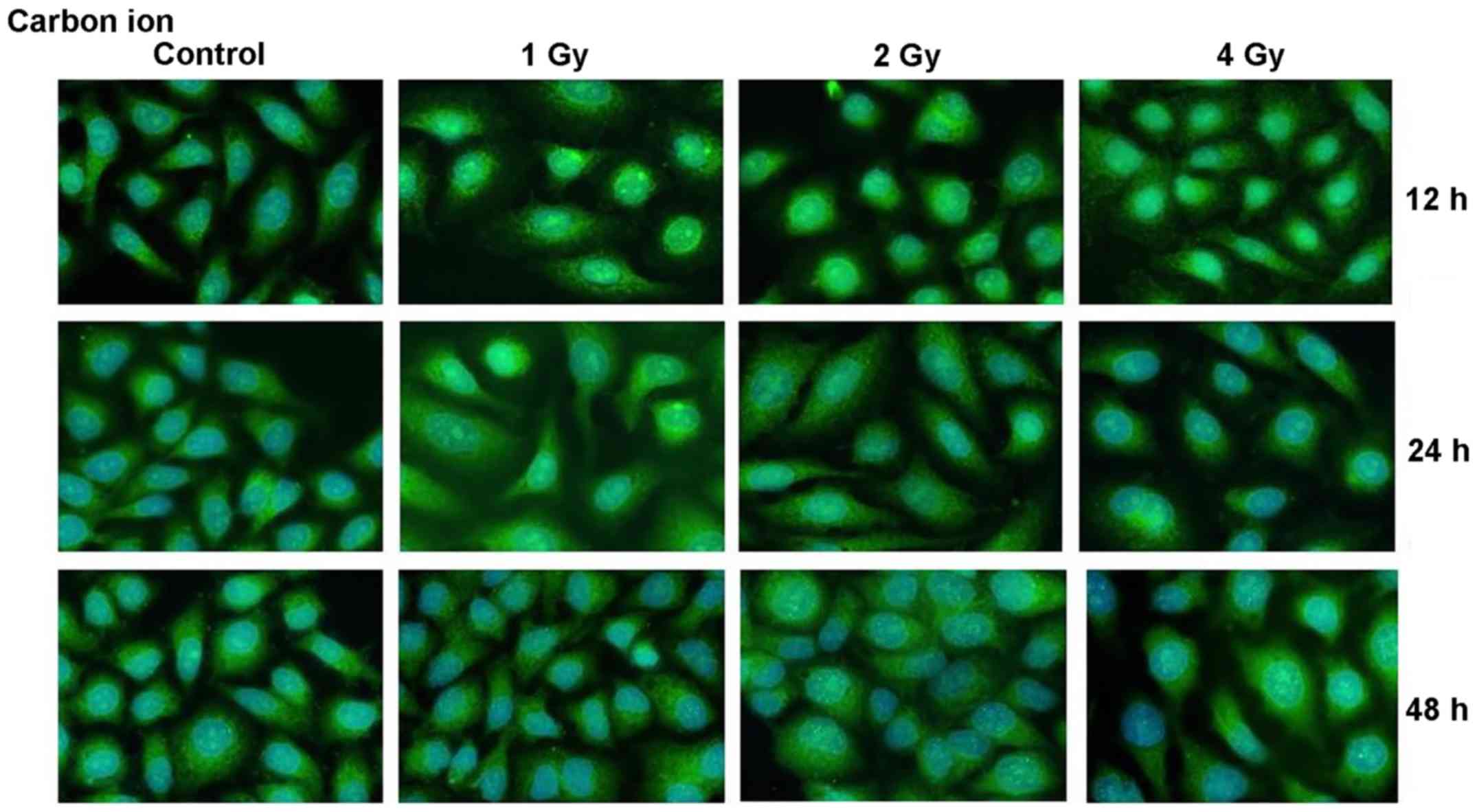
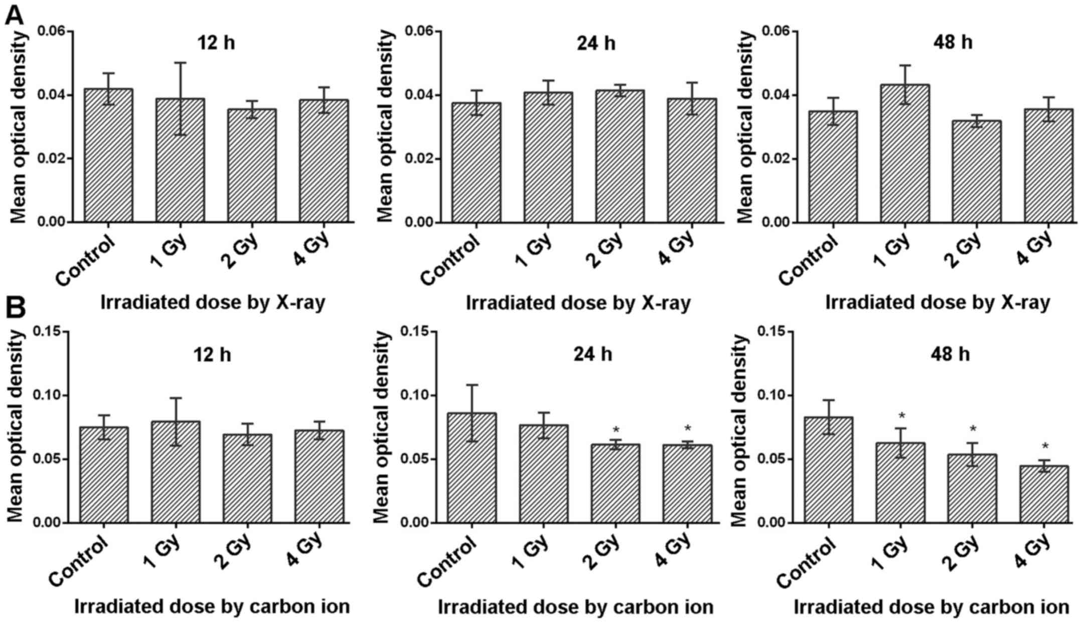
Effects of irradiation on VEGF
expression
The expression of VEGF in Tca8113 cells irradiated
with carbon ions and X-rays was evaluated by immunofluorescence
(Figs. 2 and 3). Compared with the control cells
(sham-irradiated), there was no significant change in VEGF
expression at 12, 24 or 48 h after X-ray irradiation with different
doses. However, VEGF was significantly downregulated at 24 h after
carbon ion irradiation at a dose of 2 Gy, and significantly
decreased in a dose-dependent manner at 48 h, as indicated in
Fig. 4 (P<0.05).
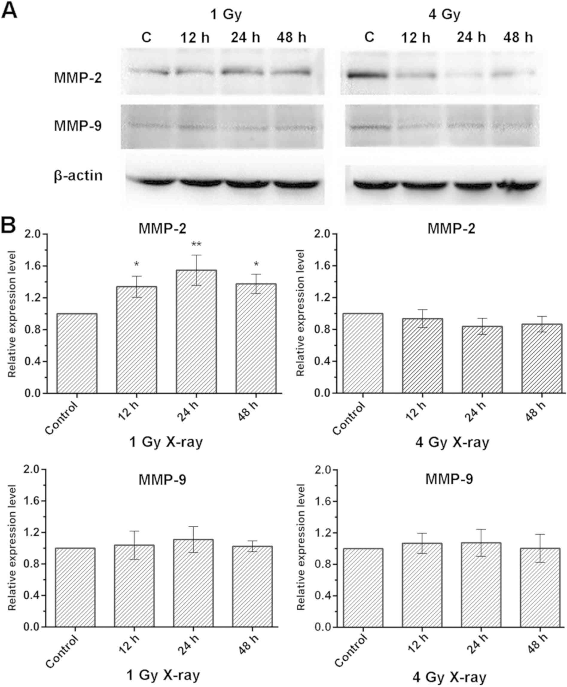
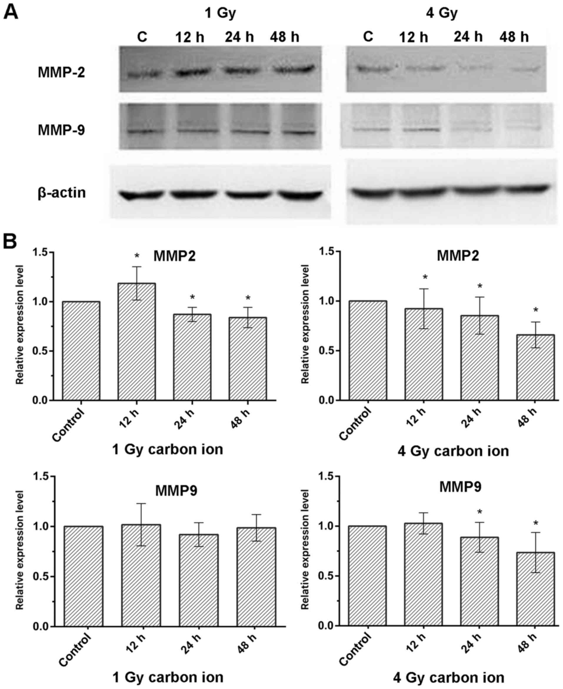
Effects of irradiation on MMP-2 and
MMP-9 protein expression
MMP-2 and MMP-9 expression were detected by western
blot. Compared with the control cells (sham-irradiated), there was
a significant increase in MMP-2 expression in Tca8113 cells treated
with X-rays at a low dose (1 Gy; P<0.05; Fig. 5A and B). However, there was no
significant difference in MMP-9 expression among cells irradiated
with the same dose of X-ray irradiation. In Tca8113 cells
irradiated with carbon ions, MMP-2 expression was downregulated at
the low dose (1 Gy) except 12 h post-irradiation and also decreased
at the high dose (4 Gy) over time post-irradiation. Similar to
MMP-2 expression, the level of MMP-9 expression also decreased in
Tca8113 cells irradiated at the high dose (4 Gy), although no
significant change was observed at the low dose (1 Gy; P>0.05;
Fig. 6A and B).
Discussion
Successful treatment of TSCC is frequently
complicated by invasion and metastasis, which may severely impact
survival and quality of life in patients (1,16–18).
Radiotherapy provides an auxiliary treatment strategy for this
disease. Although it is an effective strategy for local control of
the primary tumor, recurrence and metastasis are frequently
observed in patients with TSCC (19). Therefore, the suppression of invasion
and metastasis are considered to be critical therapeutic targets
for TSCC (20–22). Previous studies have demonstrated
that high-LET particles, including carbon ions, are more effective
compared with low-LET radiation, including X-rays, in suppressing
tumor cell invasion (12,23). However, the responses of tumor cells
to radiation are different and complex (24,25). The
invasion of a malignant tumor is a complicated phenomenon involving
angiogenesis, adhesion of the cell to the ECM, degradation of the
ECM and invasion of the cell into the surrounding tissues (26,27). The
metastatic potential of the non-small cell lung cancer A549 cell
line and the lung squamous cell carcinoma EBC-1 cell line has been
reported to be inhibited by heavy ions (12), therefore we hypothesized that
high-LET heavy ions may inhibit the metastatic potential of Tca8113
cells. Therefore, in the present study, the biological effects of
heavy ion irradiation on cells were investigated, including
invasion and the expression levels of molecules associated with
metastasis, including MMP-2, MMP-9 and VEGF.
There are numerous factors associated with
metastatic potential. During the process of tumor invasion and
metastasis, the basement membrane, which mainly consists of type IV
collagen, is degraded by numerous types of enzyme, including serine
proteinases, cysteine proteinases and MMPs (13). Among the secreted MMPs, MMP-2 and
MMP-9 serve important roles in tumor invasion and metastasis
(28,29). Therefore, the aim of the present
study was to determine whether MMP-2 or MMP-9 expression was
altered in response to irradiation in malignant tumor cells.
Wild-Bode et al (30)
demonstrated that irradiating glioma cells with sub-lethal X-ray
doses resulted in an increase in MMP-2 activity and promoted
invasiveness of the cells. Similar findings have been reported for
other cancer cell types, including those of the liver, lung and
pancreas (31–33). The results of the present study
additionally demonstrated that the expression level of MMP-2
increased following X-ray irradiation of 1 Gy in Tca8113 cells.
Notably, for Tca8113 cells irradiated with heavy ions, the invasive
ability significantly decreased in a dose-dependent manner, and the
expression of MMP-2 protein was downregulated at both low dose (1
Gy) and a high doses (4 Gy) over time, in accordance with the
findings by Ogata et al (23). There was no significant difference in
MMP-9 expression in Tca8113 cells irradiated with different doses
of X-rays (1, 2 and 4 Gy) and a low dose of carbon ions (1 Gy) at
the same time points, but the expression of MMP-9 was decreased in
Tca8113 cells exposed to a high dose of carbon ions (4 Gy). These
results are consistent with a previous study (11), suggesting that high-LET heavy ions
may suppress the invasiveness of Tca8113 cells via downregulating
MMP-2 and MMP-9 expression.
VEGF, a heparin-binding glycoprotein, has five known
isoforms, and is an important growth factor in the progression and
angiogenesis of malignant tumors (34). It was demonstrated that radiation
induces VEGF expression in a range of tumor cell lines (35), and that certain tumor cells protect
themselves against radiotherapy via the release of VEGF (36). In the present study, there was no
notable difference in the expression of VEGF in Tca8113 cells
irradiated with X-rays compared with non-irradiated cells. By
contrast, VEGF levels significantly decreased in cells irradiated
with carbon ions compared with non-irradiated cells supporting the
results of a previous study by Liu et al (37), who reported that carbon ion radiation
decreased VEGF secretion in lung adenocarcinoma A549 cells. As the
immunofluorescence assay can only show the location of the target
protein and cannot detect the molecular weight of the protein, the
effect of irradiation on VEGF expression needs to be confirmed
using western blot analysis in future experiments. One possible
explanation for the carbon ion radiation mediated decrease may be
that radiation with carbon ions may cause more complex and
irreparable clustered DNA damage compared with X-rays. DNA repair
is inefficient following high-LET particle irradiation, which may
lead to chromosomal damage and eventually, cell death (38,39).
Further studies are required in order to improve understanding of
the mechanism of action of carbon ions on organisms.
In summary, the present study indicated that
high-LET carbon ion radiation has the potential to decrease Tca8113
cell invasion in a dose-dependent manner and this effect may be via
the inhibition of MMP-2, MMP-9 and VEGF. These findings provide
evidence that heavy-ion radiation therapy may be superior to
radiotherapy with conventional X-rays for the clinical management
of TSCC.
Acknowledgements
The authors would like to thank Dr Xiaodong Jin
(Institute of Modern Physics, Chinese Academy of Sciences) for his
advice with regards to conducting this experiment.
Funding
This study was supported by the National Natural
Science Foundation of China (grant no. 81260403) and the Gansu
Provincial Youth Science and Technology Fund Project (grant no.
17JR5RA275).
Availability of data and materials
The datasets used and/or analyzed during the present
study are available from the corresponding author on reasonable
request.
Authors' contributions
ZF and CL designed the experiments and wrote the
draft manuscript. QZ, WM, TL and LX participated in the experiments
and analysis, and prepared the background research. QL designed the
experiments and revised the final manuscript. All authors read and
approved the final manuscript.
Ethics approval and consent to
participate
Not applicable.
Patient consent for publication
Not applicable.
Competing interests
The authors declare that that they have no competing
interests.
Glossary
Abbreviations
Abbreviations:
|
TSCC
|
tongue squamous cell carcinoma
|
|
VEGF
|
vascular endothelial growth factor
|
|
LET
|
linear energy transfer
|
|
MMP
|
matrix metalloproteinase
|
|
ECM
|
extracellular matrix
|
References
|
1
|
Jemal A, Bray F, Center MM, Ferlay J, Ward
E and Forman D: Global cancer statistics. CA Cancer J Clin.
61:69–90. 2011. View Article : Google Scholar : PubMed/NCBI
|
|
2
|
Chandler K, Vance C, Budnick S and Muller
S: Muscle invasion in oral tongue squamous cell carcinoma as a
predictor of nodal status and local recurrence: Just as effective
as depth of invasion? Head Neck Pathol. 5:359–363. 2011. View Article : Google Scholar : PubMed/NCBI
|
|
3
|
Mücke T, Mitchell DA, Wagenpfeil S,
Ritschl LM, Wolff KD and Kanatas A: Incidence and outcome for
patients with occult lymph node involvement in T1 and T2 oral
squamous cell carcinoma: A prospective study. BMC Cancer.
14:3462014. View Article : Google Scholar : PubMed/NCBI
|
|
4
|
Ganly I, Patel S and Shah J: Early stage
squamous cell cancer of the oral tongue-clinicopathologic features
affecting outcome. Cancer. 118:101–111. 2012. View Article : Google Scholar : PubMed/NCBI
|
|
5
|
Vissink A, Jansma J, Spijkervet FK,
Burlage FR and Coppes RP: Oral sequelae of head and neck
radiotherapy. Crit Rev Oral Biol Med. 14:199–212. 2003. View Article : Google Scholar : PubMed/NCBI
|
|
6
|
Wong HM: Oral complications and management
strategies for patients undergoing cancer therapy.
ScientificWorldJournal. 2014:5817952014. View Article : Google Scholar : PubMed/NCBI
|
|
7
|
Silveira MH, Dedivitis RA, Queija DS and
Nascimento PC: Quality of life in swallowing disorders after
nonsurgical treatment for head and neck cancer. Int Arch
Otorhinolaryngol. 19:46–54. 2015. View Article : Google Scholar : PubMed/NCBI
|
|
8
|
Kraft G: The radiobiological and physical
basis for radiotherapy with protons and heavier ions. Strahlenther
Onkol. 166:10–13. 1990.PubMed/NCBI
|
|
9
|
Halperin EC: Particle therapy and
treatment of cancer. Lancet Oncol. 7:676–685. 2006. View Article : Google Scholar : PubMed/NCBI
|
|
10
|
Kamada T, Tsujii H, Blakely EA, Debus J,
De Neve W, Durante M, Jäkel O, Mayer R, Orecchia R, Pötter R, et
al: Carbon ion radiotherapy in Japan: An assessment of 20 years of
clinical experience. Lancet Oncol. 16:e93–e100. 2015. View Article : Google Scholar : PubMed/NCBI
|
|
11
|
Takahashi Y, Teshima T, Kawaguchi N,
Hamada Y, Mori S, Madachi A, Ikeda S, Mizuno H, Ogata T, Nojima K,
et al: Heavy ion irradiation inhibits in vitro angiogenesis even at
sublethal dose. Cancer Res. 63:4253–4257. 2003.PubMed/NCBI
|
|
12
|
Akino Y, Teshima T, Kihara A,
Kodera-Suzumoto Y, Inaoka M, Higashiyama S, Furusawa Y and Matsuura
N: Carbon-ion beam irradiation effectively suppresses migration and
invasion of human non-small-cell lung cancer cells. Int J Radiat
Oncol Biol Phys. 75:475–481. 2009. View Article : Google Scholar : PubMed/NCBI
|
|
13
|
Koontongkaew S: The tumor microenvironment
contribution to development, growth, invasion and metastasis of
head and neck squamous cell carcinomas. J Cancer. 4:66–83. 2013.
View Article : Google Scholar : PubMed/NCBI
|
|
14
|
Kim SH, Cho NH, Kim K, Lee JS, Koo BS, Kim
JH, Chang JH and Choi EC: Correlations of oral tongue cancer
invasion with matrix metalloproteinases (MMPs) and vascular
endothelial growth factor (VEGF) expression. J Surg Oncol.
93:330–337. 2006. View Article : Google Scholar : PubMed/NCBI
|
|
15
|
Matsui T, Shigeta T, Umeda M and Komori T:
Vascular endothelial growth factor C (VEGF-C) expression predicts
metastasis in tongue cancer. Oral Surg Oral Med Oral Pathol Oral
Radiol. 120:436–442. 2015. View Article : Google Scholar : PubMed/NCBI
|
|
16
|
Liu WH, Li X, Zhu XL, Hou ML and Zhao W:
CD63 inhibits the cell migration and invasion ability of tongue
squamous cell carcinoma. Oncol Lett. 15:9033–9042. 2018.PubMed/NCBI
|
|
17
|
Sano D and Myers JN: Metastasis of
squamous cell carcinoma of the oral tongue. Cancer Metastasis Rev.
26:645–662. 2007. View Article : Google Scholar : PubMed/NCBI
|
|
18
|
Olaleye O, Ekrikpo U, Lyne O and Wiseberg
J: Incidence and survival trends of lip, intra-oral cavity and
tongue base cancers in south-east England. Ann R Coll Surg Engl.
97:229–234. 2015. View Article : Google Scholar : PubMed/NCBI
|
|
19
|
Sun C, Wang X, Zhong Z, Wang W, Luo J,
Chen X, Chu H, Tao X and Yang A: Differences in clinicopathological
characteristics and prognosis between primary and postirradiation
tongue squamous cell carcinoma. J Oral Maxillofac Surg.
75:2235–2241. 2017. View Article : Google Scholar : PubMed/NCBI
|
|
20
|
Roy R, Yang J and Moses MA: Matrix
metalloproteinases as novel biomarkers and potential therapeutic
targets in human cancer. J Clin Oncol. 27:5287–5297. 2009.
View Article : Google Scholar : PubMed/NCBI
|
|
21
|
Jiang L, Liu X, Kolokythas A, Yu J, Wang
A, Heidbreder CE, Shi F and Zhou X: Downregulation of the Rho
GTPase signaling pathway is involved in the microRNA-138-mediated
inhibition of cell migration and invasion in tongue squamous cell
carcinoma. Int J Cancer. 127:505–512. 2010. View Article : Google Scholar : PubMed/NCBI
|
|
22
|
Shang B, Cao Z and Zhou Q: Progress in
tumor vascular normalization for anticancer therapy: Challenges and
perspectives. Front Med. 6:67–78. 2012. View Article : Google Scholar : PubMed/NCBI
|
|
23
|
Ogata T, Teshima T, Kagawa K, Hishikawa Y,
Takahashi Y, Kawaguchi A, Suzumoto Y, Nojima K, Furusawa Y and
Matsuura N: Particle irradiation suppresses metastatic potential of
cancer cells. Cancer Res. 65:113–120. 2005.PubMed/NCBI
|
|
24
|
Hill RP: The changing paradigm of tumour
response to irradiation. Br J Radiol. 90:201604742017. View Article : Google Scholar : PubMed/NCBI
|
|
25
|
Matsuda S, Furuya K, Ikura M, Matsuda T
and Ikura T: Absolute quantification of acetylation and
phosphorylation of the histone variant H2AX upon ionizing radiation
reveals distint cellular responses in two cancer cell lines. Radiat
Environ Biophys. 54:403–411. 2015. View Article : Google Scholar : PubMed/NCBI
|
|
26
|
Chambers AF, Groom AC and MacDonald IC:
Dissemination and growth of cancer cells in metastatic sites. Nat
Rev Cancer. 2:563–572. 2002. View
Article : Google Scholar : PubMed/NCBI
|
|
27
|
Joyce JA and Pollard JW:
Microenvironmental regulation of metastasis. Nat Rev Cancer.
9:239–252. 2009. View
Article : Google Scholar : PubMed/NCBI
|
|
28
|
Xue Q, Cao L, Chen XY, Zhao J, Gao L, Li
SZ and Fei Z: High expression of MMP9 in glioma affects cell
proliferation and is associated with patient survival rates. Oncol
Lett. 13:1325–1330. 2017. View Article : Google Scholar : PubMed/NCBI
|
|
29
|
Fan HX, Li HX, Chen D, Gao ZX and Zheng
JH: Changes in the expression of MMP2, MMP9, and ColIV in stromal
cells in oral squamous tongue cell carcinoma: Relationships and
prognostic implications. J Exp Clin Cancer Res. 31:902012.
View Article : Google Scholar : PubMed/NCBI
|
|
30
|
Wild-Bode C, Weller M, Rimner A, Dichgans
J and Wick W: Sublethal irradiation promotes migration and
invasiveness of glioma cells: Implications for radiotherapy of
human glioblastoma. Cancer Res. 61:2744–2750. 2001.PubMed/NCBI
|
|
31
|
Araya J, Maruyama M, Sassa K, Fujita T,
Hayashi R, Matsui S, Kashii T, Yamashita N, Sugiyama E and
Kobayashi M: Ionizing radiation enhances matrix metalloproteinase-2
production in human lung epithelial cells. Am J Physiol Lung Cell
Mol Physiol. 280:L30–L38. 2001. View Article : Google Scholar : PubMed/NCBI
|
|
32
|
Qian LW, Mizumoto K, Urashima T, Nagai E,
Maehara N, Sato N, Nakajima M and Tanaka M: Radiation-induced
increase in invasive potential of human pancreatic cancer cells and
its blockade by a matrix metalloproteinase inhibitor, CGS27023.
Clin Cancer Res. 8:1223–1227. 2002.PubMed/NCBI
|
|
33
|
Cheng JC, Chou CH, Kuo ML and Hsieh CY:
Radiation-enhanced hepatocellular carcinoma cell invasion with
MMP-9 expression through PI3K/Akt/NF-kappaB signal transduction
pathway. Oncogene. 25:7009–7018. 2006. View Article : Google Scholar : PubMed/NCBI
|
|
34
|
Flickinger I, Rütgen BC, Gerner W, Calice
I, Tichy A, Saalmüller A and Kleiter M: Radiation up-regulates the
expression of VEGF in a canine oral melanoma cell line. J Vet Sci.
14:207–214. 2013. View Article : Google Scholar : PubMed/NCBI
|
|
35
|
Gorski DH, Beckett MA, Jaskowiak NT,
Calvin DP, Mauceri HJ, Salloum RM, Seetharam S, Koons A, Hari DM,
Kufe DW and Weichselbaum RR: Blockage of the vascular endothelial
growth factor stress response increases the antitumor effects of
ionizing radiation. Cancer Res. 59:3374–3378. 1999.PubMed/NCBI
|
|
36
|
Park JS, Qiao L, Su ZZ, Hinman D,
Willoughby K, McKinstry R, Yacoub A, Duigou GJ, Young CS, Grant S,
et al: Ionizing radiation modulates vascular endothelial growth
factor (VEGF) expression through multiple mitogen activated protein
kinase dependent pathways. Oncogene. 20:3266–3280. 2001. View Article : Google Scholar : PubMed/NCBI
|
|
37
|
Liu Y, Liu Y, Zhang H, Sun C, Zhao Q, Di
C, Li H, Gan L and Wang Y: Effects of carbon-ion beam irradiation
on the angiogenic response in lung adenocarcinoma A549 cells. Cell
Biol Int. 38:1304–1310. 2014. View Article : Google Scholar : PubMed/NCBI
|
|
38
|
Mohamad O, Sishc BJ, Saha J, Pompos A,
Rahimi A, Story MD, Davis AJ and Kim DWN: Carbon ion radiotherapy:
A review of clinical experiences and preclinical research, with an
emphasis on DNA damage/repair. Cancers (Basel). 9(pii): E662017.
View Article : Google Scholar : PubMed/NCBI
|
|
39
|
Hagiwara Y, Niimi A, Isono M, Yamauchi M,
Yasuhara T, Limsirichaikul S, Oike T, Sato H, Held KD, Nakano T and
Shibata A: 3D-structured illumination microscopy reveals clustered
DNA double-strand break formation in widespread γH2AX foci after
high LET heavy-ion particle radiation. Oncotarget. 8:109370–109381.
2017. View Article : Google Scholar : PubMed/NCBI
|