Introduction
Lung cancer remains a major cause of
cancer-associated morbidity and mortality worldwide, particularly
in China (1,2). Non-small cell lung cancer (NSCLC) is a
major histopathological subtype that accounts for 80–85% of all
lung cancer cases (3). Despite
significant improvements in clinical strategies for NSCLC,
including surgery, targeted therapy, immunotherapy, chemotherapy
and radiotherapy, the prognosis of patients with NSCLC remains poor
(1). Considerable evidence has
demonstrated that the poor prognosis of NSCLC is attributed to
tumor metastasis (4), drug
resistance (5) and lack of early
sensitivity and specificity diagnostic markers (6). Therefore, it is urgent to find novel
therapeutic targets for NSCLC.
Long non-coding RNAs (lncRNAs) are a member of the
non-coding RNA family, >200 nucleotides in length and without
protein translation capacity (7). An
increasing number of studies (8,9) have
demonstrated that lncRNAs serve an important role in human
diseases, including cancer (10).
lncRNAs are involved in multiple physiological and pathological
processes, including cell proliferation, apoptosis and invasion, as
well as tumor metastasis and tumorigenesis (10,11).
Notably, lncRNAs are abundantly found in the serum, plasma, saliva
and urine, and can serve as potential biomarkers for the early
diagnosis and prognosis of NSCLC (12,13).
Previous studies (8,13) have indicated that the dysregulation
of certain lncRNAs has been identified in NSCLC and serves an
important role in the tumorigenesis and progression of NSCLC,
providing a novel potential therapeutic target for NSCLC. For
example, HOX transcript antisense RNA is overexpressed in NSCLC and
promotes tumorigenesis and metastasis by regulating microRNA
(miR)-613 (14). Distal-less homebox
6 antisense RNA 1 (DLX6-AS1) is upregulated in various solid
tumors, including NSCLC (15–17),
renal cell carcinoma (18),
hepatocellular carcinoma (19–21),
colorectal cancer (22), pancreatic
cancer (23,24), glioma (25) and osteosarcoma (26,27),
acting as an early diagnostic and prognostic biomarker. However,
the expression and function of circulating DLX6-AS1 in NSCLC have
not been widely demonstrated.
The present study revealed that DLX6-AS1 was
overexpressed not only in tumor tissues compared with in adjacent
normal tissues, but also in the serum of patients with NSCLC
compared with that of healthy donors. In addition, the circulating
DLX6-AS1 expression was significantly associated with advanced
disease stage and lymph node metastasis in patients with NSCLC.
Furthermore, exosomal DLX6-AS1 was upregulated in patients with
NSCLC and had a higher sensitivity and specificity for NSCLC
diagnosis than CYFRA21-1, which is a serum diagnostic marker for
NSCLC (28), suggesting it could be
a potential novel marker for the early diagnosis and metastasis of
NSCLC.
Materials and methods
Patient samples and healthy
donors
A total of 72 patients who were pathologically
confirmed with NSCLC and 64 healthy donors were enrolled at the
First Affiliated Hospital of Huzhou University between October 2016
and December 2018. The NSCLC patient group consisted of 39 men and
33 women (age range, 28–81 years; mean age, 61 years). Healthy
controls comprised 31 men and 33 women (age range, 29–89 years;
mean age, 62 years). Serum samples were obtained from patients on
pre-operative day 1 and post-operative day 3, and from healthy
controls, and stored at −80°C prior to use. Furthermore, 27 pairs
of tumor tissues and matched adjacent normal tissues (>5 cm away
from the tumor) were collected immediately after resection and
stored in liquid nitrogen until further use. No patients had
received adjuvant therapy prior to surgical resection. The
pathological diagnosis was confirmed and classified by two
certified pathologists based on the 7th Edition of the Union for
International Cancer Control (29).
Clinical information, including age, sex, smoking history, tumor
size, tumor differentiation, lymph node metastasis and TNM stage,
was collected. The present study was approved by the Ethics
Committee of the First Affiliated Hospital of Huzhou University,
and written informed consent was provided by all patients and
healthy controls.
Gene Expression Profiling Interactive
Analysis (GEPIA)
GEPIA (http://gepia.cancer-pku.cn/) is a public web server
used for evaluation of cancer and normal gene expression profiling
and interactive analysis according to The Cancer Genome Atlas
(TCGA) and the Genotype-Tissue Expression (GTEx) database (30). DLX6-AS1 expression levels in lung
adenocarcinoma (LUAD, n=483) and lung squamous cell carcinoma
(LUSC, n=486) were compared with their normal cohorts (n=347;
n=338). Association between DLX6-AS1 expression and TNM stage was
investigated in LUAD and LUSC cohort. Additionally, patients with
LUAD and LUSC were divided into two groups according to DLX6-AS1
expression. Overall survival and disease-free survival were
performed using the Kaplan-Meier method.
Cell lines and culture
The human NSCLC A549, H1299 and 95D, and normal
human epithelial BEAS-2B cell lines were obtained from The Cell
Bank of Type Culture Collection of the Chinese Academy of Sciences,
and cultured in DMEM (HyClone; GE Healthcare Life Sciences)
containing 10% fetal bovine serum (FBS; Gibco; Thermo Fisher
Scientific, Inc.), 100 U/ml penicillin and 100 µg/ml streptomycin
(Sigma-Aldrich; Merck KGaA) at 37°C in a humidified incubator with
5% CO2.
Cell transfection
The two specific small interfering RNAs (siRNAs) of
lncRNA DLX6-AS1 (20 µM) and scrambled siRNA (20 µM) were designed
and synthesized by Guangzhou RiboBio Co., Ltd.. Once they reached
50–70% confluence, the A549 and H1299 cells were transfected with
Lipofectamine® 2000 reagent (Invitrogen; Thermo Fisher
Scientific, Inc.), according to the manufacturer's protocol. At 2
days after transfection, cells were harvested and the DLX6-AS1
expression was measured by reverse transcription-quantitative PCR
(RT-qPCR) to confirm the transfection efficiency. The siRNA
sequences used in the experiment were as follows: si-DLX6-AS1#1
forward, 5′-GGCUAACACAUCCAUGGAAdTdT-3′ and reverse,
5′-UUCCAUGGAUGUGUUAGCCdTdT-3′; si-DLX6-AS1#2 forward,
5′-GCCGCUUGUCUUACUUAAAdTdT-3′ and reverse,
5′-UUUAAGUAAGACAAGCGGCdTdT-3′; and scrambled siRNA forward,
5′-UUCUCCGAACGUGUCACGUTT-3′ and reverse,
5′-ACGUGACACGUUCGGAGAATT-3′.
RNA extraction and RT-qPCR
Total RNA from tissues, cells and exosomes was
extracted using TRIzol® reagent (Invitrogen; Thermo
Fisher Scientific, Inc.), as described previously (31). Complementary DNA was reverse
synthesized using the PrimeScript RT reagent kit (Takara
Biotechnology Co., Ltd.), according to the manufacturer's protocol.
RNA (500 ng), 2 µl PrimeScript™ RT Master Mix (Perfect Real Time)
and RNase free water (up to 10 µl), were mixed together and
incubated at 37°C for 15 min and 85°C for 5 sec. qPCR was performed
with SYBR Green PCR Master Mix using the 7500 system (Applied
Biosystems; Thermo Fisher Scientific, Inc.). The qPCR reaction
system (10 µl) included the following: 5 µl 2X SYBR Green PCR
Master Mix, 0.3 µl forward primer (10 µM), 0.3 µl reverse primer
(10 µM), 50 ng cDNA and RNase free water. The thermocycling
conditions used for the qPCR were as follows 95°C for 10 min,
followed by 40 cycles of 95°C for 15 sec and 60°C for 1 min. Gene
expression was normalized to 18S ribosomal RNA (18sRNA), and the
relative expression level was calculated using the
2−ΔΔCq method (32). The
primer sequences used in the present study were as follows:
DLX6-AS1 forward, 5′-AGTTTCTCTCTAGATTGCCTT-3′ and reverse,
5′-ATTGACATGTTAGTGCCCTT-3′; 18sRNA forward,
5′-GTAACCCGTTGAACCCCATT-3′ and reverse,
5′-CCATCCAATCGGTAGTAGCG-3′.
Western blotting
Exosomes were lysed in RIPA buffer (cat. no. P0013B;
Beyotime Institute of Biotechnology) containing protease and
phosphatase inhibitor (Beyotime Institute of Biotechnology) and the
total protein concentration was measured using a bicinchoninic acid
assay (Beyotime Institute of Biotechnology). An equal amount of
protein (30 µg) was loaded onto 10–15% SDS-PAGE gels. The proteins
were then transferred onto 0.45-µm PVDF membranes (EMD Millipore).
The membranes were blocked with 5% non-fat milk at room temperature
for 1 h, and incubated with specific primary antibodies at 4°C
overnight, followed by incubation with horseradish
peroxidase-conjugated goat-anti-mouse secondary antibody (cat. no.
A0286; dilution, 1:1,000; Beyotime Institute of Biotechnology) at
room temperature for 1 h. Bands were visualized using enhanced
chemiluminescence (Beyotime Institute of Biotechnology). Relative
expression level of proteins was normalized to β-actin using ImageJ
v1.6.0 software (National Institutes of Health). The primary
antibodies used in the present study were as follows: Anti-CD81
(cat. no. sc-166029; dilution, 1:200; Santa Cruz Biotechnology,
Inc.); anti-CD9 (cat. no. sc-13118; dilution, 1:200; Santa Cruz
Biotechnology, Inc.); anti-Alix (cat. no. sc-53540; dilution,
1:200; Santa Cruz Biotechnology, Inc.); and anti-β-actin (cat. no.
4967S; dilution, 1:2,000; Cell Signaling Technology, Inc.).
Exosomes isolation
Plasma (5 ml) was collected and divided into two
parts, and exosomes were isolated using ExoQuick Exosome
Precipitation solution (System Biosciences, LLC) according to the
manufacturer's protocol. Exosomes were resuspended in PBS for
transmission electron microscopy and nanoparticle tracking
analysis, in RIPA buffer for western blotting or TRIzol®
for RNA extraction.
Transmission electron microscopy
Exosomes were fixed with 4% paraformaldehyde for 4 h
at 22°C, followed by 1% glutaraldehyde for 30 min at 22°C. The
samples were dropped onto formvar carbon-coated grids and allowed
to absorb for 10 min, followed by fixation with 4% paraformaldehyde
at room temperature for 10 min. Following PBS washing, exosomes
were stained with 2% uranyl acetate solution for 1 min at room
temperature. Images were observed under a Tecnai G2 spititi
electron microscopy.
Nanoparticle tracking analysis
(NTA)
The size distribution and concentration of exosomes
were analyzed by NTA, according to the manufacturer's instructions.
Briefly, exosomes were resuspended and diluted at 1:200 in PBS, and
the particle concentration of exosomes was assessed using a
NanoSight NS500 Instrument (Malvern Instruments, Ltd.).
Real-time cell analysis (RTCA)
assays
Cell proliferation and migration were assessed using
the RTCA xCell Ligence system, as previously described (33). For cell proliferation assays, A549 or
H1299 cells were seeded at 6,000 or 10,000 cells/well density and
incubated for 30 min at room temperature. The plate was loaded and
the data were collected every 15 min for 60 h. For cell migration
assays, 165 µl DMEM containing 10% FBS (Gibco; Thermo Fisher
Scientific, Inc.) was added into the lower chamber and 30 µl
serum-free medium was added into upper chamber. A549 or H1299 cells
were seeded at 40,000 or 60,000 cells/well density in the upper
chamber. The plate was loaded and the data were collected every 15
min for 48 h.
Statistical analysis
All data are presented as the mean ± standard
deviation of three independent experiments. Comparisons between two
groups were performed by Student's t-test, and comparisons between
more than two groups were performed using one-way ANOVA followed by
a Bonferroni post hoc test. The diagnostic values of circulating
DLX6-AS1 and CYFRA21-1 for NSCLC were analyzed using receiver
operating characteristic curve (ROC) analysis. Survival curves were
analyzed using the Kaplan-Meier method with the log-rank test. Data
were analyzed using SPSS (version 21.0; IBM Corp.). P<0.05 was
considered to indicate a statistically significant difference.
Results
DLX6-AS1 is upregulated in NSCLC
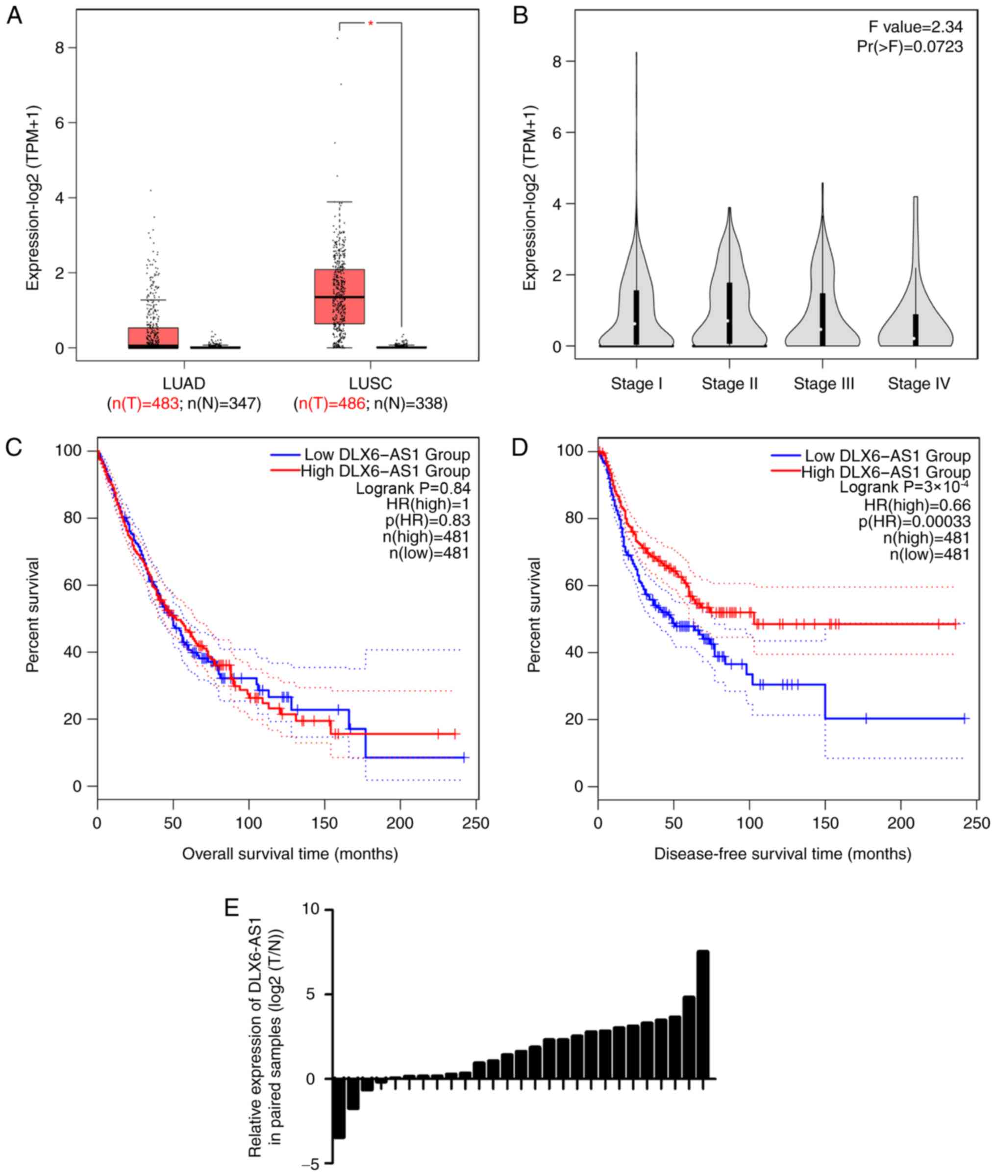
The present study first investigated DLX6-AS1
expression by using GEPIA tool (30), which provides single gene, cancer
type and multiple gene analysis. The results demonstrated that
DLX6-AS1 expression was significantly increased in lung squamous
cell carcinoma (Fig. 1A), but was
not associated with TNM stage (Fig.
1B). In addition, patients with low DLX6-AS1 expression had a
poor prognosis with a shorter disease-free survival (Fig. 1C and D). Additionally, it was found
that DLX6-AS1 was overexpressed in 23 pairs (accounting for 85% of
total samples) of tumor tissues, compared with in adjacent normal
tissues from patients with NSCLC (Fig.
1E).
DLX6-AS1 expression was studied in NSCLC cell lines,
and it was identified that DLX6-AS1 exhibited significantly higher
expression levels in A549, H1299 and 95D cells, compared with in
the normal human epithelial BEAS-2B cells (Fig. S1A). Subsequently, the effect of
DLX6-AS1 on cell proliferation and migration in A549 and H1299
cells transfected with two DLX6-AS1 siRNAs or scrambled siRNA was
investigated. The RT-qPCR results demonstrated that DLX6-AS1
expression was significantly reduced following transfection
(Fig. S1B). DLX6-AS1-knockdown
suppressed A549 and H1299 proliferation and migration in
vitro (Fig. S1C-F).
Serum DLX6-AS1 expression is increased
in NSCLC
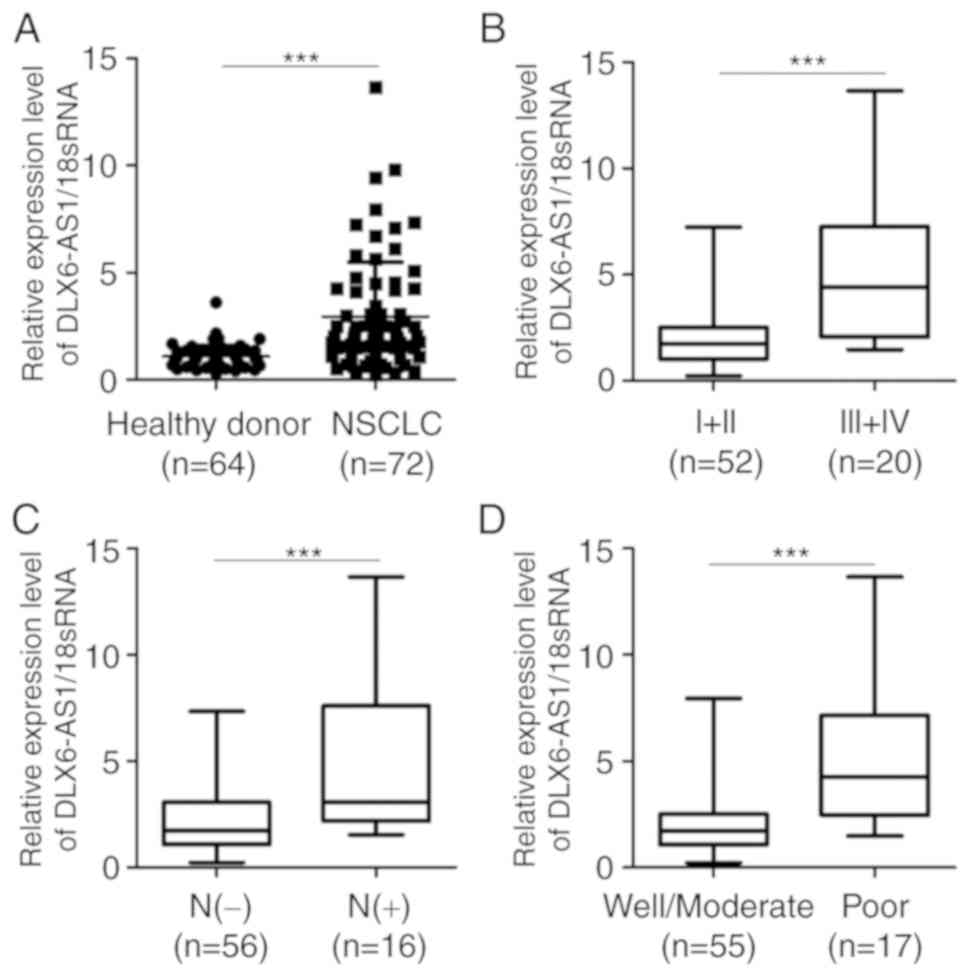
The present study investigated the serum expression
levels of DLX6-AS1 in patients with NSCLC and healthy controls. The
results demonstrated that serum DLX6-AS1 expression was
significantly increased in patients with NSCLC compared with
healthy controls (Fig. 2A). The
association between the serum expression levels of DLX6-AS1 and
clinicalpathological features of patients with NSCLC was also
investigated. The data indicated that the serum expression levels
of DLX6-AS1 were significantly higher in patients with advanced
disease stage, positive lymph node metastasis and poor tumor
differentiation compared with in patients with earlier disease
stage, negative lymph node metastasis and well/moderate tumor
differentiation, respectively (Fig.
2B-D). However, DXL6-AS1 expression was not associated with
sex, age, smoking history and tumor size (Table I).
 | Table I.Association between DLX6-AS1
expression and clinicopathological characteristics of patients with
non-small cell lung cancer. |
Table I.
Association between DLX6-AS1
expression and clinicopathological characteristics of patients with
non-small cell lung cancer.
| Features | n | DLX6-AS1 (mean ±
SD) | t | P-value |
|---|
| Sex |
|
| 1.902 | 0.065 |
|
Male | 39 | 3.15±3.00 |
|
|
|
Female | 33 | 2.00±1.31 |
|
|
| Age, years |
|
| 0.066 | 0.197 |
|
>65 | 35 | 2.92±2.95 |
|
|
|
≤65 | 37 | 2.96±2.12 |
|
|
| Smoking
history |
|
| 1.347 | 0.075 |
|
Yes | 37 | 3.33±2.88 |
|
|
| No | 35 | 2.52±2.08 |
|
|
| Tumor size, cm |
|
| 0.981 | 0.102 |
|
>3 | 30 | 3.71±3.11 |
|
|
| ≤3 | 42 | 2.69±1.90 |
|
|
| Tumor
differentiation |
|
| 5.294 | <0.001 |
|
Well/moderate | 55 | 2.08±1.46 |
|
|
|
Poor | 17 | 5.37±3.40 |
|
|
| TNM |
|
| 2.334 | 0.022 |
|
I–II | 52 | 2.52±2.26 |
|
|
|
III–IV | 20 | 4.03±2.95 |
|
|
| Lymphovascular
invasion |
|
| 6.005 | <0.001 |
|
Present | 16 | 5.69±2.40 |
|
|
|
Absent | 56 | 2.15±1.98 |
|
|
Serum DLX6-AS1 expression is reduced
in patients post-operatively
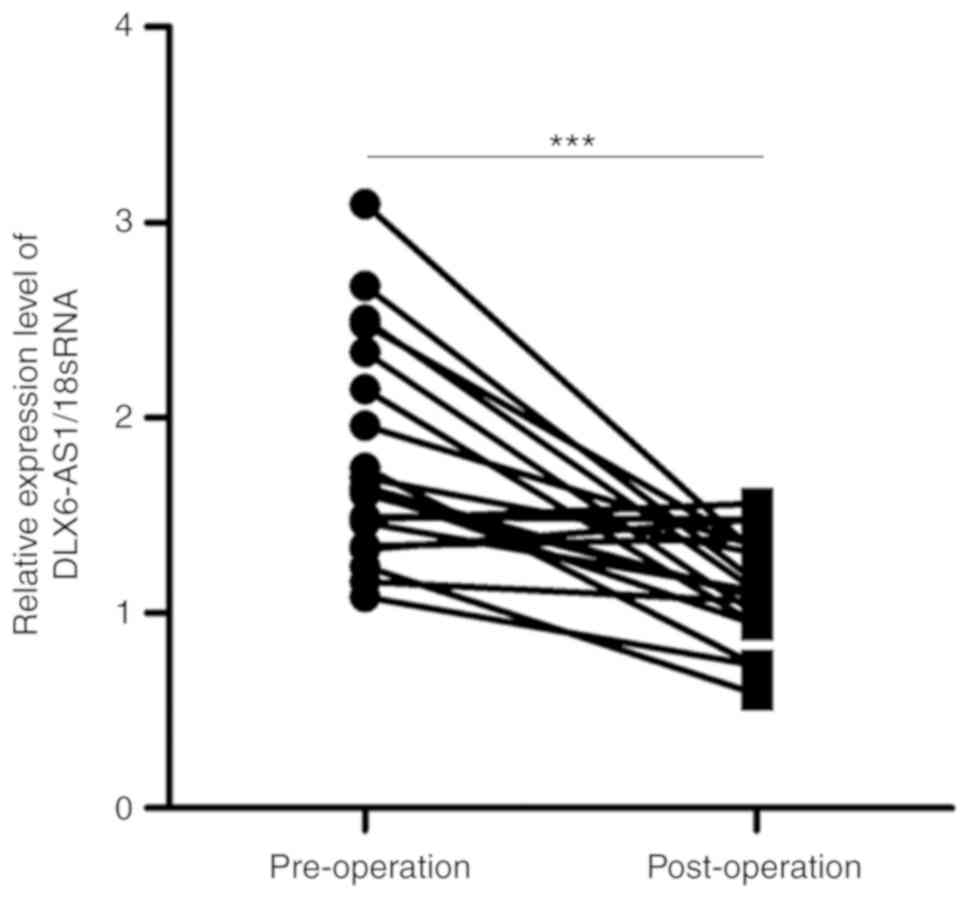
To investigate the effect of surgery on the serum
DLX6-AS1 expression, alterations in DLX6-AS1 expression were
compared between pre- and post-operative serum samples from a set
of 20 matched patients, who were selected to our follow-up study.
The data demonstrated that the serum DLX6-AS1 expression was
significantly lower in post-operative samples than in pre-operative
samples (Fig. 3). These results
suggest that circulating DLX6-AS1 is derived from tumor tissues,
providing a potential marker for the early diagnosis of NSCLC.
Exosomal DLX6-AS1 expression in
patients with NSCLC and healthy donors
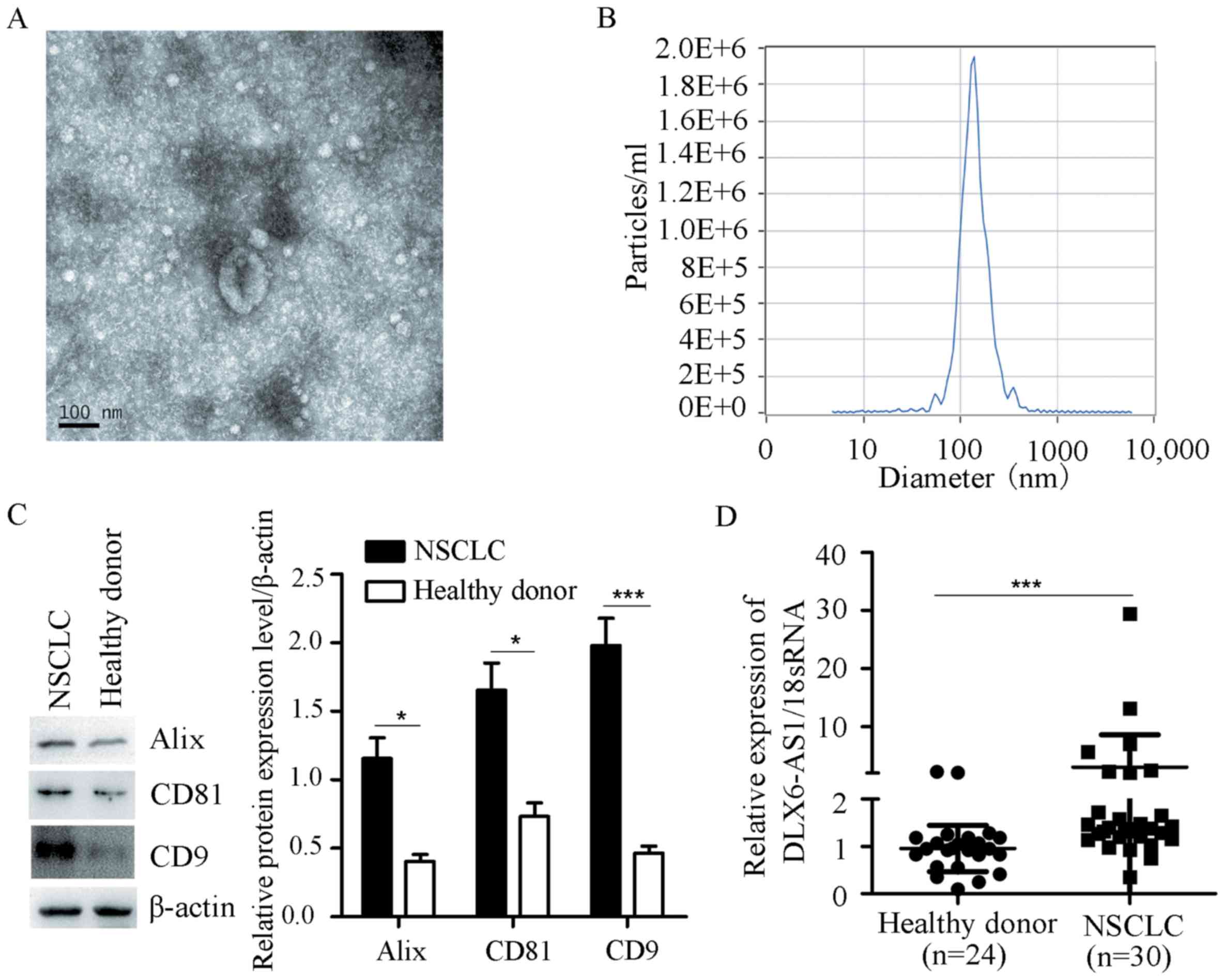
Previous studies have demonstrated that exosomes are
abundantly present in the serum, and exosomal lncRNAs are more
stable than free lncRNAs (34,35).
Therefore, the exosomal DLX6-AS1 levels were examined in patients
with NSCLC and healthy donors. The results revealed that exosomes
were abundant in the serum of patients with NSCLC and healthy
controls (Fig. 4A) according to
electron microscopy. The concentration and size of particles were
detected by NTA, and the majority of particles were distributed
between 39.8 and 136 nm (Fig. 4B).
Exosomal markers (CD9, CD81 and Alix) were measured by western blot
analysis (Fig. 4C). In addition, the
exosomal DLX6-AS1 expression was significantly increased in
patients with NSCLC compared with in healthy controls (Fig. 4D).
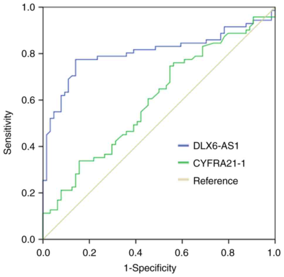
Diagnostic value of circulating
DLX6-AS1 in patients with NSCLC
To further explore the diagnostic value of DLX6-AS1
in NSCLC, ROC curve analysis was performed (Fig. 5). The results indicated that the area
under curve of DLX6-AS1 was 0.806 and the sensitivity and
specificity were 0.775 and 0.859, respectively. However, the area
under curve of CYFRA21-1 was 0.600. The sensitivity reached 0.606
and specificity was 0.547. These results suggested that circulating
DLX6-AS1 could serve as a novel biomarker for the early diagnosis
of NSCLC.
Discussion
In recent years, an increasing number of functional
lncRNAs have been identified through next generation sequencing
technology or gene microarrays. Accumulating evidence has
demonstrated the effect of lncRNAs on cell proliferation,
migration, invasion and metastasis, which can be regarded as an
effective biomarker for the early diagnosis and prognosis of NSCLC
(36,37). Notably, lncRNAs are abundantly
present in bodily fluids, including serum, plasma, urine and saliva
(35). Circulating lncRNAs have
great potential for diagnosis in liquid biopsy due to being easily
and non-invasively obtained from patients (35). For instance, three circulating lncRNA
signatures are significantly elevated in patients with NSCLC
compared with in healthy donors, demonstrating their critical role
in the tumorigenesis of NSCLC (38).
The present study investigated the expression of circulating
DLX6-AS1 and its role in the clinical diagnosis of NSCLC.
DLX6-AS1 has been identified as an oncogene in
various solid tumors, including NSCLC (15–17),
renal cell carcinoma (18),
hepatocellular carcinoma (19–21),
colorectal cancer (22), pancreatic
cancer (23,24), glioma (25) and osteosarcoma (26,27).
Previous studies have demonstrated that DLX6-AS1 expression is
upregulated in NSCLC (15,16). DLX6-AS1-knockdown significantly
suppresses cell proliferation, migration and invasion, and promotes
cell apoptosis via the miR-144/proline rich 11 signaling pathway
(15), suggesting that DLX6-AS1 can
be regarded as a promising target for NSCLC therapy.
The present study revealed that DLX6-AS1 was highly
expressed in NSCLC, particularly in squamous cell lung carcinoma
(data from GEPIA database and study cohort), which was consistent
with previous studies (15–17). Patients with lower DLX6-AS1
expression exhibited a poorer prognosis than those with higher
expression, since the relative expression of DLX6-AS1 in stage
III+IV disease was lower than in stage I+II disease. Recent studies
have demonstrated that lncRNAs are easily detected in the serum or
plasma of patients and provide a non-invasive target for NSCLC
(36,37). Therefore, the present study
investigated the circulating DLX6-AS1 expression in NSCLC and
revealed that the serum DLX6-AS1 expression was significantly
higher in patients with NSCLC compared with in healthy controls.
The higher expression levels of DLX6-AS1 were associated with
advanced disease stage, positive lymph node metastasis and poor
tumor differentiation, suggesting that DLX6-AS1 may be a promising
therapeutic target for NSCLC. In addition, the serum expression of
DLX6-AS1 was reduced in patients post-operatively.
An increasing number of studies have indicated that
exosomes containing lncRNAs stably exist in various conditions and
serve a critical role in metastasis, drug resistance and
immunosuppression, providing a novel therapeutic target for NSCLC
(36,39–42).
Therefore, exosomal DLX6-AS1 expression in NSCLC and healthy
control samples was investigated. The results indicated that
exosomal DLX6-AS1 levels were significantly increased in NSCLC
samples compared with in samples from healthy controls. In
addition, ROC analysis results demonstrated that circulating
DLX6-AS1 had a higher sensitivity and specificity for NSCLC
diagnosis than CYFRA21-1. These results suggest that exosomal
DLX6-AS1 may serve as a potential diagnostic marker for NSCLC.
However, further investigation is required to determine the
underlying mechanisms of circulating DLX6-AS1 on NSCLC
progression.
Supplementary Material
Supporting Data
Acknowledgements
The authors would like to thank Professor Qilin Shi
(Department of Pathology, the First People's Hospital of Huzhou,
Huzhou, China) and Professor Hui Xia (Department of Pathology, the
First People's Hospital of Huzhou, Huzhou, China) for their help in
pathological diagnosis.
Funding
This work was supported by grants from Zhejiang
Province Science and Technology Department of Public Welfare
Project (grant no. LGF18H160019), the Scientific Technology
Projects of Health and Medicine of Zhejiang Province (grant nos.
WKJ-ZJ-1830, 2017KY642 and 2019KY207), and Huzhou Science and
Technology Fund (grant nos. 2017GY33, 2017GY32 and 2018GY04).
Availability of data and materials
The datasets used and/or analyzed during the current
study are available from the corresponding author on reasonable
request.
Authors' contributions
DX and XW designed and conceived the study. XZ, HG
and HY performed the experiments. XZ, YB and XW analyzed the data.
XZ, DX and XW wrote the manuscript. All authors have read the
manuscript.
Ethics approval and consent to
participate
The study was undertaken on agreement of the Ethics
Committee of the First Affiliated Hospital of Huzhou University
(approval no. 2017014), and written informed consent was provided
by all patients or healthy donors.
Patient consent for publication
Not applicable.
Competing interests
The authors declare that they have no competing
interests.
References
|
1
|
Siegel RL, Miller KD and Jemal A: Cancer
statistics, 2019. CA Cancer J Clin. 69:7–34. 2019. View Article : Google Scholar : PubMed/NCBI
|
|
2
|
Chen W, Zheng R, Baade PD, Zhang S, Zeng
H, Bray F, Jemal A, Yu XQ and He J: Cancer statistics in China,
2015. CA Cancer J Clin. 66:115–132. 2016. View Article : Google Scholar : PubMed/NCBI
|
|
3
|
Bray F, Ferlay J, Soerjomataram I, Siegel
RL, Torre LA and Jemal A: Global cancer statistics 2018: GLOBOCAN
estimates of incidence and mortality worldwide for 36 cancers in
185 countries. CA Cancer J Clin. 68:394–424. 2018. View Article : Google Scholar : PubMed/NCBI
|
|
4
|
Hsu PC, Tian B, Yang YL, Wang YC, Liu S,
Urisman A, Yang CT, Xu Z, Jablons DM and You L: Cucurbitacin E
inhibits the Yes-associated protein signaling pathway and
suppresses brain metastasis of human nonsmall cell lung cancer in a
murine model. Oncol Rep. 42:697–707. 2019.PubMed/NCBI
|
|
5
|
Imakita T, Matsumoto H, Hirano K, Morisawa
T, Sakurai A and Kataoka Y: Impact on prognosis of rebiopsy in
advanced non-small cell lung cancer patients after epidermal growth
factor receptor-tyrosine kinase inhibitor treatment: A systematic
review. BMC Cancer. 19:1052019. View Article : Google Scholar : PubMed/NCBI
|
|
6
|
Hamard C, Mignard X, Pecuchet N, Mathiot
N, Blons H, Laurent-Puig P, Leroy K, Lupo A, Chapron J, Giraud F,
et al: IHC, FISH, CISH, NGS in non-small cell lung cancer: What
changes in the biomarker era? Rev Pneumol Clin. 74:327–338.
2018.(In French). View Article : Google Scholar : PubMed/NCBI
|
|
7
|
Bhan A, Soleimani M and Mandal SS: Long
noncoding RNA and cancer: A new paradigm. Cancer Res. 77:3965–3981.
2017. View Article : Google Scholar : PubMed/NCBI
|
|
8
|
Jiang L, Li Z and Wang R: Long noncoding
RNAs in lung cancer: Regulation patterns, biologic function and
diagnosis implications (Review). Int J Oncol. Jul 29–2019.(Epub
ahead of print). View Article : Google Scholar
|
|
9
|
Lim LJ, Wong SYS, Huang F, Lim S, Chong
SS, Ooi LL, Kon OL and Lee CG: Roles and regulation of long
non-coding RNAs in hepatocellular carcinoma. Cancer Res. Jul
23–2019.(Epub ahead of print). View Article : Google Scholar
|
|
10
|
Huarte M: The emerging role of lncRNAs in
cancer. Nat Med. 21:1253–1261. 2015. View
Article : Google Scholar : PubMed/NCBI
|
|
11
|
Fatica A and Bozzoni I: Long non-coding
RNAs: New players in cell differentiation and development. Nat Rev
Genet. 15:7–21. 2014. View
Article : Google Scholar : PubMed/NCBI
|
|
12
|
Dai SP, Jin J and Li WM: Diagnostic
efficacy of long non-coding RNA in lung cancer: A systematic review
and meta-analysis. Postgrad Med J. 94:578–587. 2018. View Article : Google Scholar : PubMed/NCBI
|
|
13
|
Lu T, Wang Y, Chen D, Liu J and Jiao W:
Potential clinical application of lncRNAs in non-small cell lung
cancer. Onco Targets Ther. 11:8045–8052. 2018. View Article : Google Scholar : PubMed/NCBI
|
|
14
|
Jiang C, Yang Y, Yang Y, Guo L, Huang J,
Liu X, Wu C and Zou J: Long noncoding RNA (lncRNA) HOTAIR affects
tumorigenesis and metastasis of non-small cell lung cancer by
upregulating miR-613. Oncol Res. 26:725–734. 2018. View Article : Google Scholar : PubMed/NCBI
|
|
15
|
Huang Y, Ni R, Wang J and Liu Y: Knockdown
of lncRNA DLX6-AS1 inhibits cell proliferation, migration and
invasion while promotes apoptosis by downregulating PRR11
expression and upregulating miR-144 in non-small cell lung cancer.
Biomed Pharmacother. 109:1851–1859. 2019. View Article : Google Scholar : PubMed/NCBI
|
|
16
|
Li J, Li P, Zhao W, Yang R, Chen S, Bai Y,
Dun S, Chen X, Du Y, Wang Y, et al: Expression of long non-coding
RNA DLX6-AS1 in lung adenocarcinoma. Cancer Cell Int. 15:482015.
View Article : Google Scholar : PubMed/NCBI
|
|
17
|
Sun W, Zhang L, Yan R, Yang Y and Meng X:
LncRNA DLX6-AS1 promotes the proliferation, invasion, and migration
of non-small cell lung cancer cells by targeting the
miR-27b-3p/GSPT1 axis. Onco Targets Ther. 12:3945–3954. 2019.
View Article : Google Scholar : PubMed/NCBI
|
|
18
|
Zeng X, Hu Z, Ke X, Tang H, Wu B, Wei X
and Liu Z: Long noncoding RNA DLX6-AS1 promotes renal cell
carcinoma progression via miR-26a/PTEN axis. Cell Cycle.
16:2212–2219. 2017. View Article : Google Scholar : PubMed/NCBI
|
|
19
|
Zhang L, He X, Jin T, Gang L and Jin Z:
Long non-coding RNA DLX6-AS1 aggravates hepatocellular carcinoma
carcinogenesis by modulating miR-203a/MMP-2 pathway. Biomed
Pharmacother. 96:884–891. 2017. View Article : Google Scholar : PubMed/NCBI
|
|
20
|
Li D, Tang X, Li M and Zheng Y: Long
noncoding RNA DLX6-AS1 promotes liver cancer by increasing the
expression of WEE1 via targeting miR-424-5p. J Cell Biochem.
120:12290–12299. 2019. View Article : Google Scholar : PubMed/NCBI
|
|
21
|
Wu DM, Zheng ZH, Zhang YB, Fan SH, Zhang
ZF, Wang YJ, Zheng YL and Lu J: Down-regulated lncRNA DLX6-AS1
inhibits tumorigenesis through STAT3 signaling pathway by
suppressing CADM1 promoter methylation in liver cancer stem cells.
J Exp Clin Cancer Res. 38:2372019. View Article : Google Scholar : PubMed/NCBI
|
|
22
|
Zhou FR, Pan ZP, Shen F, Huang LQ, Cui JH,
Cai K and Guo XL: Long noncoding RNA DLX6-AS1 functions as a
competing endogenous RNA for miR-577 to promote malignant
development of colorectal cancer. Eur Rev Med Pharmacol Sci.
23:3742–3748. 2019.PubMed/NCBI
|
|
23
|
An Y, Chen XM, Yang Y, Mo F, Jiang Y, Sun
DL and Cai HH: LncRNA DLX6-AS1 promoted cancer cell proliferation
and invasion by attenuating the endogenous function of miR-181b in
pancreatic cancer. Cancer Cell Int. 18:1432018. View Article : Google Scholar : PubMed/NCBI
|
|
24
|
Yang J, Ye Z, Mei D, Gu H and Zhang J:
Long noncoding RNA DLX6-AS1 promotes tumorigenesis by modulating
miR-497-5p/FZD4/FZD6/Wnt/β-catenin pathway in pancreatic cancer.
Cancer Manag Res. 11:4209–4221. 2019. View Article : Google Scholar : PubMed/NCBI
|
|
25
|
Li X, Zhang H and Wu X: Long noncoding RNA
DLX6-AS1 accelerates the glioma carcinogenesis by competing
endogenous sponging miR-197-5p to relieve E2F1. Gene. 686:1–7.
2019. View Article : Google Scholar : PubMed/NCBI
|
|
26
|
Zhang RM, Tang T, Yu HM and Yao XD: LncRNA
DLX6-AS1/miR-129-5p/DLK1 axis aggravates stemness of osteosarcoma
through Wnt signaling. Biochem Biophys Res Commun. 507:260–266.
2018. View Article : Google Scholar : PubMed/NCBI
|
|
27
|
Zhang N, Meng X, Mei L, Zhao C and Chen W:
LncRNA DLX6-AS1 promotes tumor proliferation and metastasis in
osteosarcoma through modulating miR-641/HOXA9 signaling pathway. J
Cell Biochem. Mar 6–2019.(Epub ahead of print).
|
|
28
|
Wei L, Wu W, Han L, Yu W and Du Y: A
quantitative analysis of the potential biomarkers of non-small cell
lung cancer by circulating cell-free DNA. Oncol Lett. 16:4353–4360.
2018.PubMed/NCBI
|
|
29
|
Jhun BW, Lee KJ, Jeon K, Suh GY, Chung MP,
Kim H, Kwon OJ, Sun JM, Ahn JS, Ahn MJ, et al: Clinical
applicability of staging small cell lung cancer according to the
seventh edition of the TNM staging system. Lung Cancer. 81:65–70.
2013. View Article : Google Scholar : PubMed/NCBI
|
|
30
|
Tang Z, Li C, Kang B, Gao G, Li C and
Zhang Z: GEPIA: A web server for cancer and normal gene expression
profiling and interactive analyses. Nucleic Acids Res. 45:W98–W102.
2017. View Article : Google Scholar : PubMed/NCBI
|
|
31
|
Zhang X, Hu J, Zhong L, Wang N, Yang L,
Liu CC, Li H, Wang X, Zhou Y, Zhang Y, et al: Quercetin stabilizes
apolipoprotein E and reduces brain Aβ levels in amyloid model mice.
Neuropharmacology. 108:179–192. 2016. View Article : Google Scholar : PubMed/NCBI
|
|
32
|
Livak KJ and Schmittgen TD: Analysis of
relative gene expression data using real-time quantitative PCR and
the 2(-Delta Delta C(T)) method. Methods. 25:402–408. 2001.
View Article : Google Scholar : PubMed/NCBI
|
|
33
|
Yan G, Du Q, Wei X, Miozzi J, Kang C, Wang
J, Han X, Pan J, Xie H, Chen J and Zhang W: Application of
real-time cell electronic analysis system in modern pharmaceutical
evaluation and analysis. Molecules. 23:E32802018. View Article : Google Scholar : PubMed/NCBI
|
|
34
|
Jakobsen KR, Paulsen BS, Baek R, Varming
K, Sorensen BS and Jorgensen MM: Exosomal proteins as potential
diagnostic markers in advanced non-small cell lung carcinoma. J
Extracell Vesicles. 4:266592015. View Article : Google Scholar : PubMed/NCBI
|
|
35
|
Wang L, Duan W, Yan S, Xie Y and Wang C:
Circulating long non-coding RNA colon cancer-associated transcript
2 protected by exosome as a potential biomarker for colorectal
cancer. Biomed Pharmacother. 113:1087582019. View Article : Google Scholar : PubMed/NCBI
|
|
36
|
Li C, Lv Y, Shao C, Chen C, Zhang T, Wei
Y, Fan H, Lv T, Liu H and Song Y: Tumor-derived exosomal lncRNA
GAS5 as a biomarker for early-stage non-small-cell lung cancer
diagnosis. J Cell Physiol. 234:20721–20727. 2019. View Article : Google Scholar : PubMed/NCBI
|
|
37
|
Cheng Y, Dai X, Yang T, Zhang N, Liu Z and
Jiang Y: Low long noncoding RNA growth arrest-specific transcript 5
expression in the exosomes of lung cancer cells promotes tumor
angiogenesis. J Oncol. 2019:24761752019. View Article : Google Scholar : PubMed/NCBI
|
|
38
|
Tang Q, Ni Z, Cheng Z, Xu J, Yu H and Yin
P: Three circulating long non-coding RNAs act as biomarkers for
predicting NSCLC. Cell Physiol Biochem. 37:1002–1009. 2015.
View Article : Google Scholar : PubMed/NCBI
|
|
39
|
Zhang R, Xia Y, Wang Z, Zheng J, Chen Y,
Li X, Wang Y and Ming H: Serum long non coding RNA MALAT-1
protected by exosomes is up-regulated and promotes cell
proliferation and migration in non-small cell lung cancer. Biochem
Biophys Res Commun. 490:406–414. 2017. View Article : Google Scholar : PubMed/NCBI
|
|
40
|
Zhang W, Cai X, Yu J, Lu X, Qian Q and
Qian W: Exosome-mediated transfer of lncRNA RP11838N2.4 promotes
erlotinib resistance in non-small cell lung cancer. Int J Oncol.
53:527–538. 2018.PubMed/NCBI
|
|
41
|
Lei Y, Guo W, Chen B, Chen L, Gong J and
Li W: Tumor-released lncRNA H19 promotes gefitinib resistance via
packaging into exosomes in nonsmall cell lung cancer. Oncol Rep.
40:3438–3446. 2018.PubMed/NCBI
|
|
42
|
Mathieu M, Martin-Jaular L, Lavieu G and
Théry C: Specificities of secretion and uptake of exosomes and
other extracellular vesicles for cell-to-cell communication. Nat
Cell Biol. 21:9–17. 2019. View Article : Google Scholar : PubMed/NCBI
|