Introduction
Gastric cancer (GC) is the fourth most common
malignancy in the world and remains the second leading cause of
death for all malignancies worldwide (738,000 deaths per year)
(1,2). China is a large country with a high
incidence of gastric cancer. As the early symptoms of gastric
cancer are relatively hidden, most patients are diagnosed with
advanced gastric cancer, and the treatment effect is generally poor
(3). The data show that the 5-year
relative survival rate of gastric cancer in China is relatively low
(4). Therefore, it is of great
significance to search for molecular biomarkers related to the
diagnosis of gastric cancer, explore the regulatory mechanism of
molecular signals during the occurrence and development of gastric
cancer, and identify therapeutic targets of biological markers that
affect its occurrence and development, so as to improve the early
diagnosis and prognosis of gastric cancer patients (5,6).
miRNA is a non-coding RNA with a length of 19–25
nucleotides. It is a novel gene expression regulatory molecule that
can participate in the occurrence and development of tumors by
regulating the expression of many oncogenes or tumor suppressor
genes (7). It has been previously
shown that some miRNAs are closely related to the occurrence and
development of gastric cancer. For example, Zhu et al
(8) showed that plasma microRNAs
were potential new biomarkers for early detection of early gastric
cancer. Li et al (9) found
microRNA-28 promotes the proliferation and invasion of gastric
cancer cells through the PTEN/PI3K/AKT signaling pathway. In
addition, Peng et al (10)
found that microRNA-494 increases the chemosensitivity of
doxorubicin in gastric cancer cells by targeting phosphodiesterase
4D. microRNA-23a (miR-23a) has been shown to be upregulated in
non-small cell lung cancer (11) and
laryngeal cancer (12). Previous
findings have shown that microRNA-135 (miR-135) is
under-represented in non-small cell lung cancer (13). However, there are few studies on the
expression of miR-23a and miR-135 in gastric cancer and the
significance of gastric cancer diagnosis.
At present, serum tumor markers such as CEA and
carbohydrate antigen 199 (CA199) are mainly used in the clinical
diagnosis of gastric cancer, which has the advantages of short
detection cycle and small trauma. However, no tumor marker with
strong specificity and high sensitivity has been found to be able
to detect tumors early (14–16). Therefore, the aim of this study was
to compare the serum levels of miR-23a and miR-135 in patients with
gastric cancer and normal controls, and explored the correlation
between the expression level and cancer markers CEA and CA199 in
patients with gastric cancer and their diagnostic significance, so
as to provide a reference for clinical search for potential
molecular markers of gastric cancer.
Patients and methods
General information
A total of 78 patients with gastric cancer admitted
to Dongying People's Hospital from July 2015 to June 2017 were
selected as the observation group, and 80 healthy individuals in
the same period were selected as the control group. There were 52
males and 26 females in the gastric cancer group, aged 41–76 years,
with an average age of 51.57±9.19 years. In the control group,
there were 55 males and 25 females, aged 43–68 years, with an
average age of 51.39±10.17 years. According to the Borrmann
classification (17), there were 23
cases of type I, 22 cases of type II, 16 cases of type III, and 19
cases of type IV in the observation group. The inclusion criteria
were: diagnosis of gastric cancer was confirmed by pathology, and
the clinical, imaging and histopathological data were complete; the
healthy group was examined in the physical examination center of
Dongying People's Hospital and the results were normal, without
other types of tumors, heart, liver, kidney and other important
organ diseases, and no family members had a history of cancer. The
exclusion criteria were: patients with immune system diseases;
long-term bedridden patients; patients with other malignant tumors;
severe hypertension, diabetic, mental and cognitive dysfunction;
pregnant or lactation women; using glucocorticoids and antibiotics
for up to 2 weeks. Clinical data, including BMI (kg/m2),
heart rate(time/min), urea nitrogen, age, tumor stage, smoking
history, alcohol abuse history, of the two groups were collected
and compared.
The study was approved by the Ethics Committee of
Dongying People's Hospital. All the patients agreed to participate
in the experiment and signed informed consent.
Main instruments and reagents
CA-199 ELISA kit [Shanghai Jing Kang Bioengineering
Co., Ltd., JK-(a)-5894]; CEARIA and ELISA kit [Shanghai Jing Kang
Bioengineering Co., Ltd., JK-(a)-6071]; Multi-function microplate
reader (BioTek Berten, DLK0001622); desktop high-speed centrifuge
(Sichuan Yanke Instrument Co., Ltd., TG-16); PCR instrument
(Applied Biosystems; Thermo Fisher Scientific, Inc. 7500); total
RNA extraction kit Easy Pure miRNA kit, PCR + reverse transcription
kit TransScript Green miRNA Two-Step qRT-PCR Super Mix
(TransGenBiotech Co., Ltd., Beijing, China); UV spectrophotometer
(Thermo Fisher Scientific, Inc.; Multiskan Sky); qmiR-23a, miR-135
and U6 internal reference primers were synthesized by Shanghai
Harling Biotechnology Co., Ltd. (Table
I).
 | Table I.miR-23a, miR-135 and U6 primer
sequences. |
Table I.
miR-23a, miR-135 and U6 primer
sequences.
| Gene | Forward primer | Reverse primer |
|---|
| miR-23a |
5′-GGGGATCACATTGCCAGG-3′ |
5′-AGTGCGTGTCGTGGAGTC-3′ |
| miR-135 |
5′-ACAUAGGAAUAAAAAGCCAUAtt-3′ |
5′-CUAUGGCUUUUUAUUCCUAUGUGA-3′ |
| U6 |
5′-CTCGCTTCGGCAGCACA-3′ |
5′-AACGCTTCACGAATTTGCGT-3′ |
Detection method
Venous blood (5 ml) of two groups collected in the
morning was placed in the vacuum collecting vessel and centrifuged
for routine separation at a speed of 2,600 × g for 10 min at 4°C.
The expression levels of CEA and CA199 in the collected serum
samples were determined by ELISA. Blank, sample and standard wells
were set up, respectively. The samples were added to the the sample
well to be tested, and the standard sample with different
concentrations was added to the standard well. The samples were
added to the bottom of the well of the enzyme label plate, avoiding
touching the wall of the well, then gently shaken and mixed. After
sealing the plate with the sealing plate membrane, the samples were
incubated at 37°C for 30 min. The 30-fold concentrated detergent
was diluted with distilled water 30 times for later use. The
sealing plate film was carefully removed, the liquid was discarded,
and shaken dry. Each well was filled with the washing solution, and
left to stand for 30 sec, then discarded, and repeated 5 times, and
patted dry. Enzyme-labeled reagent was added to each well and
placed on the sealing plate at 37°C for 30 min, except the blank
wells. The sealing film was carefully removed, the liquid was
discarded and the film dried. Color reagent A (50 µl) was added in
each well, followed by color reagent B 50 µl, gently shaken and
mixed at 37°C in the dark for 15 min to develop the color.
Termination liquid (50 µl) was added to each well to terminate the
reaction. The OD of each well was measured in sequence at 0 and 450
nm wavelength of blank air conditioner, and the determination was
carried out within 15 min after the addition of terminating liquid.
The calibration curve was drawn and the linear regression equation
was obtained. The OD value of the sample was substituted into the
equation and the concentration of the sample was calculated.
miR-23a and miR-135 detected by
RT-qPCR
Total RNA in serum was extracted according to the
instruction of total RNA extraction kit. The purity of the
extracted total RNA was detected by ultraviolet spectrophotometer
and the concentration was calculated. Total RNA (2 µl) and reverse
transcription were detected as per the instruction manual of the
kit for cDNA synthesis. The reaction system was: 42°C for 60 min,
95°C for 5 min, and the synthesized cDNA sample was stored at −20°C
for later use. The total volume of 20 µl reaction system was:
SYBR-Green PCRP remix 10 µl, upstream primer (10X) 2 µl, downstream
primer (10X) 2 µl, dd water (Rnase- and Dnase-free) 5 µl. RT-qPCR
reaction conditions were: 90°C for 5 min, 90°C for 5 sec, 60°C for
30 sec, 72°C for 5 sec, total of 40 cycles. U6 was the internal
reference gene, and 2−∆∆Cq was used to analyze the data
(18).
Statistical analysis
SPSS 22.0 (SPSS Inc.) was used to analyze the data.
Enumeration data were described by [n (%)], and Chi-square test was
used for comparison between groups. Measurement data were expressed
as mean ± standard deviation. The t-test was used to compare the
measurements between groups. The receiver operating characteristic
(ROC) curve was used to evaluate the diagnostic efficacy of miR-23a
and miR-135 for gastric cancer. Pearson's correlation analysis was
used to test the correlation between miR-23a and miR-135 and CEA
and CA-199. P<0.05 was considered to indicate a statistically
significant difference.
Results
Comparison of general clinical
data
The clinical data of the two groups were collected
for comparison, and the results showed that there was no
statistical difference between the observation and control groups
in BMI (kg/m2), heart rate (time/min), urea nitrogen,
age, tumor stage, smoking history, alcohol abuse history
(P>0.05), as shown in Table
II.
| Table II.Comparison of general clinical data
between the two groups (mean ± SD) [n (%)]. |
Table II.
Comparison of general clinical data
between the two groups (mean ± SD) [n (%)].
| Clinical factors | Control group
(n=80) | Observation group
(n=78) | t/χ2
value | P-value |
|---|
| BMI
(kg/m2) | 23.98±1.26 | 24.36±1.57 | 1.68 | 0.095 |
| Heart rate
(times/points) | 105.61±11.53 | 105.79±11.66 | 0.098 | 0.922 |
| Urea nitrogen
(mmol/l) | 12.97±3.51 | 13.16±3.71 | 0.331 | 0.741 |
| Age | 49.67±5.68 | 50.45±4.71 | 0.938 | 0.35 |
| Tumor staging |
|
| 0.239 | 0.625 |
| I+II | 39 (48.75) | 35 (44.87) |
|
|
|
IIIa+IIIb | 41 (51.25) | 43 (55.13) |
|
|
| Borrmann
classification |
|
| 0.534 | 0.465 |
| I+II | 37 (46.25) | 32 (41.03) |
|
|
|
III+IV | 43 (53.75) | 46 (58.97) |
|
|
| Sex |
|
| 0.143 | 0.378 |
| Male | 45 (56.25) | 42 (53.85) |
|
|
|
Female | 35 (43.75) | 36 (46.15) |
|
|
| Drinking history |
|
| 0.38 | 0.617 |
| Yes | 51 (63.75) | 47 (60.26) |
|
|
| No | 29 (36.25) | 31 (39.74) |
|
|
| Degree of
differentiation |
|
| 0.355 | 0.837 |
|
Differentiated type | 21 (26.25) | 21 (26.92) |
|
|
| Poorly
differentiated | 32 (40) | 34 (43.59) |
|
|
|
Undifferentiated type | 27 (33 75) | 23 (29.49) |
|
|
| Lymph node
metastasis |
|
| 0.894 | 0.345 |
|
Yes | 46 (57.5) | 39 (50) |
|
|
| No | 34 (42.5) | 39 (50) |
|
|
| Degree of
differentiation |
|
| 0.261 | 0.61 |
| High
school and below | 11 (13.75) | 13 (16.67) |
|
|
| Over
Senior High School | 69 (86.25) | 65 (83.33) |
|
|
| History of
smoking |
|
| 0.23 | 0.632 |
| No | 41 (51.25) | 37 (47.44) |
|
|
|
Yes | 39 (48.75) | 41 (52.56) |
|
|
Comparison of serum miR-23a and
miR-135 expression between the observation and control groups
The expression levels of serum miR-23a and miR-135
in the observation group were significantly higher than those in
the control group, with statistically significant differences
(P<0.05) (Table III).
| Table III.Comparison of serum miR-23a and
miR-135 expression between the two groups (mean ± SD). |
Table III.
Comparison of serum miR-23a and
miR-135 expression between the two groups (mean ± SD).
| Group | n | miR-23a | miR-135 |
|---|
| Observation | 78 | 13.72±3.27 | 4.50±1.41 |
| Control | 80 | 10.15±2.26 | 3.03±0.74 |
| t value | – | 8.000 | 8.234 |
| P-value | – | <0.001 | <0.001 |
Comparison of the expression of serum
CA199 and CEA between the observation and control groups
The levels of serum CA199 and CEA in the observation
group were significantly higher than those in the control group
(P<0.05) (Table IV).
| Table IV.Comparison of serum CA199 and CEA
expression between the two groups (mean ± SD). |
Table IV.
Comparison of serum CA199 and CEA
expression between the two groups (mean ± SD).
| Group | n | CA199 (ng/ml) | CEA (ng/ml) |
|---|
| Observation | 78 | 53.07±38.27 | 16.58±11.18 |
| Control | 80 | 11.34±3.5 |
1.8±1.40 |
| t value | – | 0.877 | 11.730 |
| P-value | – | <0.001 | <0.001 |
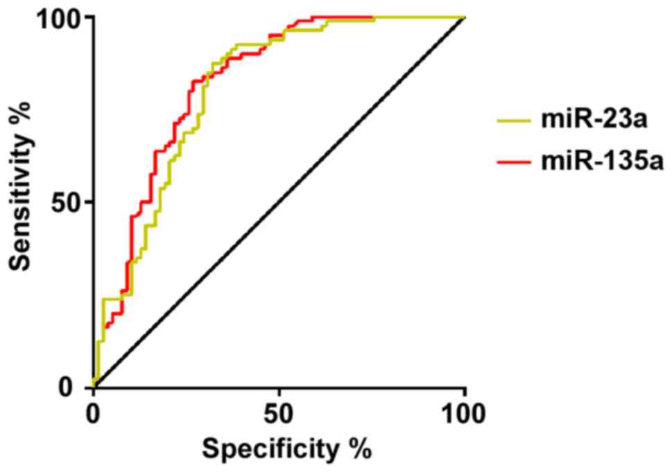
Diagnostic value of serum miR-23a and
miR-135 in gastric cancer
The ROC curve was drawn for the diagnosis of gastric
cancer by the expression levels of miR-23a and miR-135. The results
showed that the AUC for the diagnosis of gastric cancer by miR-23a
was 0.805 (95% CI: 0.735–0.874), the specificity was 67.95%, the
sensitivity was 87.50%, and the cut-off value was 12.500. The AUC
of gastric cancer diagnosed by miR-135 was 0.824 (95% CI:
0.758–0.890), the specificity was 73.08%, the sensitivity was
82.50%, and the cut-off value was 3.677 (Table V, Fig.
1).
| Table V.Diagnostic value of serum miR-23a and
miR-135 in gastric cancer. |
Table V.
Diagnostic value of serum miR-23a and
miR-135 in gastric cancer.
| Indicators | AUC | 95% CI | Specificity
(%) | Sensitivity
(%) | Cut-off |
|---|
| miR-23a | 0.805 | 0.735–0.874 | 67.95 | 87.50 | 12.500 |
| miR-135 | 0.824 | 0.758–0.890 | 73.08 | 82.50 |
3.677 |
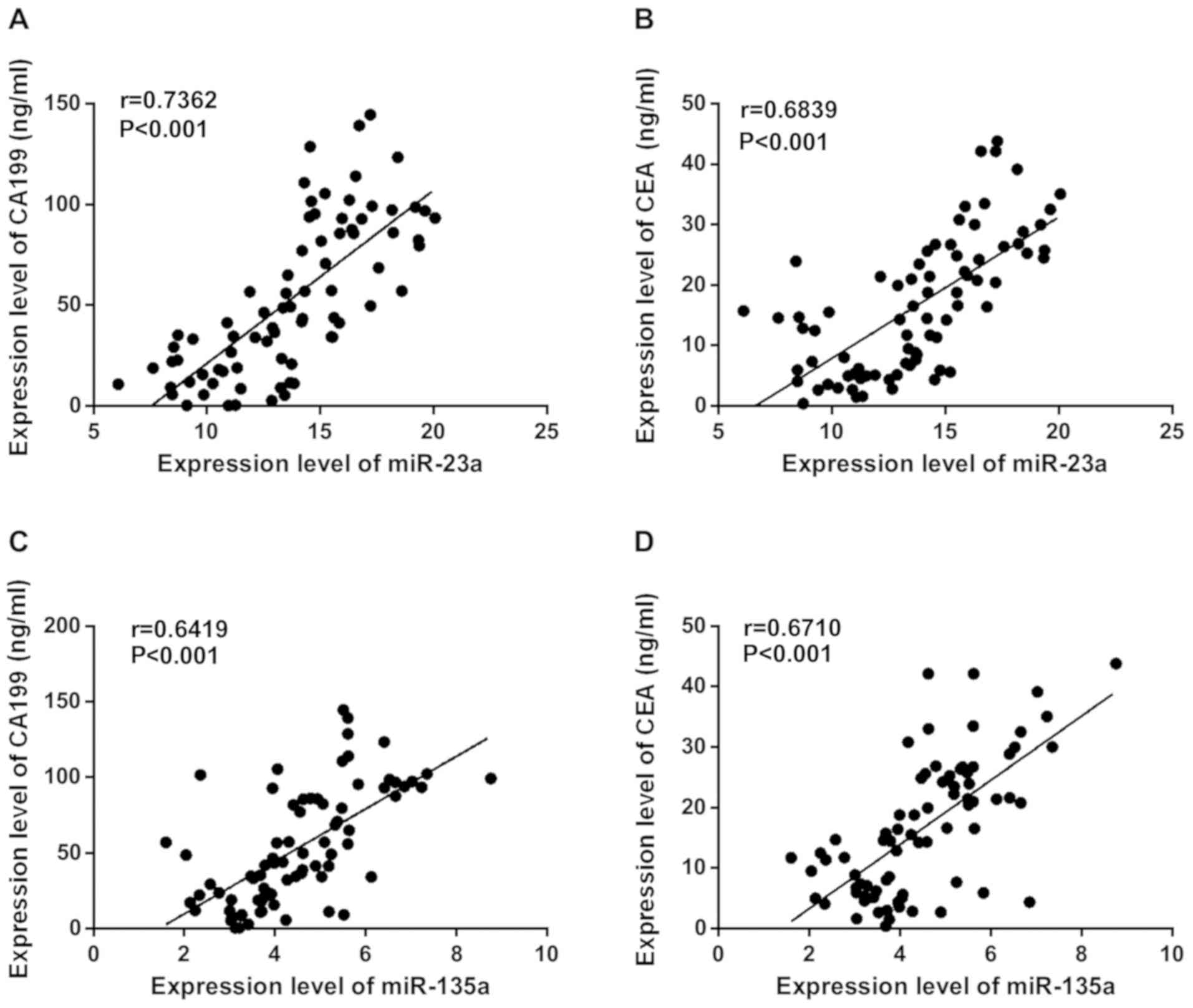
Correlation analysis between serum
miR-23a, miR-135 and CA199, CEA in patients with gastric
cancer
The relationship between miR-23a and miR-135 in
serum and CA199 and CEA was analyzed by Pearson's correlation
analysis. It was found that miR-23a was positively correlated with
the expression of CA199 (r=0.7362, P<0.001) and CEA (r=0.6839,
P<0.001); miR-135 was positively correlated with the expression
of CA199 (r=0.6419, P<0.001) and CEA (r=0.6710, P<0.001)
(Fig. 2).
Discussion
Gastric cancer is a relatively common malignant
tumor in clinical practice, and its pathogenesis has not been
clarified at present (19). Many
gastric cancer patients have a lack of specificity in the early
stage of the disease, and screening for gastric cancer requires
biopsy pathology under gastroscopy. This invasive examination can
make patients feel more uncomfortable, which leads to a low
diagnostic rate of gastric cancer (20). Many patients are in the middle and
late stages of treatment, and it is often difficult to achieve the
desired therapeutic effect (21).
Therefore, searching for biological indicators that have a strong
diagnostic value for gastric cancer is of great significance for
improving the prognosis of patients and increasing the survival
rate (22).
miRNA is a class of non-coding small RNAs with a
wide range of biological effects, which participates in the
regulation of the expression of various proto-oncogenes and
anti-oncogenes in vivo, thereby affecting the biological
behavior of cell proliferation, invasion and angiogenesis (23,24).
Although the current understanding of miRNA function is not very
clear, relevant studies have shown that its abnormal expression or
mutation is closely related to the occurrence and development of
many tumors and the efficacy of some antitumor drugs (25), which is a new highlight in the field
of tumor biotherapy research (26).
The levels of serum miR-23a and miR-135 in the observation group
were significantly higher than those in the control group. Previous
studies have shown that miR-23a is abnormally expressed in gastric
cancer. Zhu et al (27) found
that miR-23a is up-regulated in gastric adenocarcinoma and may
promote the growth of gastric cancer tissues. Studies on gastric
cancer cell line MGC80 showed that the expression of interleukin-6
receptor protein can be reduced by the expression of mRNA-23a,
which can promote the growth of gastric adenocarcinoma cell line
MGC803. The expression of mRNA-23a in peripheral blood and tumor
cells of patients with cancer is increased. Zhou et al
(28) found that miR-135 can promote
the growth and invasion of colorectal cancer in vitro by
transferring inhibitors, while Golubovskaya et al (29) showed that miR-135 can directly target
adhesive plaque kinase, inhibit cell invasion and improve the
sensitivity of cancer cells to chemotherapy. However, its specific
mechanism in gastric cancer is still unclear, but it indicates that
miR-23a and miR-135 are very likely to be involved in the
occurrence, development, migration and metastasis of gastric
cancer, and they are expected to become biological markers for the
diagnosis of gastric cancer.
Our further investigation showed that the
specificity of serum miR-23a in the diagnosis of gastric cancer was
67.95%, and the sensitivity was 87.50%; the specificity of serum
miR-135 in the diagnosis of gastric cancer was 73.08%, and the
sensitivity was 82.50%. These results suggest that both can be used
as biological indicators with better sensitivity and specificity in
the diagnosis of gastric cancer. At the end of the study, Pearson's
correlation analysis was used to detect the correlation between the
expression levels of miR-21 and miR-124 in serum of patients with
gastric cancer and the expression levels of CA199 and CEA. It was
found that miR-23a and miR-135 were positively correlated with the
expression of CA199 and CEA, but the specific relationship was not
explored in depth.
In this study, we compared the expression levels of
miR-23a and miR-135 in the serum of patients with gastric cancer
and normal controls, and explored the diagnostic value of the
detection for gastric cancer. However, the efficacy and prognosis
of these markers were not observed, thus, futher studies are still
required.
In conclusion, miR-23a and miR-135 are highly
expressed in gastric cancer patients and positively correlated with
tumor markers. The detection of serum miR-23a and miR-135 has good
sensitivity and specificity for the diagnosis of gastric cancer,
and show potential as biomarkers for the diagnosis of gastric
cancer.
Acknowledgements
Not applicable.
Funding
The present study was supported by the Hospital
level Scientific Research Project ‘Application of microRNA-203
combined with TSGF and CEA in diagnosis of early gastric cancer’
(no. 2016DYYZ20).
Availability of data and materials
The datasets used and/or analyzed during the present
study are available from the corresponding author on reasonable
request.
Authors' contributions
LY and GX conceived and designed the study, and
drafted the manuscript. LY, GX, YZ and YW collected, analyzed and
interpreted the experimental data. YZ and YW performed PCR. LY
revised the manuscript for important intellectual content and
assisted with statistical analysis. All the authors read and
approved the final manuscript.
Ethics approval and consent to
participate
The study was approved by the Ethics Committee of
Dongying People's Hospital. Signed informed consents were obtained
from the patients or the guardians.
Patient consent for publication
Not applicable.
Competing interests
The authors declare that they have no competing
interests.
References
|
1
|
Sitarz R, Skierucha M, Mielko J, Offerhaus
GJA, Maciejewski R and Polkowski WP: Gastric cancer: Epidemiology,
prevention, classification, and treatment. Cancer Manag Res.
10:239–248. 2018. View Article : Google Scholar : PubMed/NCBI
|
|
2
|
Jemal A, Bray F, Center MM, Ferlay J, Ward
E and Forman D: Global cancer statistics. CA Cancer J Clin.
61:69–90. 2011. View Article : Google Scholar : PubMed/NCBI
|
|
3
|
Shen L, Li J, Xu J, Pan H, Dai G, Qin S,
Wang L, Wang J, Yang Z, Shu Y, et al: Bevacizumab plus capecitabine
and cisplatin in Chinese patients with inoperable locally advanced
or metastatic gastric or gastroesophageal junction cancer:
Randomized, double-blind, phase III study (AVATAR study). Gastric
Cancer. 18:168–176. 2015. View Article : Google Scholar : PubMed/NCBI
|
|
4
|
Zeng H, Zheng R, Guo Y, Zhang S, Zou X,
Wang N, Zhang L, Tang J, Chen J, Wei K, et al: Cancer survival in
China, 2003–2005: A population-based study. Int J Cancer.
136:1921–1930. 2015. View Article : Google Scholar : PubMed/NCBI
|
|
5
|
Buckland G, Travier N, Huerta JM,
Bueno-de-Mesquita HB, Siersema PD, Skeie G, Weiderpass E, Engeset
D, Ericson U, Ohlsson B, et al: Healthy lifestyle index and risk of
gastric adenocarcinoma in the EPIC cohort study. Int J Cancer.
137:598–606. 2015. View Article : Google Scholar : PubMed/NCBI
|
|
6
|
Massarrat S and Stolte M: Development of
gastric cancer and its prevention. Arch Iran Med. 17:514–520.
2014.PubMed/NCBI
|
|
7
|
Rao SA, Santosh V and Somasundaram K:
Genome-wide expression profiling identifies deregulated miRNAs in
malignant astrocytoma. Mod Pathol. 23:1404–1417. 2010. View Article : Google Scholar : PubMed/NCBI
|
|
8
|
Zhu XL, Ren LF, Wang HP, Bai ZT, Zhang L,
Meng WB, Zhu KX, Ding FH, Miao L, Yan J, et al: Plasma microRNAs as
potential new biomarkers for early detection of early gastric
cancer. World J Gastroenterol. 25:1580–1591. 2019. View Article : Google Scholar : PubMed/NCBI
|
|
9
|
Li L, Zhu X, Shou T, Yang L, Cheng X, Wang
J, Deng L and Zheng Y: MicroRNA-28 promotes cell proliferation and
invasion in gastric cancer via the PTEN/PI3K/AKT signalling
pathway. Mol Med Rep. 17:4003–4010. 2018.PubMed/NCBI
|
|
10
|
Peng QP, Du DB, Ming Q, Hu F, Wu ZB and
Qiu S: MicroRNA 494 increases chemosensitivity to doxorubicin in
gastric cancer cells by targeting phosphodiesterases 4D. Cell Mol
Biol. 64:62–66. 2018. View Article : Google Scholar : PubMed/NCBI
|
|
11
|
Qu WQ, Liu L and Yu Z: Clinical value of
microRNA-23a upregulation in non-small cell lung cancer. Int J Clin
Exp Med. 8:13598–13603. 2015.PubMed/NCBI
|
|
12
|
Zhang XW, Liu N, Chen S, Wang Y, Zhang ZX,
Sun YY, Qiu GB and Fu WN: High microRNA-23a expression in laryngeal
squamous cell carcinoma is associated with poor patient prognosis.
Diagn Pathol. 10:222015. View Article : Google Scholar : PubMed/NCBI
|
|
13
|
Wang N and Zhang T: Downregulation of
MicroRNA-135 promotes sensitivity of non-small cell lung cancer to
Gefitinib by targeting TRIM16. Oncol Res. 26:1005–1014. 2018.
View Article : Google Scholar : PubMed/NCBI
|
|
14
|
Virgilio E, Proietti A, D'Urso R, Cardelli
P, Giarnieri E, Montagnini M, Giovagnoli MR, Mercantini P, Balducci
G and Cavallini M: Measuring intragastric tumor markers in gastric
cancer patients: A systematic literature review on significance and
reliability. Anticancer Res. 37:2817–2821. 2017.PubMed/NCBI
|
|
15
|
Xiao S, Feng F, Sun L, Cai L, Liu Z, Liu
S, Fan D and Zhang H: Blood type AB predicts promising prognosis in
gastric cancer patients with positive preoperative serum CEA.
Medicine (Baltimore). 96:e84962017. View Article : Google Scholar : PubMed/NCBI
|
|
16
|
Tu H, Sun L, Dong X, Gong Y, Xu Q, Jing J,
Bostick RM, Wu X and Yuan Y: A serological biopsy using five
stomach-specific circulating biomarkers for gastric cancer risk
assessment: A multi-phase study. Am J Gastroenterol. 112:704–715.
2017. View Article : Google Scholar : PubMed/NCBI
|
|
17
|
Wang L, Wang X H, Kou H J, et al:
Comparing single oral contrast-enhanced ultrasonography and double
contrast-enhanced ultrasonography in the preoperative Borrmann
classification of advanced gastric cancer. Oncotarget.
9:87162018.PubMed/NCBI
|
|
18
|
Livak KJ and Schmittgen TD: Analysis of
relative gene expression data using real-time quantitative PCR and
the 2(-Delta Delta C(T)) method. Methods. 25:402–408. 2001.
View Article : Google Scholar : PubMed/NCBI
|
|
19
|
Okines A, Verheij M, Allum W, Cunningham D
and Cervantes A; ESMO Guidelines Working Group, : Gastric cancer:
ESMO Clinical Practice Guidelines for diagnosis, treatment and
follow-up. Ann Oncol. 21 (Suppl 5):v50–v54. 2010. View Article : Google Scholar : PubMed/NCBI
|
|
20
|
Deng G, Qu J, Zhai S, Shi Y and Wang X:
Effect of neoadjuvant chemotherapy on nutritional status of locally
advanced gastric cancer. Zhonghua Wei Chang Wai Ke Za Zhi.
21:331–335. 2018.(In Chinese). PubMed/NCBI
|
|
21
|
Shen L, Shan YS, Hu HM, Price TJ, Sirohi
B, Yeh KH, Yang YH, Sano T, Yang HK, Zhang X, et al: Management of
gastric cancer in Asia: Resource-stratified guidelines. Lancet
Oncol. 14:e535–e547. 2013. View Article : Google Scholar : PubMed/NCBI
|
|
22
|
Harada K, Mizrak Kaya D, Shimodaira Y,
Song S, Baba H and Ajani JA: Proteomics approach to identify
biomarkers for upper gastrointestinal cancer. Expert Rev
Proteomics. 13:1041–1053. 2016. View Article : Google Scholar : PubMed/NCBI
|
|
23
|
Qian B, Katsaros D, Lu L, Preti M, Durando
A, Arisio R, Mu L and Yu H: High miR-21 expression in breast cancer
associated with poor disease-free survival in early stage disease
and high TGF-beta1. Breast Cancer Res Treat. 117:131–140. 2009.
View Article : Google Scholar : PubMed/NCBI
|
|
24
|
Zhou J, Tian Y, Li J, Lu B, Sun M, Zou Y,
Kong R, Luo Y, Shi Y, Wang K, et al: miR-206 is down-regulated in
breast cancer and inhibits cell proliferation through the
up-regulation of cyclinD2. Biochem Biophys Res Commun. 433:207–212.
2013. View Article : Google Scholar : PubMed/NCBI
|
|
25
|
Baranwal S and Alahari SK: miRNA control
of tumor cell invasion and metastasis. Int J Cancer. 126:1283–1290.
2010.PubMed/NCBI
|
|
26
|
Rupaimoole R, Calin GA, Lopez-Berestein G
and Sood AK: miRNA deregulation in cancer cells and the tumor
microenvironment. Cancer Discov. 6:235–246. 2016. View Article : Google Scholar : PubMed/NCBI
|
|
27
|
Zhu LH, Liu T, Tang H, Tian RQ, Su C, Liu
M and Li X: MicroRNA-23a promotes the growth of gastric
adenocarcinoma cell line MGC803 and downregulates interleukin-6
receptor. FEBS J. 277:3726–3734. 2010. View Article : Google Scholar : PubMed/NCBI
|
|
28
|
Zhou W, Li X, Liu F, Xiao Z, He M, Shen S
and Liu S: MiR-135a promotes growth and invasion of colorectal
cancer via metastasis suppressor 1 in vitro. Acta Biochim Biophys
Sin (Shanghai). 44:838–846. 2012. View Article : Google Scholar : PubMed/NCBI
|
|
29
|
Golubovskaya VM, Sumbler B, Ho B, Yemma M
and Cance WG: MiR-138 and MiR-135 directly target focal adhesion
kinase, inhibit cell invasion, and increase sensitivity to
chemotherapy in cancer cells. Anticancer Agents Med Chem. 14:18–28.
2014. View Article : Google Scholar : PubMed/NCBI
|