Introduction
Triple-negative breast cancer (TNBC) is one of the
most frequently diagnosed malignancies among females (1–3), and is
characterized by a lack of estrogen and progesterone receptors, and
the absence of the human epidermal growth factor receptor (2). Chemotherapy is commonly used to treat
TNBC (4), as due to its
heterogeneity, the availability of other treatment options is
limited (5). Although a number of
molecular therapeutic targets have been identified, including poly
ADP ribose polymerase, epidermal growth factor receptor, fibroblast
growth factor receptor and the angiogenic pathway [some of which
are currently being tested in clinical trials (3)], no reliable outcomes have been observed
(6,7). Therefore identification of novel
molecular targets is required.
Runt-related transcription factor 2 (RUNX2) is a
transcription factor that participates in the regulation of cell
proliferation by influencing the expression of a large set of
downstream genes (8). A growing body
of literature has demonstrated the critical role of RUNX2 in a
number of human cancer types, including different types of breast
cancer (9,10). RUNX2 expression is now considered a
promising therapeutic target for cancer treatment (11).
Long non-coding RNAs (lncRNAs) are a group of
non-protein coding RNAs involved in physiological and pathological
processes (12,13). RUNX2 influences cancer biology, not
only by affecting protein production, but also by its interaction
with lncRNAs (12,13). lncRNA HAND2-AS1, a transcribed
antisense (AS) lncRNA adjacent to heart and neural crest
derivatives expressed 2 (HAND2), has been characterized as a tumor
suppressor lncRNA in various types of malignancy (14–16).
lncRNA HAND2-AS1 is involved in cancer biology through interactions
with multiple signaling molecules, including microRNAs (miRNAs),
hypoxia-inducible factor 1α (HIF1α) and neuromedin (14–16).
However, its functions in TNBC are yet to be elucidated.
In the present study, the role of lncRNA HAND2-AS1
in TNBC was investigated, and revealed to be downregulated.
Additionally, lncRNA HAND2-AS1 may inhibit the proliferation of
cancer cells by reducing RUNX2 expression in TNBC, providing a
potential therapeutic target for the disease.
Materials and methods
Cell lines and patient samples
Two human TNBC cell lines, MDA-MB-231 and BT-20,
were purchased from the American Type Culture Collection (ATCC,
Manassas, VA, USA) and cutured with ATCC-formulated Eagle's minimum
essential medium (cat. no. 30-2003) with 10% fetal bovine serum
(cat. no. F2442-50ML, Sigma-Aldrich, Merck KGaA, Darmstadt,
Germany) at 37°C in a 5% CO2 incubtaor.
The study included 63 female patients with TNBC and
43 healthy females. Participants were admitted to the International
Peace Maternity and Child Health Hospital (Shanghai, China) between
January 2016 and January 2018. The inclusion criteria were as
follows: i) A diagnosis of TNBC through pathological examinations;
ii) TNBC of American Joint Committee on Cancer stages (17) I and II at presentation; and iii)
willingness to donate biopsies of tumor tissues and adjacent
healthy tissues within 2 cm around the tumor site. Exclusion
criteria: i) Patients suffering from multiple diseases; ii)
treatment prior to admission; and iii) patients at advanced cancer
stages. Biopsies of tumor and adjacent healthy tissues were
confirmed by histopathological examination. Tissues were fixed in
4% formaldehyde overnight at 4°C. Subsequently, paraffin-embedded
(8 µm) tissue sections were stained with hematoxylin and eosin at
37°C for 2 h and visualized using a light microscope (×40
magnification). Plasma samples derived from the blood of patients
and healthy controls were also collected by centrifuging blood
samples in EDTA tubes for 10 min at 1,200 × g. All samples were
stored in liquid nitrogen before use. The 43 healthy females
(control group) received a routine physical examination at the
International Peace Maternity and Child Health Hospital during the
same time period. The age range of the patient group was 30–69
years, with a mean age of 45.5±6.1 years, while that of the control
group was 28–66 years, with a mean age of 43.9±5.7 years. The 2
groups had similar age distributions (revealed using Mann-Whitney U
test). The present study was approved by the ethics committee of
the International Peace Maternity and Child Health Hospital, and
all patients and healthy volunteers provided written informed
consent.
Reverse transcription-quantitative
polymerase chain reaction (RT-qPCR)
Following total RNA extraction using RNAzol
RT® (Sigma-Aldrich; Merck KGaA, Darmstadt, Germany)
according to the manufacturer's protocol, cDNA was obtained through
RT using the QuantiTect RT kit (Qiagen GmbH, Hilden, Germany). The
SuperScript III Platinum One-Step RT-qPCR kit (SYBR; Thermo Fisher
Scientific, Inc., Waltham, MA, USA) was used to prepare all
reactions. Thermocycling conditions were as follows: 95°C for 48
sec, 95°C for 16 sec and 56.5°C for 28 sec for 40 cycles. The
sequences of the primers used were: HAND2-AS1 forward,
5′-GGGTGTTTACGTAGACCAGAACC-3′ and reverse,
5′-CTTCCAAAAGCCTTCTGCCTTAG-3′; RUNX2 forward,
5′-GTTATGAAAAACCAAGTAGCCAGGTC-3′ and reverse,
5′-GTAATCTGACTCTGTCCTTGTGGAT-3′; and β-actin forward,
5′-GACCTCTATGCCAACACAGT3′ and reverse, 5′-AGTACTTGCGCTCAGGAGGA3′.
Data were normalized using the 2−ΔΔCq method (18).
Vectors and transfection
The HAND2-AS1 and RUNX2 expression vectors were
synthesized by Shanghai GenePharma Co., Ltd. (Shanghai, China).
Cells were cultured overnight to achieve 80–90% confluence, and
Lipofectamine 2000® reagent (cat. no. 11668-019;
Invitrogen; Thermo Fisher Scientific, Inc.) was used to transfect
10 nM of each vector into 5×10⁵ cells. To verify the
overexpression of HAND2-AS1 and RUNX2, untransfected cells and
those transfected with empty vectors were used as controls. A
HAND2-AS1 expression rate of >175% was confirmed prior to
subsequent experimentation. The interval between transfection and
following experimentation was 24 h.
Cell proliferation assay
Following transfection, cell proliferation was
measured using Cell Counting Kit-8 (CCK-8) (Beyotime Institute of
Biotechnology, Haimen, China). Briefly, cells were harvested and
single cell suspensions (4×10⁴ cells/ml) were prepared;
0.1 ml cell suspension was added to each well of a 96-well plate.
Cells were cultured in a 5% CO2 incubator at 37°C, and
10 µl CCK-8 solution was added at 24, 48, 72 and 96-h time points.
Cells were cultured for an additional 4 h, and the Fisherbrand™
accuSkan™ GO UV/Vis Microplate Spectrophotometer (Thermo Fisher
Scientific, Inc.) was used to measure absorbance at 450 nm.
Western blotting
The ReadyPrep™ Protein Extraction kit (Total
Protein; Bio-Rad Laboratories, Inc., Hercules, CA, USA) was used to
extract total protein from cell lines and patient samples. Protein
concentration was measured using bicinchoninic acid assay
(Sigma-Aldrich; Merck KGaA). The lysates were denatured, and 20 µg
protein per lane was separated using SDS-PAGE with a 12% gel.
Following gel transfer to PVDF membranes, blocking was performed in
5% skimmed milk (PBS) at room temperature for 2 h. The membranes
were subsequently incubated with primary rabbit anti-human
antibodies against RUNX2 (1:1,200; cat. no. ab23981; Abcam,
Cambridge, UK) and GAPDH (1:1,400; cat. no. ab9485; Abcam)
overnight at 4°C. Membranes were subsequently washed twice using
PBS at room temperature, 15 min each time, followed by further
incubation with immunoglobulin G-horseradish peroxidase-conjugated
secondary antibody (1:1,000; cat. no. MBS435036; MyBioSource, San
Diego, CA, USA) at room temperature for 2 h. Signals were developed
using enhanced chemiluminescence (Sigma-Aldrich, Merck KGaA), and
ImageJ v1.6 software (National Institutes of Health, Bethesda, MD,
USA) was used for data normalization.
Statistical analysis
GraphPad Prism 6 software (GraphPad Software, Inc.,
La Jolla, CA, USA) was used to process all data. All experiments
were performed in triplicate, and the data are expressed as the
mean ± standard deviation. Comparisons between 2 groups were
performed using the unpaired Student's t-test. Comparison of age
between two groups was performed using Mann-Whitney U test while
comparisons between tumor and adjacent-tumor tissues was performed
using paired t-test. Comparisons between >2 groups were
performed using one-way analysis of variance followed by Tukey's
test. Diagnostic analysis was performed using receiver operating
characteristic (ROC) curve analysis, and Pearson's correlation
coefficient was used to determine correlations between groups.
P<0.05 was considered to indicate a statistically significant
difference.
Results
Altered expression of lncRNA HAND2-AS1
and RUNX2 is observed in the tumor tissues of patients with
TNBC
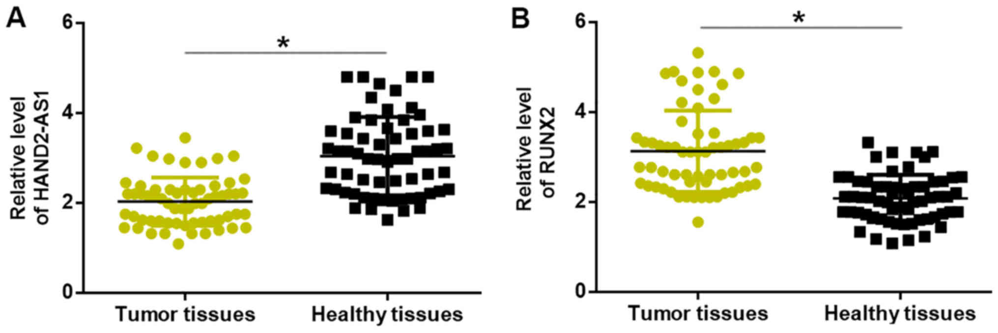
Compared with levels in adjacent healthy tissues,
expression levels of lncRNA HAND2-AS1 were significantly
downregulated (Fig. 1A), whilst
those of RUNX2 mRNA were significantly upregulated (Fig. 1B), in the cancerous tissues of
patients with TNBC.
Expression levels of lncRNA HAND2-AS1
and RUNX2 are correlated in tumor tissues, but not in paired
healthy tissues
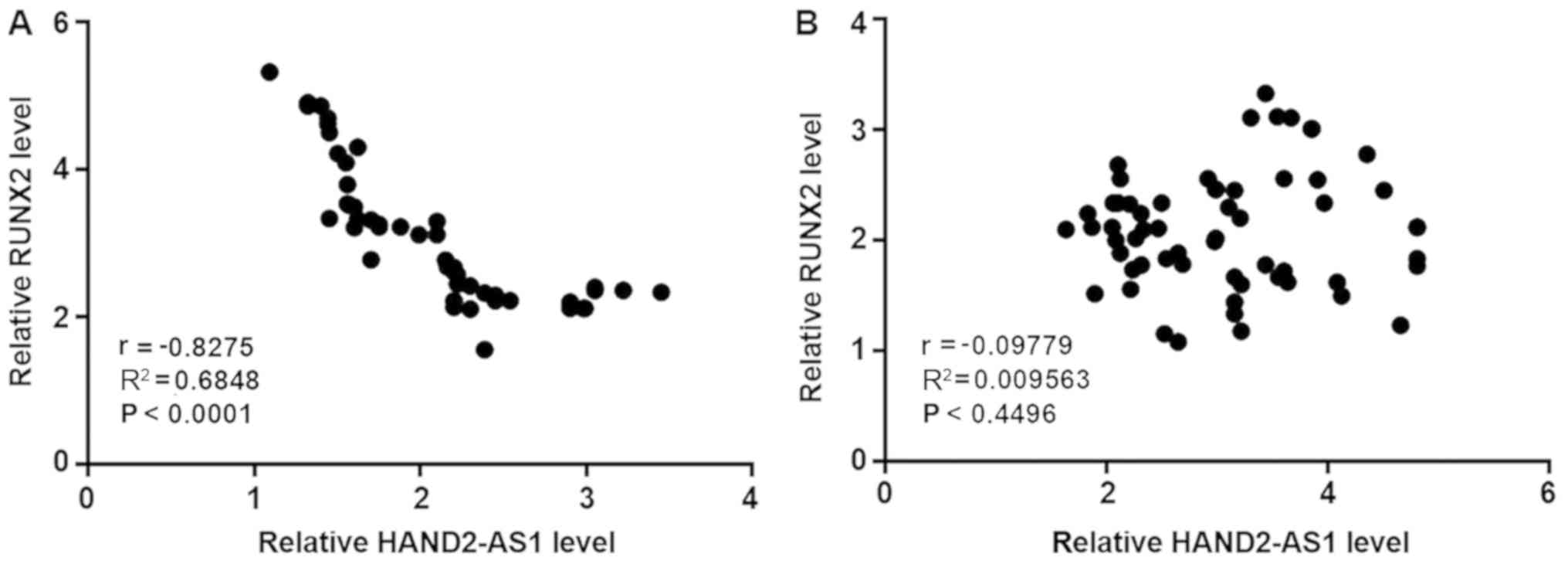
Pearson's correlation coefficient analysis revealed
an inverse correlation between the expression levels of lncRNA
HAND2-AS1 and RUNX2 in the tumor tissues of patients with TNBC
(Fig. 2A). By contrast, expression
levels of lncRNA HAND2-AS1 and RUNX2 were not significantly
correlated in paired healthy tissues (Fig. 2B).
Downregulation of lncRNA HAND2-AS1 in
the plasma distinguishes patients with TNBC from healthy
controls
Compared with healthy controls, plasma levels of
lncRNA HAND2-AS1 were significantly reduced in patients with TNBC
(Fig. 3A). ROC curve analysis was
performed to evaluate the diagnostic value of plasma lncRNA
HAND2-AS1 for TNBC. As illustrated in Fig. 3B, downregulation of plasma lncRNA
HAND2-AS1 distinguished patients with TNBC from healthy
controls.
lncRNA HAND2-AS1 overexpression
inhibits the expression of RUNX2
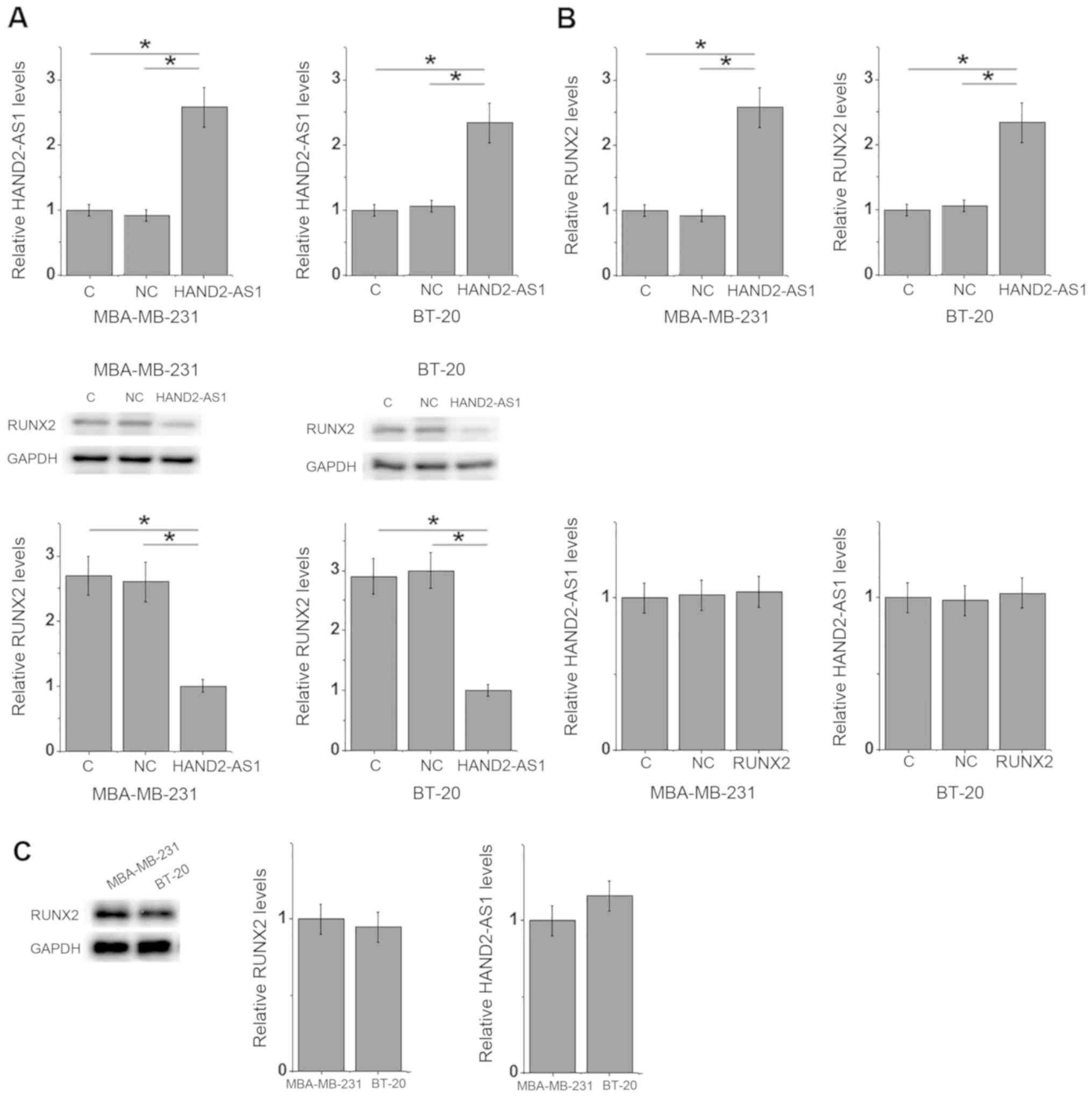
The expression level of lncRNA HAND2-AS1 was
inversely correlated with that of RUNX2 in TNBC tumor tissues.
Furthermore, compared with the negative control and control groups,
lncRNA HAND2-AS1 transfection significantly downregulated RUNX2
expression in the MDA-MB-231 and BT-20 TNBC breast cancer cell
lines (Fig. 4A). By contrast, RUNX2
transfection had no significant impact on lncRNA HAND2-AS1
expression in either cell line (Fig.
4B). No significant differences in the expression levels of
HAND2-AS1 and RUNX2 were observed between MDA-MB-231 and BT-20 TNBC
cells (Fig. 4C).
lncRNA HAND2-AS1 overexpression
reduces cancer cell proliferation, potentially by inhibiting RUNX2
expression
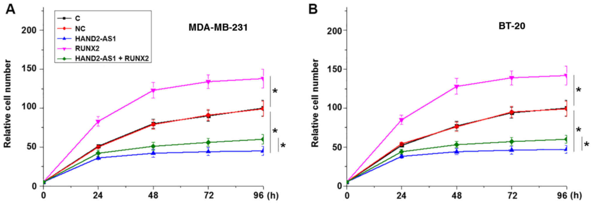
The CCK-8 assay results revealed that lncRNA
HAND2-AS1 transfection inhibited, while RUNX2 transfection
promoted, the proliferation of MDA-MB-231 (Fig. 5A) and BT-20 TNBC cells (Fig. 5B) when compared with the negative
control and control groups. In addition, RUNX2 overexpression
partially reversed the effects of lncRNA HAND2-AS1 overexpression
on cancer cells.
Discussion
Previous studies have characterized the
functionality of lncRNA HAND2-AS1, a tumor suppressor gene in
osteosarcoma (14), colorectal
cancer (15) and endometrioid
endometrial carcinoma (16).
However, the involvement of lncRNA HAND2-AS1 in TNBC, a subtype of
breast cancer, is unknown. The present study revealed that lncRNA
HAND2-AS1 may also possess a tumor suppression role in TNBC by
inhibiting cancer cell proliferation. The actions of lncRNA
HAND2-AS1 in TNBC are potentially influenced by the inhibition of
RUNX2 expression.
An early diagnosis of cancer is critical for the
successful surgical resection of tumor tissues prior to metastasis,
which is the principal cause of mortality among patients with
breast cancer (19). The present
study included TNBC patients at cancer stages I and II, which are
considered to be the early stages of cancer. Differential gene
expression in tumor and adjacent healthy tissues indicates the
involvement of certain genes in cancer. As a tumor suppressor, the
expression of lncRNA HAND2-AS1 has been reported to decrease in
osteosarcoma (14), colorectal
cancer (15) and endometrioid
endometrial carcinoma (16). In the
present study, significantly downregulated expression of lncRNA
HAND2-AS1 was observed in tumor tissues compared with that in the
adjacent healthy tissues of patients with TNBC. In addition, plasma
expression levels of lncRNA HAND2-AS1 were also significantly lower
in patients with TNBC compared with those in healthy females. The
downregulation of lncRNA HAND2-AS1 was used to distinguish patients
with TNBC from healthy controls; therefore, lncRNA HAND2-AS1 may
act as tumor suppressor in TNBC, and the detection of circulating
lncRNA HAND2-AS1 may provide guidance for the early diagnosis of
the disease.
As an oncogene, RUNX2 is frequently upregulated
during the development of human cancer (20,21).
Consistent with previous studies, the present study also
illustrated significantly upregulated expression of RUNX2 in tumor
tissues compared with that in adjacent healthy tissues in patients
with TNBC. Furthermore, an inverse correlation between the
expression level of lncRNA HAND2-AS1 and RUNX2 in tumor tissues was
observed. It was also revealed that lncRNA HAND2-AS1 may serve as
an upstream regulator of RUNX2 in cancer cell proliferation; lncRNA
HAND2-AS1 overexpression inhibited RUNX2 expression, although RUNX2
overexpression did not significantly affect HAND2-AS1 expression.
Additionally, RUNX2 overexpression partially reversed the
inhibitory effect of HAND2-AS1 overexpression in TNBC cells.
Therefore, lncRNA HAND2-AS1 may serve as a potential therapeutic
target for TNBC by inhibiting the expression of RUNX2.
It is worth noting that no correlations between
lncRNA HAND2-AS1 and RUNX2 expression levels were observed in
adjacent healthy tissues, indicating the indirect action of lncRNA
HAND2-AS1 on RUNX2 expression. In addition, lncRNA HAND2-AS1
overexpression had no significant effect on cancer cell migration
and invasion (data not shown), suggesting that lncRNA HAND2-AS1 may
specifically participate in cell proliferation in TNBC.
lncRNA HAND2-AS1 is involved in different types of
cancer through interactions with different signaling molecules,
including miRNAs, HIF1α and neuromedin (14–16),
indicating that lncRNA HAND2-AS1 may participate in different
pathological processes between diseases. However, the present study
failed to establish lncRNA HAND2-AS1 siRNA silencing in TNBC cells,
which is a consideration for future studies.
In conclusion, lncRNA HAND2-AS1 may serve as a tumor
suppressor gene in TNBC by inhibiting cancer cell proliferation
through the downregulation of RUNX2.
Acknowledgements
Not applicable.
Funding
No funding was received.
Availability of data and materials
The datasets used and/or analyzed during the current
study are available from the corresponding author on reasonable
request.
Authors' contributions
MW and ZW designed the experiments. MW and LL
performed the experiments and analyzed the data. ZW drafted the
manuscript and all authors approved the manuscript for
publication.
Ethics approval and consent to
participate
The present study was approved by the Ethics
Committee of the International Peace Maternity and Child Health
Hospital, and all patients and healthy volunteers provided written
informed consent.
Patient consent for publication
Not applicable.
Competing interests
The authors declare that they have no competing
interests.
References
|
1
|
Zubeda S, Kaipa PR, Shaik NA, Mohiuddin
MK, Vaidya S, Pavani B, Srinivasulu M, Latha MM and Hasan Q:
Her-2/neu status: A neglected marker of prognostication and
management of breast cancer patients in India. Asian Pac J Cancer
Prev. 14:2231–2235. 2013. View Article : Google Scholar : PubMed/NCBI
|
|
2
|
Podo F, Buydens L, Degani H, Hilhorst R,
Klipp E, Gribbestad IS, Van Huffel S, van Laarhoven HW, Luts J,
Monleon D, et al: Triple-negative breast cancer: Present challenges
and new perspectives. Mol Oncol. 4:209–229. 2010. View Article : Google Scholar : PubMed/NCBI
|
|
3
|
Zhang W, Wan YW, Allen GI, Pang K,
Anderson ML and Liu Z: Molecular pathway identification using
biological network-regularized logistic models. BMC Genomics. 14
(Suppl 8):S72013. View Article : Google Scholar
|
|
4
|
Lehmann BD, Jovanović B, Chen X, Estrada
MV, Johnson KN, Shyr Y, Moses HL, Sanders ME and Pietenpol JA:
Refinement of triple-negative breast cancer molecular subtypes:
Implications for neoadjuvant chemotherapy selection. PLoS One.
11:e01573682016. View Article : Google Scholar : PubMed/NCBI
|
|
5
|
Collignon J, Lousberg L, Schroeder H and
Jerusalem G: Triple-negative breast cancer: Treatment challenges
and solutions. Breast Cancer (Dove Med Press). 8:93–107.
2016.PubMed/NCBI
|
|
6
|
Denkert C, Liedtke C, Tutt A and von
Minckwitz G: Molecular alterations in triple-negative breast
cancer-the road to new treatment strategies. Lancet. 389:2430–2442.
2017. View Article : Google Scholar : PubMed/NCBI
|
|
7
|
Mayer IA, Abramson VG, Lehmann BD and
Pietenpol JA: New strategies for triple-negative breast
cancer-deciphering the heterogeneity. Clin Cancer Res. 20:782–790.
2014. View Article : Google Scholar : PubMed/NCBI
|
|
8
|
Lucero CM, Vega OA, Osorio MM, Tapia JC,
Antonelli M, Stein GS, van Wijnen AJ and Galindo MA: The
cancer-related transcription factor Runx2 modulates cell
proliferation in human osteosarcoma cell lines. J Cell Physiol.
228:714–723. 2013. View Article : Google Scholar : PubMed/NCBI
|
|
9
|
Javed A, Barnes GL, Pratap J, Antkowiak T,
Gerstenfeld LC, van Wijnen AJ, Stein JL, Lian JB and Stein GS:
Impaired intranuclear trafficking of Runx2 (AML3/CBFA1)
transcription factors in breast cancer cells inhibits osteolysis in
vivo. Proc Natl Acad Sci USA. 102:1454–1459. 2005. View Article : Google Scholar : PubMed/NCBI
|
|
10
|
Pratap J, Wixted JJ, Gaur T, Zaidi SK,
Dobson J, Gokul KD, Hussain S, van Wijnen AJ, Stein JL, Stein GS
and Lian JB: Runx2 transcriptional activation of Indian Hedgehog
and a downstream bone metastatic pathway in breast cancer cells.
Cancer Res. 68:7795–7802. 2008. View Article : Google Scholar : PubMed/NCBI
|
|
11
|
Taipaleenmäki H, Browne G, Akech J, Zustin
J, van Wijnen AJ, Stein JL, Hesse E, Stein GS and Lian JB:
Targeting of Runx2 by miR-135 and miR-203 impairs progression of
breast cancer and metastatic bone disease. Cancer Res.
75:1433–1444. 2015. View Article : Google Scholar : PubMed/NCBI
|
|
12
|
Chai J, Guo D, Ma W, Han D, Dong W, Guo H
and Zhang Y: A feedback loop consisting of
RUNX2/lncRNA-PVT1/miR-455 is involved in the progression of
colorectal cancer. Am J Cancer Res. 8:538–550. 2018.PubMed/NCBI
|
|
13
|
Yu X, Zhao J and He Y: Long non-coding RNA
PVT1 functions as an oncogene in human colon cancer through
miR-30d-5p/RUNX2 axis. J BUON. 23:48–54. 2018.PubMed/NCBI
|
|
14
|
Kang Y, Zhu X, Xu Y, Tang Q, Huang Z, Zhao
Z, Lu J, Song G, Xu H, Deng C and Wang J: Energy stress-induced
lncRNA HAND2-AS1 represses HIF1α-mediated energy metabolism and
inhibits osteosarcoma progression. Am J Cancer Res. 8:526–537.
2018.PubMed/NCBI
|
|
15
|
Zhou J, Lin J, Zhang H, Zhu F and Xie R:
lncRNA HAND2-AS1 sponging miR-1275 suppresses colorectal cancer
progression by upregulating KLF14. Biochem Biophys Res Commun.
503:1848–1853. 2018. View Article : Google Scholar : PubMed/NCBI
|
|
16
|
Yang X, Wang CC, Lee WYW, Trovik J, Chung
TKH and Kwong J: Long non-coding RNA HAND2-AS1 inhibits invasion
and metastasis in endometrioid endometrial carcinoma through
inactivating neuromedin U. Cancer Lett. 413:23–34. 2018. View Article : Google Scholar : PubMed/NCBI
|
|
17
|
Giuliano AE, Connolly JL, Edge SB,
Mittendorf EA, Rugo HS, Solin LJ, Weaver DL, Winchester DJ and
Hortobagyi GN: Breast cancer-major changes in the American joint
committee on cancer eighth edition cancer staging manual. CA Cancer
J Clin. 67:290–303. 2017. View Article : Google Scholar : PubMed/NCBI
|
|
18
|
Livak KJ and Schmittgen TD: Analysis of
relative gene expression data using real-time quantitative PCR and
the 2(-Delta Delta C(T)) method. Methods. 25:402–408. 2001.
View Article : Google Scholar : PubMed/NCBI
|
|
19
|
Cristofanilli M, Budd GT, Ellis MJ,
Stopeck A, Matera J, Miller MC, Reuben JM, Doyle GV, Allard WJ,
Terstappen LW and Hayes DF: Circulating tumor cells, disease
progression, and survival in metastatic breast cancer. N Engl J
Med. 351:781–791. 2004. View Article : Google Scholar : PubMed/NCBI
|
|
20
|
Pratap J, Imbalzano KM, Underwood JM,
Cohet N, Gokul K, Akech J, van Wijnen AJ, Stein JL, Imbalzano AN,
Nickerson JA, et al: Ectopic runx2 expression in mammary epithelial
cells disrupts formation of normal acini structure: Implications
for breast cancer progression. Cancer Res. 69:6807–6814. 2009.
View Article : Google Scholar : PubMed/NCBI
|
|
21
|
Sancisi V, Manzotti G, Gugnoni M, Rossi T,
Gandolfi G, Gobbi G, Torricelli F, Catellani F, Faria do Valle I,
Remondini D, et al: RUNX2 expression in thyroid and breast cancer
requires the cooperation of three non-redundant enhancers under the
control of BRD4 and c-JUN. Nucleic Acids Res. 45:11249–11267. 2017.
View Article : Google Scholar : PubMed/NCBI
|