Introduction
Desmoid tumors (DTs), also known as aggressive
fibromatosis, are rare, locally infiltrative mesenchymal neoplasms
that are associated with high rates of local recurrence, but do not
possess the potential to metastasize (1). DTs account for <3% of all soft
tissue tumors (2) and have an
estimated incidence rate of 2–5 individuals per million per year in
Europe (3). The exact etiology of
DTs is currently unknown, but hormonal, genetic and physical
factors all serve roles in their development and growth. Two
different types of DTs have been described: Sporadic tumors and
familial adenomatous polyposis (FAP)-associated DTs (4). Regardless of type, an important part of
the biology is the disruption of the Wnt signaling pathway, leading
to the accumulation of nuclear β-catenin and the inappropriate
stimulation of downstream genes (4).
In sporadic DT, this is often caused by a mutation in the β-catenin
gene (CTNNB1) (5), whereas in
FAP-associated DT, the loss of adenomatous polyposis coli
(APC) function serves a role (6). Recently described biallelic mutations
of MutY DNA glycosylase (MUTYH) have been demonstrated to be
responsible for adenomatous polyposis with an increased risk of
colorectal cancer and have been observed in 30–40% of adenomatous
polyposis cases in which an APC mutation was not identified;
however, no evidence has indicated thus far that MUTYH
serves a role in DT (7). Further
studies have revealed that the mutation site of APC, as well
as that of CTNNB1, may impact the incidence, severity and
prognosis of DT (8–10). This may be significant for the
prevention and treatment of DTs.
DT can occur in any location, often involving the
extremities, the trunk, including the pelvic and shoulder girdles,
and the abdomen (4). For abdominal
DTs, the clinical presentation depends on its primary site. In the
present study, according to its location, abdominal DTs are
classified into two types: Peripheral and central DT. The
clinicopathological and molecular characteristics were collected
from a group of patients with abdominal DTs and analyzed to explore
the optimal strategies for diagnosing and management of different
types of abdominal DTs.
Materials and methods
Study design, patients and
samples
Tumor samples, peripheral blood samples and matched
clinical data from 15 patients with DT were retrospectively
collected from the Zhongshan Hospital (Shanghai, China) with
informed consent. The present study was approved by the Committee
for Ethical Review of Research Involving Human Subjects of
Zhongshan Hospital Affiliated with Fudan University. All 15
patients received surgery and were diagnosed with DT consecutively
between July, 2009 and January, 2016; no patients were excluded
from the study. DTs were diagnosed by an experienced pathologist
based on tumor morphology and immunohistochemistry. The expression
levels of APC, CTNNB1 and MUTYH genes of 15 patients
were analyzed by direct sequencing of tumor and blood samples. The
clinicopathological data and follow-up outcomes were collected and
analyzed.
Data retrieved included age, sex, date of surgery,
anatomic site, tumor size and margin status. The results of the
APC, CTNNB1 and MUTYH mutational analysis were
recorded along with the type of treatments administered prior to
and/or following surgery. Patients were followed to record any
future recurrence of disease with the date of recurrence, treatment
following recurrence, date of the last follow-up and status at last
follow-up. According to the anatomic site, abdominal DTs were
classified as either peripheral (derived from the abdominal wall)
or central (derived from the retroperitoneal space and mesentery).
Tumor size was defined as the greatest DT dimension in the surgical
specimen reported by the original pathologists. Surgical excisions
were considered macroscopically complete in the absence of gross
residual disease. All macroscopically complete resections were
classified according to the closest surgical margin, which was
microscopically categorized as positive (R1, tumor within 1 mm from
the inked surface) or negative (R0, the absence of tumor within 1
mm from the inked surface). Non-surgical treatments were
administered in the primary or recurrent phase of the disease on an
individualized basis. These included radiotherapy, chemotherapy
(methotrexate and vinorelbine in the majority of cases), medical
therapy/hormone agents (tamoxifen, toremifene) and non-steroidal
anti-inflammatory drugs (cyclooxygenase-2 inhibitors).
Telephone and outpatient follow-up were used to
collect the data. The median follow-up time following the diagnosis
of DT was 60 months (range, 20–90 months), and all follow-up data
were complete.
Mutational analysis
Genomic DNA was extracted from peripheral blood
using a TIANamp Blood DNA kit (Tiangen Biotech Co., Ltd.) and tumor
samples using a TIANamp Genomic DNA kit (Tiangen Biotech Co.,
Ltd.). Primers flanking all coding exons and intron-exon boundaries
of APC, CTNNB1 and MUTYH were designed using Primer
Premier (version 5.0; Premier Biosoft) and are presented in
Table SI. Genomic DNA samples were
amplified using PCR. The thermocycling conditions were as follows:
Denaturation at 94°C for 5 min; 31 cycles of denaturation at 94°C
for 30 sec, annealing for 30 sec (temperature was set according to
the primers of each fragment) and an extension at 72°C for 1 min. A
final extension step was performed at 4°C for 5 min, and the
experiment was repeated 10–20 times. The PCR products were
evaluated by a 2% agarose gel electrophoresis and were further
purified using an AxyPrep DNA Gel Extraction kit (Corning Life
Sciences) according to the manufacturer's protocol. Sanger
sequencing was subsequently performed using an ABI PRISM 3730
automated sequencer (Applied Biosystems; Thermo Fisher Scientific,
Inc.). Sequencing results were analyzed by Geneious software
(version 5.6.7; Biomatters Ltd.). An identified mutation was
verified in the corresponding region of the unaffected parents of
the proband and 100 population-matched healthy controls. The
mutation was described by comparison with the NCBI cDNA reference
sequences (11) NM_001354897.1 for
APC, NM_001098209.1 for CTNNB1 and NM_012222.2 for MUTYH.
Statistical analysis
All statistical analyses were performed using SPSS
software (version 18.0; SPSS, Inc.). The primary endpoint was the
time to progress (TTP) and overall survival (OS), and the event
time was calculated as the time between the date of surgery and the
date of relapse or death, whichever occurred first, or was censored
at the date of last follow-up assessment in event-free patients.
TTP curves were estimated using the Kaplan-Meier method and were
compared statistically using the log-rank test. Multivariate Cox
model analyses were also performed using following characteristics:
Sex, tumor site, tumor size and margin status. A Fisher's exact
test was performed to analyze the differences between sporadic DT
and FAP-associated DT. P<0.05 was considered to indicate a
statistically significant difference.
Results
Patient demographics and disease
characteristics
Between July, 2009 and January, 2016, 15 patients,
including nine female and six male patients, were enrolled in the
present study. The demographic and disease characteristics are
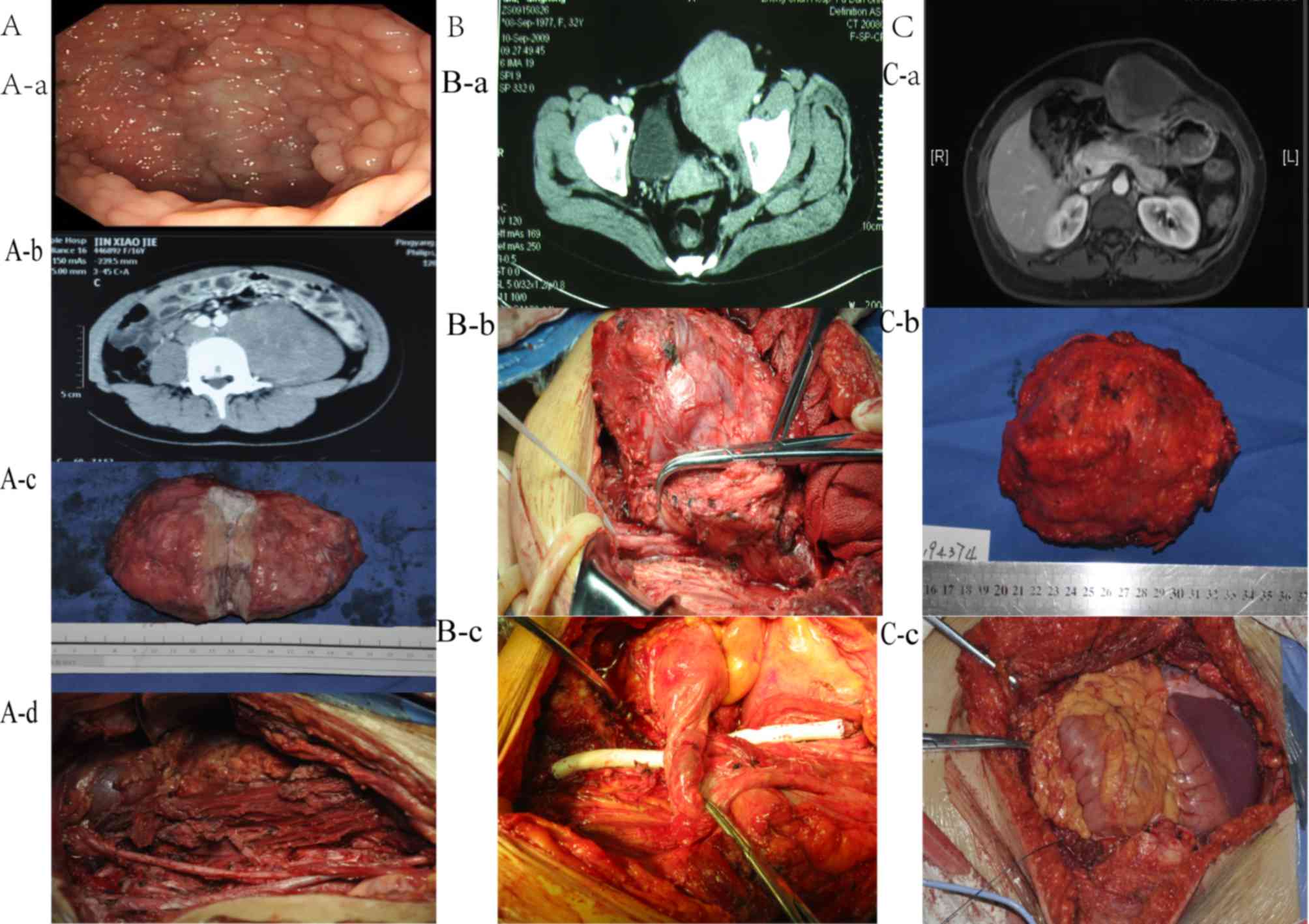
presented in Table I and Fig. 1. In total, nine patients were ≥30
years, and the median age was 32 years (range, 26–47 years).
According to the primary site of DT, the central type accounted for
80% (12/15), whereas the peripheral type accounted for 20% (3/15)
of the cases. The median size of the primary lesions was 10 cm
(range, 4–17 cm). Of the 15 cases, nine were sporadic (four male
and five female patients), whereas six patients had a family
history of FAP and a history of abdominal injury, including
surgery.
 | Table I.Clinical characteristics of 15
patients with DT. |
Table I.
Clinical characteristics of 15
patients with DT.
|
Characteristics | Cases, n (%) |
|---|
| Type of DT |
|
|
FAP-associated | 6 (42.86) |
|
Sporadic | 9 (57.14) |
| Age, years |
|
|
<30 | 6 (42.86) |
|
≥30 | 9 (57.14) |
| Sex |
|
|
Male | 6 (42.86) |
|
Female | 9 (57.14) |
| History of
abdominal surgery |
|
|
Yes | 6 (42.86) |
| No | 9 (57.14) |
| Site |
|
|
Central | 12 (80) |
|
Peripheral | 3
(20) |
| Tumor size |
|
|
<10 | 5 (33.33) |
| ≥10
cm | 10 (66.67) |
| Margin status for
primary surgery |
|
|
R0/R1 | 9 (60) |
| R2 | 6 (40) |
| Recurrence or
progression after primary surgery |
|
| No | 3 (20) |
|
Yes | 12 (80) |
| Treatment after
primary surgery |
|
|
Radiotherapy | 2 (13.33) |
|
Chemotherapy | 6 (40) |
| Other
(COX-2I, TAM or wait and see) | 7 (46.67) |
| Type of treatment
for recurrence |
|
|
Surgery | 6 |
|
Radiotherapy | 3 |
|
Chemotherapy | 6 |
| Other
(COX-2I, TAM or none) | 8 |
All the patients accepted surgical resection as a
treatment course, with a number of patients undergoing multiple
surgeries due to recurrence. Overall, nine patients underwent
complete surgical excision (R0/R1), and six patients underwent R2
resection. Due to variations in the anatomic site of the tumor, the
extent of surgery was different for each case. The three patients
with peripheral-type sporadic DTs underwent R0/R1 resection without
combined organ resection. Among the 12 patients with central type
DTs, 50% (6/12) underwent R0/R1 resection, whereas 50% (6/12)
underwent R2 resection. For the central type group, combined
evisceration was performed in 75% (9/12) of patients, and the
resected organs included the small intestine (66.67%, 6/9), colon
(33.33%, 3/9), ureter (22.22%, 2/9), appendix (11.11%, 1/9), ovary
(11.11%, 1/9) and pancreas (11.11%, 1/9). Following primary
surgery, only three patients did not experience recurrence or
progression. In addition, six patients who underwent R2 resection
received chemotherapy, two patients received radiotherapy and seven
patients received Tamoxifen, a non-steroid anti-inflammatory drug
(NSAID; COX-2 inhibitor) and/or waited for the outcome. Of patients
who experienced recurrence and progression following primary
resection, six cases underwent additional surgery, three received
radiotherapy and six received chemotherapy; eight patients received
Tamoxifen, NSAID (COX2 inhibitor) or waited for the outcome.
Difference between sporadic and
FAP-associated DT
To reveal the differences between sporadic and
FAP-associated DT, clinicopathological characteristics between the
two groups were compared. The results revealed a significant
difference between the two groups in past history of abdominal
surgery, with patients with FAP-associated DT exhibiting a higher
rate of abdominal surgery. This may suggest an increased
susceptibility to the development of DT in patients with FAP that
had a history of abdominal surgery. No significant differences were
identified in the other characteristics (Table II).
| Table II.Differences between sporadic and
FAP-associated desmoid tumors. |
Table II.
Differences between sporadic and
FAP-associated desmoid tumors.
|
Characteristics | FAP-associated,
n | Sporadic, n | Fisher's exact test
P-value |
|---|
| Sex |
|
|
|
|
Male | 2 | 4 | >0.999 |
|
Female | 4 | 5 |
|
| Age, years |
|
|
|
|
20–30 | 3 | 3 | 0.622 |
|
≥30 | 3 | 6 |
|
| History of
abdominal surgery |
|
|
|
|
Yes | 5 | 2 | 0.011a |
| No | 1 | 7 |
|
| Site |
|
|
|
|
Central | 6 | 6 | 0.229 |
|
Peripheral | 0 | 3 |
|
| Tumor size, cm |
|
|
|
|
<10 | 1 | 4 | 0.580 |
|
≥10 | 5 | 5 |
|
| Margin status |
|
|
|
|
R0/R1 | 4 | 7 | 0.136 |
| R2 | 2 | 2 |
|
| Recurrence after
R0/R1 resection |
|
|
|
|
Yes | 4 | 4 | 0.236 |
| No | 0 | 3 |
|
Mutations in the study cohort
Among the 15 patients with DT, only one patient did
not exhibit any mutations in CTNNB1, APC or MUTYH. Of
the six patients with FAP-associated DT, one patient harbored a
MUTYH gene mutation, whereas the others possessed APC
gene mutations. In the nine patients with sporadic DT, analysis of
the tumor samples revealed six mutations in the CTNNB1 gene,
four mutations in the APC gene and one mutation in the
MUTYH gene. In the group of patients with sporadic DT, one
harbored mutations in CTNNB1, APC and MUTYH genes,
whereas another carried CTNNB1 and APC mutations
(Table III).
| Table III.Mutations identified in APC,
CTNNB1 and MUTYH in the present study. |
Table III.
Mutations identified in APC,
CTNNB1 and MUTYH in the present study.
| No. | Incidence | Gene | Location | Mutation | Mutation type |
|---|
| 1 | Familial | APC | E15 |
c.1875_1878delGACA | Frameshift |
| 2 | Familial | APC | E09 |
c.904C>T:p.R302X | Missense |
| 3 | Familial | MUTYH | E15 |
c.1005G>C:p.Q335H | Missense |
| 4 | Familial | APC | E16-9 |
c.3927_3931delAAAGA | Frameshift |
| 5 | Familial | APC | E16 |
c.3927-3931delAAAGA | Frameshift |
| 6 | Familial | APC | E16-10 |
c.4429C>T:p.Q1477X | Missense |
| 7 | Sporadic | APC | E16 |
c.5465T>A:p.V1822D, c.1635G>A | Missense,
synonymous |
| 8 | Sporadic | APC |
| c.1458T>C,
c.1635G>A | Synonymous |
| 9 | Sporadic | CTNNB1 | E3 |
c.133T>C:p.S45P | Missense |
| 10 | Sporadic | APC |
| c.1458T>C,
c.1488A>T, c.1635G>A | Synonymous |
|
|
| CTNNB1 | E15 | c.2340C>T | Synonymous |
|
|
| MUTYH | E12 | c.1005 G>C | Synonymous |
| 11 | Sporadic |
|
| None |
|
| 12 | Sporadic | APC |
| c.1458T>C,
c.1635G>A | Synonymous |
|
|
| CTNNB1 | E15 | c.2340 C>T | Synonymous |
| 13 | Sporadic | CTNNB1 | E3 |
c.134C>T:p.S45F | Missense |
| 14 | Sporadic | CTNNB1 | E3 |
c.121:A>G:p.T41A | Missense |
| 15 | Sporadic | CTNNB1 | E3 |
c.121:A>G:p.T41A | Missense |
Survival analysis
In the follow-up to December 2016, two patients with
FAP-associated DT succumbed to disease, six patients survived with
progression and seven exhibited disease-free survival. The median
TTP was 13 months, and median OS time was 57 months. Survival
analysis revealed that patients with FAP-associated DT exhibited
shorter TTP (P=0.030). Log-rank test revealed a tendency for
shorter TTP in the cases that underwent R2 resection (P=0.072) and
poor prognosis in the cases with FAP-associated DT (P=0.087)
(Table IV; Fig. 2). In the multivariate Cox regression
analysis, FAP-associated DT was revealed to be associated with a
higher risk of recurrence following resection (Table V).
| Table IV.Univariate analysis of TPP and
OS. |
Table IV.
Univariate analysis of TPP and
OS.
| A, Univariate
analysis of TTP |
|---|
|
|---|
|
| Log-rank test |
|---|
|
|
|
|---|
| Variables | χ2 | P-value |
|---|
| Sex (male vs.
female) | 1.401 | 0.236 |
| Age, years (20–30
vs. ≥30) | 0.242 | 0.623 |
| Tumor size, cm
(<10 vs. ≥10) | 0.787 | 0.375 |
| Site (central vs.
peripheral) | 2.658 | 0.103 |
| Margin status
(R0/R1 vs. R2) | 3.227 | 0.072a |
| FAP-associated (yes
vs. no) | 4.684 | 0.030a |
| Systematic therapy
(yes vs. no) | 0.003 | 0.959 |
|
| B, Univariate
analysis of OS |
|
|
| Log-rank
test |
|
|
|
|
Variables |
χ2 | P-value |
|
| Sex (male vs.
female) | 1.606 | 0.205 |
| Age, years (20–30
vs. ≥30) | 0.072 | 0.788 |
| Tumor size, cm
(<10 vs. ≥10) | 0.143 | 0.705 |
| Site (central vs.
peripheral) | 0.573 | 0.449 |
| Margin status
(R0/R1 vs. R2) | 0.072 | 0.788 |
| FAP-associated (yes
vs. no) | 2.925 | 0.087 |
| Systematic therapy
(yes vs. no) | 1.180 | 0.277 |
| Table V.Cox regression analysis of risk
factors for recurrence. |
Table V.
Cox regression analysis of risk
factors for recurrence.
| Risk factor | HR | 95% CI | P-value |
|---|
| Sex | 1.404 | 0.188–10.474 | 0.740 |
| Age | 1.015 | 0.151–6.821 | 0.998 |
| Site | 0.090 | 0.003–2.297 | 0.145 |
| FAP-associated | 0.086 | 0.006–1.283 | 0.075a |
| Margin status | 0.410 | 0.062–2.703 | 0.354 |
| Systematic
therapy | 15.367 | 0.325–726.452 | 0.165 |
Discussion
DTs are heterogenous borderline lesions that
originate from mesenchymal fibroblastic proliferation with an
incidence rate of 0.03% of all neoplasms and 3.00% of soft tissue
tumors (12). It is a rare condition
that typically infiltrates surrounding structures and tends to
recur with minimal metastatic potential (12). The majority of DTs occur
sporadically, but a number of DTs are associated with FAP syndrome
(13). In the present study, when
clinicopathological characteristics were compared between the
sporadic and FAP-associated groups, the history of abdominal
surgery and prognosis were notably different. Among the 15
patients, all patients with FAP-associated DTs had a history of
abdominal surgery. Clark et al (14) reported that 82% of patients with
FAP-associated DTs had predisposing surgery. It has been
hypothesized that surgical trauma and the process of tissue repair
by fibroblasts contribute to DT development following injury. The
risk of tumor development among patients with FAP following injury
or surgical trauma remains unknown.
Among patients with FAP, DT is one of the most
common causes of death after colorectal cancer (15). Due to the high local recurrence rate,
recurrence and survival are of equal importance in the management
of DT. In the present study, 2 patients with FAP-associated DTs
succumbed to complications associated with the tumor. Survival
analysis revealed that patients with FAP-associated DT exhibited
shorter TTP and a relatively poor prognosis compared with those
with sporadic DTs. These results suggest a more aggressive
pathology and poor prognosis in patients with FAP-associated DT,
but more evidence is required to support this hypothesis.
Alterations in the Wnt/β-catenin signaling pathway
have been reported as the likely driving mechanism of tumorigenesis
in the development of the disease; mutations in the β-catenin gene
occur in the majority of sporadic cases, as well as mutations in
APC, which regulates β-catenin degradation in FAP-associated
DTs (16). In the present study,
APC, CTNNB1 and MUTYH mutational analyses were
performed in 15 patients, and only one patient did not exhibit any
mutations in these genes. Of the six patients with FAP-associated
DT, five harbored APC mutations, of which 80% were located
downstream from codon 1444. Previous studies have reported that
FAP-associated DTs are associated with mutations toward the 3′ end
of the gene, distal to codon 1444, compared with the general FAP
population, where 90% of mutations are located upstream from codon
1444 (17,18). A novel finding from the present study
was that one patient with FAP-associated DT exhibited MUTYH
mutations within the tumor tissues. Mutations in the MUTYH gene
have been previously demonstrated in a type of colorectal polyposis
referred to as MUTYH-associated polyposis (MAP), which has a
relatively mild phenotype, exhibiting <100 adenomas per patient
and an average age at diagnosis of 45 years (19). In certain patients with MAP, the
extracolonic manifestations can also be demonstrated, but DTs are
not part of the MAP tumor spectrum (20). To date, MUTYH mutations in
FAP-associated DTs have not been reported in published literature
or in any database. The mutation c.1005G>C has also not yet been
reported. Whether it is meaningless or functionally associated with
the development of DT will require further investigation. In tumors
of the nine patients with sporadic DT, six mutations in the
CTNNB1 gene, four mutations in the APC gene and one
mutation in the MUTYH gene were identified. Three particular
mutations in the CTNNB1 gene have been reported (and
associated) with DT: T41A, S45F and S45P (6). A number of studies have demonstrated a
significantly increased risk of disease recurrence in patients
harboring an S45F mutation compared with those who had either
tumors with a T41A mutation of WT tumors (21,22);
however, the association between genotype and phenotype has not yet
been determined. In the present study, two T41A, one S45F and one
S45P mutations were identified in CTNNB1. However, due to
the small sample size of the present study, the association between
particular gene mutations and prognosis could not be confirmed.
Due to the lack of prospective cohort studies for
rare conditions such as DTs, the optimal treatment remains to be
elucidated. Surgery is the primary treatment for patients with
resectable DTs (23). According to
the results of the present study, resection of central-type DTs is
more difficult compared with that of peripheral-type DTs due to
organ involvement. Although margin status remains a controversial
topic in the management of DTs (24,25), the
present study demonstrated a tendency towards shorter TTP in the
patients that underwent R2 resection, which may be due to the small
sample size. DTs behave in an unpredictable manner, as they may
remain stable, grow aggressively or even regress without
intervention (26,27). Due to the heterogeneity,
unpredictable natural biology and high recurrence rate of DTs, the
benefit of aggressive treatment modalities remains unclear. A move
towards conservative management strategies has emerged over the
past decade, including hormonal therapy, NSAIDs, chemotherapy and a
‘wait and see’ approach. In the present study, following primary
surgery, seven patients who accepted R0/R1 or R2 resection,
received Tamoxifen, NSAID (COX2 inhibitor) and/or waited for the
outcome remained alive by the end of the follow-up period. Bonvalot
et al (28) revealed that
patients with extra-abdominal DT with a microscopically complete
surgery had a similar outcome compared with patients in a
no-surgery group. However, abdominal DT has different
characteristics, particularly for retroperitoneal and
intra-abdominal cases, and may lead to severe morbidity, functional
impairment and even mortality due to its anatomic site (12). In certain cases, resectable tumors
may progress to be unresectable or involve additional viscera
(12). Therefore, it is currently
unknown when a ‘wait and see’ approach may be the best treatment
strategy. More prospective trials are required to provide a basis
for clinical management. A number of systemic therapies have been
used for DT, ranging from those with low toxicity such as hormonal
therapy or NSAIDs (29,30) to combination chemotherapy (31,32). In
the present study, hormonal therapy, NSAIDs and chemotherapy were
used alone or in combinations to treat patients with primary or
recurrent DTs, but the effect was difficult to evaluate. Potential
complications should be carefully considered when systemic therapy
is used as, In the present study, two patients experienced
gastrointestinal perforation due to chemotherapy (33).
For abdominal DTs, which are a rare condition, the
management remains controversial due to the high number of
variations, such as the incisional margin, site of tumor and
genotype. However, surgery remains the preferred treatment for DTs.
Although the present study comprised a small sample size, the
analysis revealed a number of meaningful results, such as mutations
in APC, CTNNB1 and MUTYH. To the best of our
knowledge, the present study is the first to report a patient with
MUTYH-associated FAP and an abdominal DT; abdominal DT was
prone to occur in patients with FAP who had undergone abdominal
surgery, and the prognosis of patients with FAP-associated DT was
poor. The present study may contribute to the future development of
optimal strategies for diagnosing and management of different types
of abdominal DTs.
Supplementary Material
Supporting Data
Acknowledgements
Not applicable.
Funding
No funding was received.
Availability of data and materials
The datasets used and analyzed during the present
study are available from the corresponding author on reasonable
request.
Authors' contributions
JYW, NJ, QWL, YH, YZ and HXT conceived and designed
the study. JYW and HXT drafted the manuscript. QWL, JLL, QJ and WSL
performed the mutation analysis. YFH and JL collected the samples
and performed pathological analysis. YH performed the enteroscopy.
NJ, JX and ML collected clinical data and assisted in the
follow-up. WQL and YHZ assisted in data analysis and
interpretation. YZ revised the manuscript critically. All authors
read and approved the final manuscript.
Ethics approval and consent to
participate
The study was approved by the Committee for Ethical
Review of Research Involving Human Subjects of Zhongshan Hospital
affiliated with Fudan University. Informed consent was acquired
from all patients.
Patient consent for publication
Not applicable.
Competing interests
The authors declare that they have no competing
interests.
Glossary
Abbreviations
Abbreviations:
|
DT
|
desmoid tumor
|
|
FAP
|
familial adenomatous polyposis
|
|
TTP
|
time to progress
|
|
OS
|
overall survival
|
References
|
1
|
Wu C, Nik-Amini S, Nadesan P, Stanford WL
and Alman BA: Aggressive fibromatosis (desmoid tumor) is derived
from mesenchymal progenitor cells. Cancer Res. 70:7690–7698. 2010.
View Article : Google Scholar : PubMed/NCBI
|
|
2
|
Desmoid-type fibromatoses, ; Fletcher CDM,
Bridge JA, Hogendoorn PCW and Mertens F: WHO Classification of
Tumours of Soft Tissue and Bone. 4th. IARC Press; Lyon: pp. 72–73.
2013
|
|
3
|
Penel N, Coindre JM, Bonvalot S, Italiano
A, Neuville A, Le Cesne A, Terrier P, Ray-Coquard I, Ranchere-Vince
D, Robin YM, et al: Management of desmoid tumours: A nationwide
survey of labelled reference centre networks in France. Eur J
Cancer. 58:90–96. 2016. View Article : Google Scholar : PubMed/NCBI
|
|
4
|
Lips DJ, Barker N, Clevers H and Hennipman
A: The role of APC and beta-catenin in the aetiology of aggressive
fibromatosis (desmoid tumors). Eur J Surg Oncol. 35:3–10. 2009.
View Article : Google Scholar : PubMed/NCBI
|
|
5
|
Hartley JE, Church JM, Gupta S, mcGannon E
and Fazio VW: Significance of incidental desmoids identified during
surgery for familial adenomatous polyposis. Dis Colon Rectum.
47:334–340. 2004. View Article : Google Scholar : PubMed/NCBI
|
|
6
|
Miyoshi Y, Iwao K, Nawa G, Yoshikawa H,
Ochi T and Nakamura Y: Frequent mutations in the beta-catenin gene
in desmoid tumors from patients without familial adenomatous
polyposis. Oncol Res. 10:591–594. 1998.PubMed/NCBI
|
|
7
|
Sereno M, Merino M, López-Gómez M,
Gómez-Raposo C, Zambrana Tébar F, Moreno Rubio J, Espinós J,
Martín-Algarra S and Casado Sáenz E: MYH polyposis syndrome:
Clinical findings, genetics issues and management. Clin Transl
Oncol. 16:675–679. 2014. View Article : Google Scholar : PubMed/NCBI
|
|
8
|
Couture J, Mitri A, Lagace R, Smits R,
Berk T, Bouchard HL, Fodde R, Alman B and Bapat B: A germline
mutation at the extreme 3′ end of the APC gene results in a severe
desmoids phenotype and is associated with overexpression of
beta-catenin in the desmoid tumor. Clin Genet. 57:205–212. 2000.
View Article : Google Scholar : PubMed/NCBI
|
|
9
|
Colombo C, Miceli R, Lazar AJ, Perrone F,
Pollock RE, Le Cesne A, Hartgrink HH, Cleton-Jansen AM, Domont J,
Bovée JV, et al: CTNNB1 45F mutation is a molecular prognosticator
of increased postoperative primary desmoids tumor recurrence: An
independent, multicenter validation study. Cancer. 119:3696–3702.
2013. View Article : Google Scholar : PubMed/NCBI
|
|
10
|
Van Broekhoven DL, Verhoef C, Grünhagen
DJ, van Gorp JM, den Bakker MA, Hinrichs JW, de Voijs CM and van
Dalen T: Prognostic value of CTNNB1 gene mutation in primary
sporadic aggressive fibromatosis. Ann Surg Oncol. 22:1464–1470.
2015. View Article : Google Scholar : PubMed/NCBI
|
|
11
|
CCDS Database, . https://www.ncbi.nlm.nih.gov/CCDS/CcdsBrowse.cgi
|
|
12
|
Sakorafas GH, Nissotakis C and Peros G:
Abdominal desmoid tumors. Surg Oncol. 16:131–142. 2007. View Article : Google Scholar : PubMed/NCBI
|
|
13
|
Clark SK and Phillips RK: Desmoids in
familial adenomatous polyposis. Br J Surg. 83:1494–1504. 1996.
View Article : Google Scholar : PubMed/NCBI
|
|
14
|
Clark SK, Neale KF, Landgrebe JC and
Phillips RK: Desmoid tumours complicating familial adenomatous
polyposis. Br J Surg. 86:1185–1189. 1999. View Article : Google Scholar : PubMed/NCBI
|
|
15
|
de Campos FG, Peres RO, Imperiale AR, Seid
VE, Nahas SC and Cecconello I: Evaluating causes of death in
familial adenomatous polyposis. J Gastrointest Surg. 14:1943–1949.
2010. View Article : Google Scholar : PubMed/NCBI
|
|
16
|
Skubitz KM: Biology and treatment of
aggressive fibromatosis or desmoid tumor. Mayo Clin Proc.
92:947–964. 2017. View Article : Google Scholar : PubMed/NCBI
|
|
17
|
Bertario L, Russo A, Sala P, Eboli M,
Giarola M, D'amico F, Gismondi V, Varesco L, Pierotti MA and Radice
P; Hereditary Colorectal Tumours Registry, : Hereditary Colorectal
Tumours Registry: Genotype and phenotype factors as determinants of
desmoid tumors in patients with familial adenomatous polyposis. Int
J Cancer. 95:102–107. 2001. View Article : Google Scholar : PubMed/NCBI
|
|
18
|
Lefevre JH, Parc Y, Kerneis S, Goasguen N,
Benis M, Parc R and Tiret E: Risk factors for development of
desmoid tumours in familial adenomatous polyposis. Br J Surg.
95:1136–1139. 2008. View
Article : Google Scholar : PubMed/NCBI
|
|
19
|
Out AA, Tops CM, Nielsen M, Weiss MM, van
Minderhout IJ, Fokkema IF, Buisine MP, Claes K, Colas C, Fodde R,
et al: Leiden open variation database of the MUTYH gene. Hum Mutat.
31:1205–1215. 2010. View Article : Google Scholar : PubMed/NCBI
|
|
20
|
Mazzei F, Viel A and Bignamia M: Role of
MUTYH in human cancer. Mutat Res. 743-744:33–43. 2013. View Article : Google Scholar : PubMed/NCBI
|
|
21
|
Colombo C, Miceli R, Lazar AJ, Perrone F,
Pollock RE, Le Cesne A, Hartgrink HH, Cleton-Jansen AM, Domont J,
Bovée JV, et al: CTNNB1 45F mutation is a molecular prognosticator
of increased postoperative primary desmoid tumor recurrence: An
independent, multicenter validation study. Cancer. 119:3696–3702.
2013. View Article : Google Scholar : PubMed/NCBI
|
|
22
|
Lazar AJ, Tuvin D, Hajibashi S, Habeeb S,
Bolshakov S, Mayordomo-Aranda E, Warneke CL, Lopez-Terrada D,
Pollock RE and Lev D: Specific mutations in the beta-catenin gene
(CTNNB1) correlate with local recurrence in sporadic desmoid
tumors. Am J Pathol. 173:1518–1527. 2008. View Article : Google Scholar : PubMed/NCBI
|
|
23
|
Lev D, Kotilingam D, Wei C, Ballo MT,
Zagars GK, Pisters PW, Lazar AA, Patel SR, Benjamin RS and Pollock
RE: Optimizing treatment of desmoid tumors. J Clin Oncol.
25:1785–1791. 2007. View Article : Google Scholar : PubMed/NCBI
|
|
24
|
Mullen JT, Delaney TF, Kobayashi WK,
Szymonifka J, Yeap BY, Chen YL, Rosenberg AE, Harmon DC, Choy E,
Yoon SS, et al: Desmoid tumor: Analysis of prognostic factors and
outcomes in a surgical series. Ann Surg Oncol. 19:4028–4035. 2012.
View Article : Google Scholar : PubMed/NCBI
|
|
25
|
Crago AM, Denton B, Salas S, Dufresne A,
Mezhir JJ, Hameed M, Gonen M, Singer S and Brennan MF: Prognostic
nomogram for prediction of recurrence in desmoid fibromatosis. Ann
Surg. 258:347–353. 2013. View Article : Google Scholar : PubMed/NCBI
|
|
26
|
Stoeckle E, Coindre JM, Longy M, Binh MB,
Kantor G, Kind M, de Lara CT, Avril A, Bonichon F and Bui BN: A
critical analysis of treatment strategies in desmoid tumours: A
review of a series of 106 cases. Eur J Surg Onco. 35:129–134. 2009.
View Article : Google Scholar
|
|
27
|
Briand S, Barbier O, Biau D,
Bertrand-Vasseur A, Larousserie F, Anract P and Gouin F:
Wait-and-see policy as a first-line management for extra-abdominal
desmoid tumors. J Bone Joint Surg Am. 96:631–638. 2014. View Article : Google Scholar : PubMed/NCBI
|
|
28
|
Bonvalot S, Eldweny H, Haddad V, Rimareix
F, Missenard G, Oberlin O, Vanel D, Terrier P, Blay JY, Le Cesne A
and Le Péchoux C: Extra-abdominal primary fibromatosis: Aggressive
management could be avoided in a subgroup of patients. Eur J Surg
Oncol. 34:462–468. 2008. View Article : Google Scholar : PubMed/NCBI
|
|
29
|
Hansmann A, Adolph C, Vogel T, Unger A and
Moeslein G: High-dose tamoxifen and sulindac as first-line
treatment for desmoid tumors. Cancer. 100:612–620. 2004. View Article : Google Scholar : PubMed/NCBI
|
|
30
|
Waddell WR and Kirsch WM: Testolactone,
sulindac, warfarin, and vitamin K1 for unresectable desmoid tumors.
Am J Surg. 161:416–421. 1991. View Article : Google Scholar : PubMed/NCBI
|
|
31
|
Patel SR and Benjamin RS: Desmoid tumors
respond to chemotherapy: Defying the dogma in oncology. J Clin
Oncol. 24:11–12. 2006. View Article : Google Scholar : PubMed/NCBI
|
|
32
|
Patel SR, Evans HL and Benjamin RS:
Combination chemotherapy in adult desmoid tumors. Cancer.
72:3244–3247. 1993. View Article : Google Scholar : PubMed/NCBI
|
|
33
|
Li W, Zhou Y, Li Q, Tong H and Lu W:
Intestinal perforation during chemotherapeutic treatment of
intra-abdominal desmoid tumor in patients with Gardner's syndrome:
Report of two cases. World J Surg Oncol. 14:1782016. View Article : Google Scholar : PubMed/NCBI
|