Introduction
Colorectal cancer is one of the most commonly
diagnosed types of cancer worldwide, and was the second and third
most prevalent cancer in women and men in 2014, respectively
(1). The global colorectal cancer
incidence, particularly in China, has continued to increase by 2-
to 4-fold in recent years (2,3). To
date, the treatment of colorectal cancer consists of a combination
of surgery, chemotherapy, radiotherapy and/or immunotherapy. For
example, treatment with 5-fluorouracil (5-FU) was shown to improve
the survival of patients with various types of cancer, including
colorectal cancer, rectal cancer, gastric cancer, breast cancer
(4–6), compared with untreated patients. 5-FU
directly inhibits the activity of the thymidylate synthetase
enzymes in tumor cells (7). Since
the introduction of 5-FU into clinical practice in the 1950s, it
remains the most widely used chemotherapeutic drug for cancer
treatment, including colorectal cancer (8). However, 5-FU resistance develops in
~50% of patients with colorectal cancer, leading to a poor
long-term outcome and prognosis (9).
Thus, the elucidation of the molecular mechanism(s) underlying 5-FU
resistance may prevent or reverse resistance in colorectal cancer,
thereby benefiting patients.
Matrine and oxymatrine are biologically active
compounds extracted from Sophora flavescens, a leguminous
plant. The two compounds exhibit similar molecular structures and
pharmacological activities, and are often converted into each other
(10). Previous studies demonstrated
the anticancer activities of Sophora flavescens (11–13).
Another study revealed that oxymatrine exerts an anticancer effect
in colorectal cancer via the inhibition of the nuclear factor κB
(NF-κB) signaling pathway (14).
Furthermore, oxymatrine reversed vincristine, paclitaxel and
doxorubicin chemoresistance in various cancer cell lines by
inhibiting the expression of lung resistance-related protein,
P-glycoprotein (P-gp) and multidrug resistance (MDR) proteins
(15). Moreover, an association
between chemotherapy resistance and the epithelial mesenchymal
transition (EMT) in tumor cells in various types of human cancer
has been documented, including lung, breast and colon cancers
(16–18). Therefore, targeting the EMT in cancer
cells may improve the effects of anticancer agents. Indeed, the EMT
is a key step in several biological processes in the human body,
including early embryonic differentiation and development, wound
healing, tissue fibrosis, and cancer invasion and metastasis
(19,20). Major EMT characteristics in tumor
cells include increased cell migratory ability, altered cellular
morphology and the generation of cancer stem cells (21). These characteristics were observed in
certain chemoresistant cancers, including colorectal cancer
(22), nasopharyngeal cancer
(23), hepatocellular carcinoma
(24) and breast cancer (25). Tumor cell chemoresistance was
associated with EMT phenotypes, including decreased expression of
the epithelial marker E-cadherin and the increased expression of
mesenchymal markers (vimentin and N-cadherin) and other associated
transcription factors [twist family bHLH transcription factor 1,
snail family transcriptional repressor 2 (SNAI2) and snail family
transcriptional repressor 1] (26).
A previous study reported that the NF-κB signaling
pathway was associated with the EMT and played an important role in
5-FU resistance in various types of cancer, includign colon, rectum
and breast cancers (27). Oxymatrine
was revealed to inhibit the EMT in colon cancer cells by targeting
the NF-κB signaling pathway (14),
whereas activation of NF-κB signaling led to P-gp upregulation,
which was associated with drug resistance (28). Therefore, the present study
investigated the effects of oxymatrine on 5-FU resistance in
colorectal cancer cells in vitro. Furthermore, the potential
synergism between oxymatrine and 5-FU was explored. The results of
the present study suggested that the combination of 5-FU and
oxymatrine may be a novel therapeutic strategy for colorectal
cancer, particularly 5-FU-resistant colon cancer.
Materials and methods
Cell culture
The HCT-8 human colon cancer cell line and its
5-FU-resistant subline HCT-8/5-FU were obtained from The iCell
Bioscience Inc. Cells were cultured in RPMI-1640 medium (Gibco;
Life Technologies Corporation) supplemented with 10% fetal bovine
serum (Invitrogen; Thermo Fisher Scientific, Inc.) and 1%
penicillin in a humidified incubator with 5% CO2 at
37°C. For drug treatment, 2×10⁴/ml HCT-8/5-FU cells were
seeded into cell culture dishes or plates and grown overnight. The
cells were subsequently treated with 2 mg/ml oxymatrine (Chengdu
Push Bio-Technology Co., Ltd.), 20 ng/ml tumor necrosis factor-α
(TNF-α; NF-κB activator; Sigma-Aldrich; Merck KGaA), alone or in
combination, for up to 24 h at 37°C. The cells were then subjected
to western blot analyses.
Flow cytometry analysis of
apoptosis
Early and late apoptosis were assessed using an
annexin V-fluorescein isothiocyanate (FITC)/propidium iodide
staining kit (cat. no. WLa001; Wanleibio Co., Ltd.) according to
the manufacturer's instructions (BD FACSCanto II; BD Biosciences).
Briefly, 5-FU and oxymatrine were dissolved in sterile distilled
water. HCT-8/5-FU cells (4×10⁵/well) were treated with 1
µg/ml 5-FU and 2 mg/ml oxymatrine, alone or in combination, for 24
h at 37°C. Cells were subsequently collected and analyzed using a
flow cytometer (BD FACSCanto II). Data were analyzed using FlowJo
software version 10.4 (Flowjo LLC).
Cell morphology
HCT-8 and HCT-8/5-FU cells in the logarithmic phase
were cultured for 24 h at 37°C. Cells were subsequently treated
with 1 µg/ml 5-FU (Push Bio-technology Co., Ltd.) for 24 h at 37°C.
Changes in tumor cell morphology were monitored (at least 3 random
microscopic fields) and images were captured using a light
microscope (AE31; Motic Incorporation, Ltd.; magnification,
×100).
Fluorescence microscopy
HCT-8/5-FU cells (5×10⁵) were seeded onto
glass coverslips, grown for 24 h at 37°C, and treated with 2 mg/ml
oxymatrine or 1 µg/ml 5-FU, alone or in combination, for 24 h at
37°C. Next, tumor cells were fixed in 4% paraformaldehyde in PBS
for 15 min and permeabilized in 0.1% Triton X-100 (Roche
Diagnostics) for 30 min at 37°C. Subsequently, cells were blocked
in 10% goat serum (cat. no. SL038; Beijing Solarbio Science &
Technology Co., Ltd.) in PBS for 50 min at room temperature and
then further incubated with anti-E-cadherin antibody (1:1,000; cat.
no. Ab1416; Abcam) or anti-vimentin antibody (1:1,000; cat. no.
Ab92547; Abcam) both from Abcam overnight at 4°C. Subsequently, the
cells were washed three times with PBS and then incubated with
FITC-(cat. no. A0568; Beyotime Institute of Biotechnology) or
Cy3-congujated secondary antibodies (cat. no. A0516; Beyotime
Institute of Biotechnology) at 37°C for 60 min. Cells were stained
with DAPI Vectashield (Wanleibio Co., Ltd.) according to the
manufacturer's instructions, covered with coverslips using a
fluorescent mounting medium (Wanleibio Co., Ltd.), and examined (at
least 3 random microscopic fields; magnification, ×400) using a
fluorescence microscope (BX53; Olympus Corporation).
Western blot analysis
HCT-8/5-FU cells were cultured and treated with 1
µg/ml 5-FU and 2 mg/ml oxymatrine, alone or in combination, or
with-20 ng/ml TNF-α or 1% DMSO for 24 h at 37°C. Total cellular
protein was extracted on ice using a cell lysis buffer containing
150 mM NaCl, 1% sodium deoxycholate, 50 mM Tris (pH 7.5), 0.1%
sodium dodecyl sulfate (SDS), 1 mM phenylmethylsulfonyl fluoride,
1% Triton X-100, 1 mM EDTA and 1 mM Na3VO4.
The cell lysates were centrifuged at 12,000 × g for 15 min at 4°C,
and the protein concentration was determined using the BCA Protein
Assay Kit (Pierce; Thermo Fisher Scientific, Inc.). Subsequently,
the protein samples (35 µg) were separated by SDS-PAGE on 5–10%
gels and transferred onto polyvinylidene fluoride membranes (EMD
Millipore). After blocking in 5% nonfat milk in PBS at 4°C for 1 h,
each membrane was incubated with primary antibodies against
E-cadherin (1:1,000; cat. no. 60335-1-1g; ProteinTech Group),
vimentin (1:500; cat. no. WL00742; Wanleibio Co., Ltd.), SNAI2
(1:500; cat. no. WL01863; Wanleibio Co., Ltd.), phosphorylated
(p)-p65 (1:500; cat. no. WL02169; Wanleibio Co., Ltd.), p65 (1:500;
cat. no. WL01273b; Wanleibio Co., Ltd.), MDR1 (1:500; cat. no.
WL02395; Wanleibio Co., Ltd.or β-actin (1:1,000; cat. no. WL01372;
Wanleibio Co., Ltd.) at 4°C overnight. The following day, the
membranes were washed three times with PBS-Tween 20 and then
incubated with a peroxidase-conjugated secondary antibody (1:5,000;
cat. no. WLA023; Wanleibio Co., Ltd.) at room temperature for 1 h.
The protein signals were detected using an enhanced
chemiluminescence kit (Pierce; Thermo Fisher Scientific, Inc.)
according to the manufacturer's protocol, and visualized using
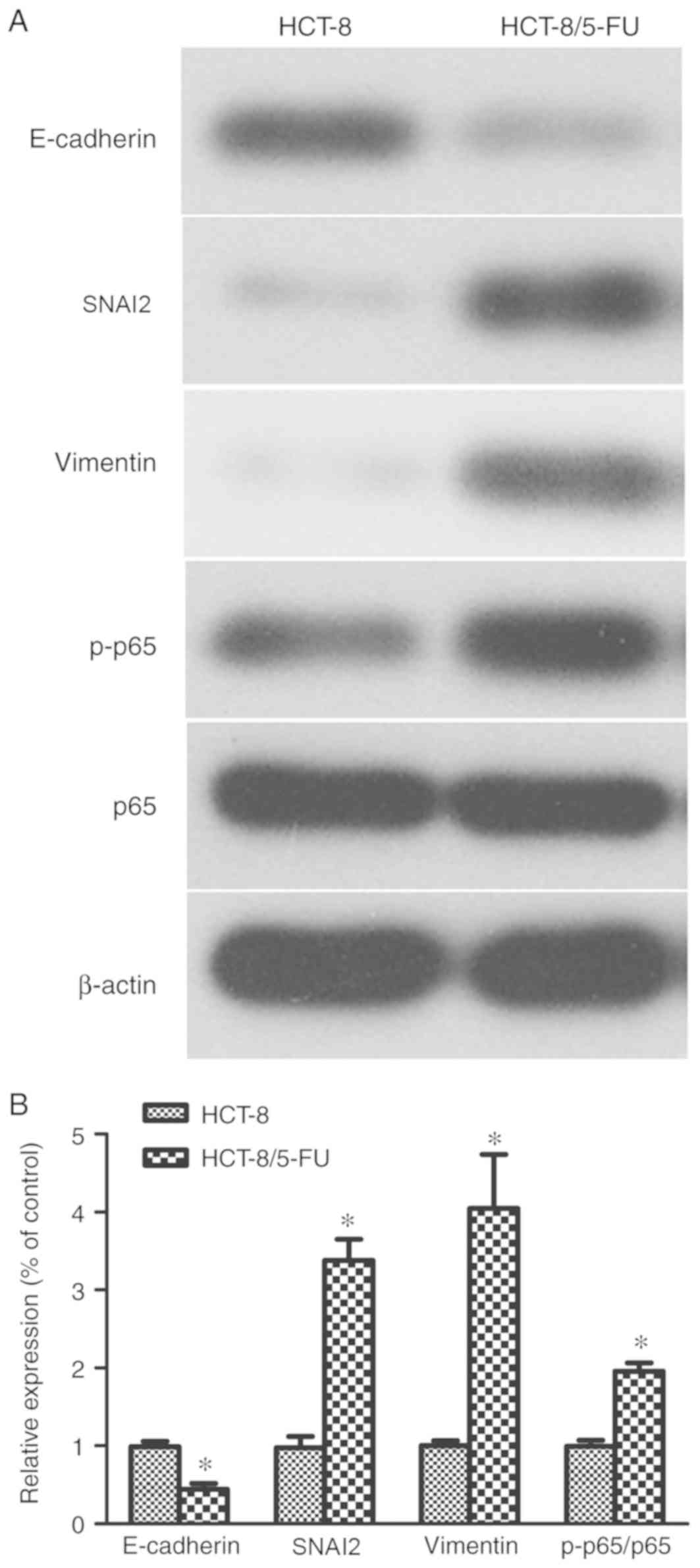
X-ray films. β-actin was used as the loading control. Changes in
EMT markers were determined in two groups: i) The negative control
cells (HCT-8 cells); and ii) the Experience group cells (HCT-8/5-FU
cells) (Fig. 2A). Changes in EMT
markers following drug treatments were assayed in four groups: i)
The negative control cells (HCT-8/5-FU cells); ii) the
oxymatrine-treated cells (HCT-8/5-FU + 2 mg/ml oxymatrine); iii)
the TNF-α-treated cells (HCT-8/5-FU + 20 ng/ml TNF-α); and iv) the
combined treatment (MIX) cells (HCT-8/5-FU + 2 mg/ml oxymatrine +20
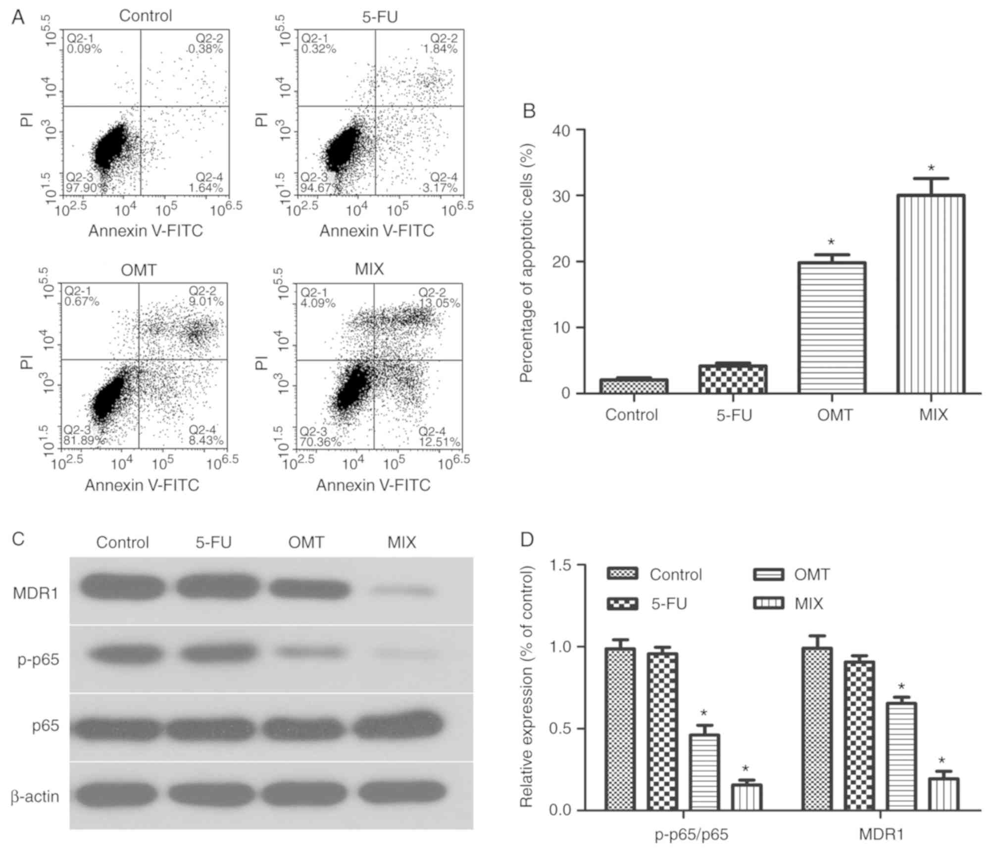
ng/ml TNF-α) (Fig. 4A). Changes in
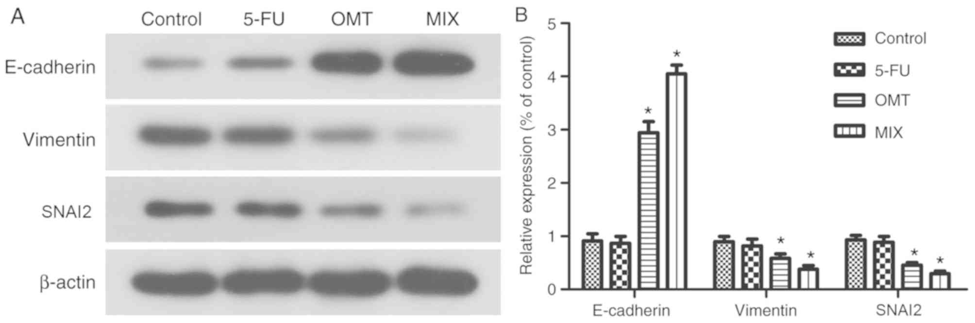
EMT markers following drug treatments were determined in four
groups: i) The negative control cells (HCT-8/5-FU cells); ii) the
5-FU-treated cells (HCT-8/5-FU + 1 µg/ml 5-FU); iii) the
oxymatrine-treated cells (HCT-8/5-FU + 2 mg/ml oxymatrine); and iv)
the combined treatment (MIX) cells (HCT-8/5-FU + 2 mg/ml oxymatrine
+1 µg/ml 5-FU) (Fig. 6A).
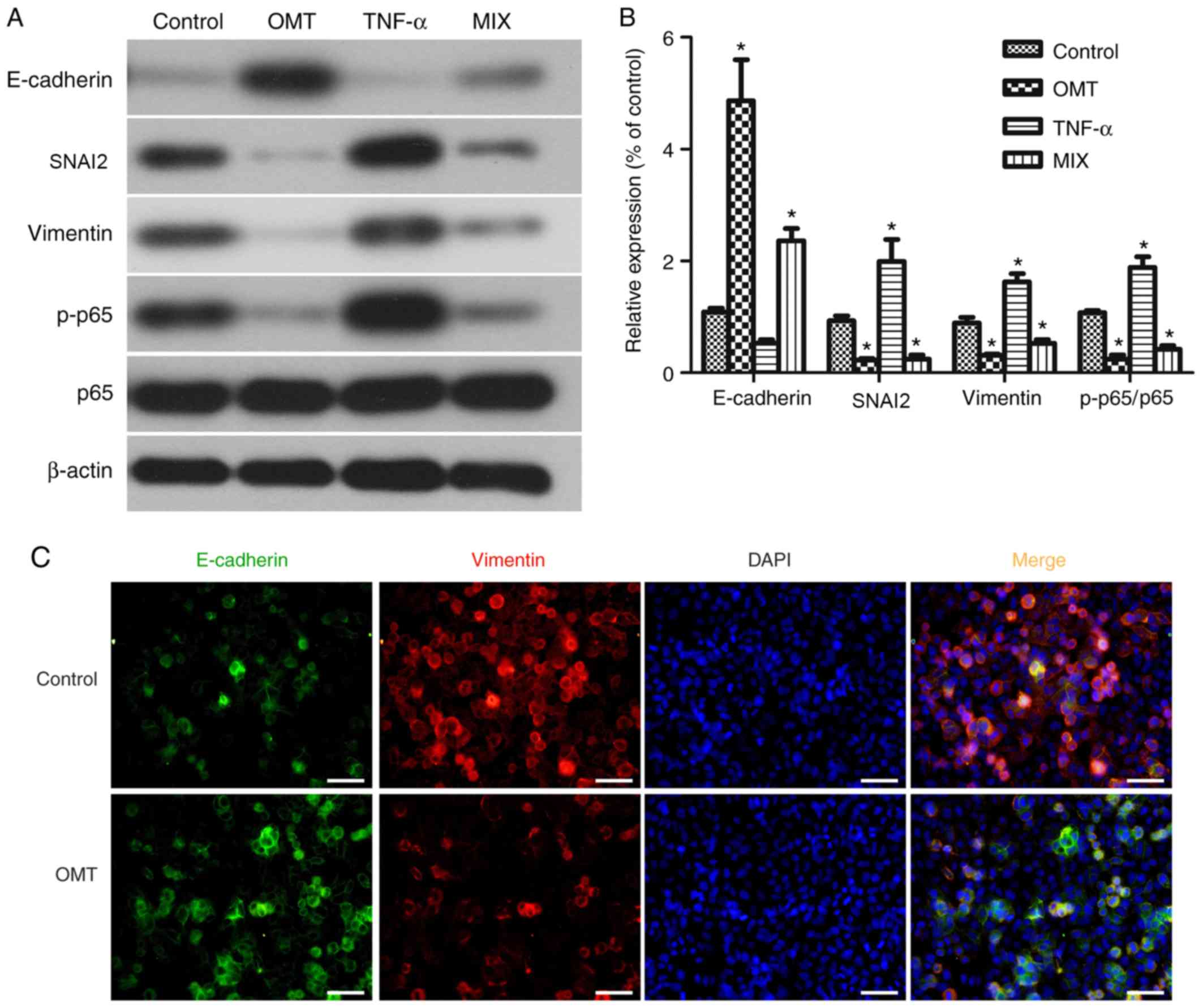 | Figure 4.Effects of oxymatrine on the
modulation of epithelial-mesenchymal transition proteins in
HCT-8/5-FU cells. (A) Western blot analysis of proteins extracted
from HCT-8/5-FU cells treated with 2 mg/ml oxymatrine and 20 ng/ml
TNF-α, alone or in combination, for 24 h. (B) Quantification of
western blot analysis. *P<0.05 vs. control cells. (C)
Immunofluorescence staining of HCT-8/5-FU cells treated with 2
mg/ml oxymatrine for 24 h, and then subjected to immunofluorescence
staining of E-cadherin (green) and vimentin (red) proteins, and
DAPI nuclei staining (blue). Scale bar, 50 µm. Magnification, ×400.
p-p65, phosphorylated p65; OMT, oxymatrine; MIX, combined
treatment; TNF-α, tumor necrosis factor-α; SNAI2, snail family
transcriptional repressor 2. |
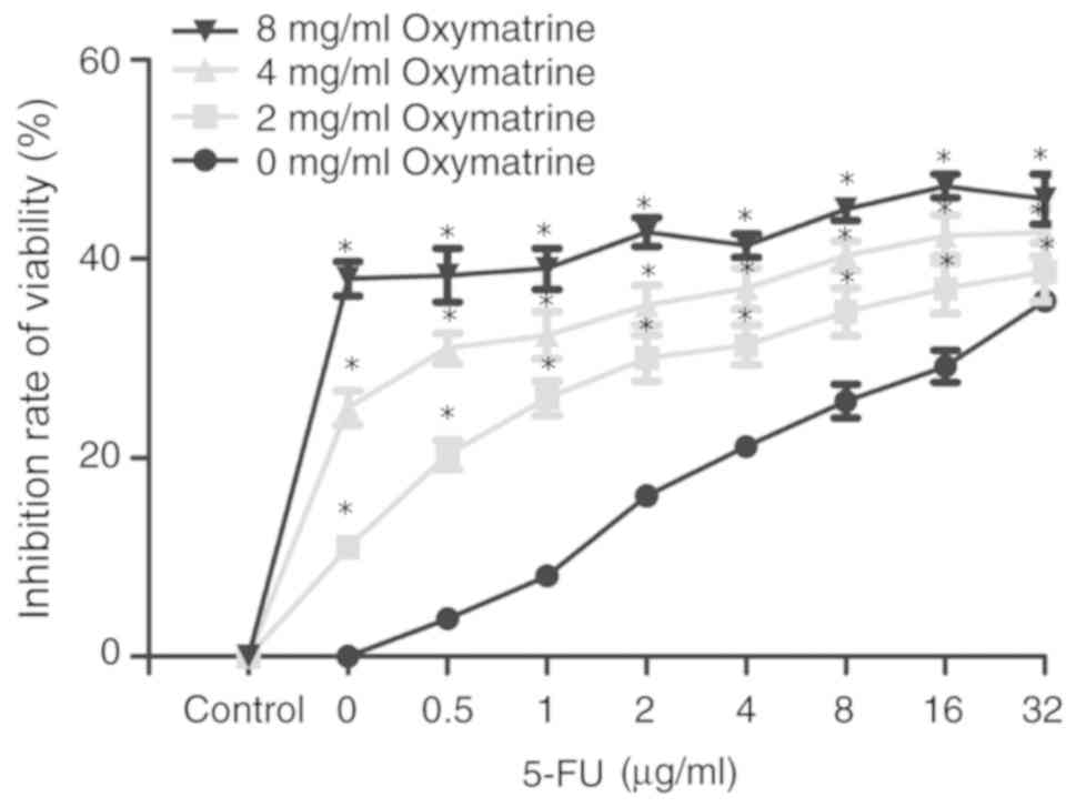
Assessing the effects of oxymatrine on
the drug resistance of HCT-8/5-FU cells
Oxymatrine (0, 2, 4 and 8 mg/ml) was added into cell
culture medium (RPMI-1640 supplemented with 10% fetal bovine serum)
containing different concentrations of 5-FU (0, 0.5, 1, 2, 4, 8, 16
and 32 µg/ml). HCT-8/5-FU cells (4×10³) were grown in
the aforementioned medium for 24 h and subjected to the MTT assay.
DMSO (150 µl/well) was used to dissolve the formzan crystals, and
the optical density was measured at 570 nm using a microplate
reader. The drug resistance index (RI) was calculated as follows:
RI=half maximal inhibitory concentration (IC50) of
drug-resistant cells/IC50 of the control cells.
Statistical analysis
The data are presented as the mean ± standard
deviation or were quantified as gray levels, where applicable, from
three independent experiments with duplicate readings. The control
and experimental groups were compared using the one-way analysis of
variance followed by the Dunnett's post hoc test. The data between
two groups were compared using the Student's t-test. All
statistical analyses were performed using SPSS software version
21.0 (IBM Corp.). P<0.05 was considered to indicate a
statistically significant difference.
Results
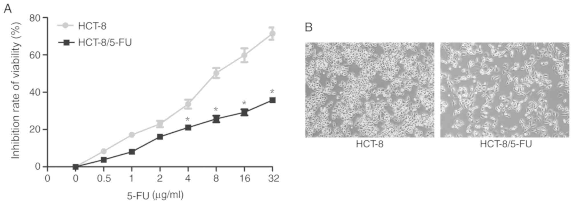
Assessment of the cancer cell RI
Compared with parental HCT-8 cells, HCT-8/5-FU cells
exhibited increased survival in culture medium containing high
doses of 5-FU (>4 µg/ml) for 24 h. In particular, the
IC50 values of HCT-8/5-FU and HCT-8 cells were
78.77±1.90 and 9.20±0.96 µg/ml, respectively (P<0.05; Fig. 1A), and the RI of HCT-8/5-FU cells was
8.56.
Changes in morphology of HCT-8 and
HCT-8/5-FU cells following 5-FU treatment
Both cell lines were treated with 1 µg/ml 5-FU for
24 h and were assessed under an inverted microscope for changes in
cell morphology. HCT-8 cells exhibited typical epithelial
morphology characterized by a round shape and distinct epithelial
clusters, whereas HCT-8/5-FU cells displayed an elongated and
irregular fibroblast-like morphology (Fig. 1B), indicating that HCT-8/5-FU cells
underwent EMT following treatment with 1 µg/ml 5-FU for 24 h.
Induction of EMT in HCT-8/5-FU and
HCT-8 cells
Differences in the protein expression levels of EMT
markers were detected in untreated HCT-8/5-FU and HCT-8 cells
(Fig. 2A). Compared with HCT-8
cells, HCT-8/5-FU cells expressed significantly increased levels of
SNAI2, vimentin and p-p65, and significantly decreased levels of
E-cadherin (P<0.05; Fig. 2B).
Effects of oxymatrine on HCT-8/5-FU
cell viability
HCT-8/5-FU cell viability was significantly reduced
following treatment with oxymatrine compared with treatment with
5-FU alone (P<0.05; Fig. 3).
However, there was no significant difference between cells treated
with any dose of 5-FU (0, 0.5, 1, 2, 4, 8, 16 and 32 µg/ml) and 4
or 8 mg/ml oxymatrine (all P>0.05; Fig. 3). The data demonstrated a favorable
inhibition of HCT-8/5-FU cell viability following treatment with 2
mg/ml oxymatrine and 1 µg/ml 5-FU; this dose combination was
therefore used for subsequent experiments.
Oxymatrine inhibits EMT in HCT-8/5-FU
cells via the NF-κB signaling pathway
In order to explore whether oxymatrine inhibits the
EMT in HCT-8/5-FU cells, the expression of EMT-associated
biomarkers was assessed in HCT-8/5-FU cells, following treatment
with 20 ng/ml TNF-α and 2 mg/ml oxymatrine, alone or in combination
for 24 h (Fig. 4A). The mesenchymal
marker vimentin and the epithelial marker E-cadherin were
downregulated and upregulated following treatment with 2 mg/ml
oxymatrine for 24 h. Moreover, oxymatrine significantly inhibited
the expression of the NF-κB signaling pathway protein p-p65
compared with the other three groups (P<0.05; Fig. 4B), suggesting that NF-κB signaling
may mediate the effects of oxymatrine in HCT-8/5-FU cells and that
oxymatrine may inhibit EMT via the NF-κB signaling pathway in
HCT-8/5-FU. Oxymatrine treatment significantly inhibited p-p65
protein expression. However, this did not exclude the fact that
oxymatrine in combination with 5-FU may also possess synergistic
antitumor activity in HCT-8 cells, or that oxymatrine alone may
exhibit antitumor effects on HCT-8/5-FU cells. In addition,
fluorescence microscopy showed that E-cadherin expression (green)
was upregulated, whereas vimentin expression (red) was
downregulated, following the incubation of HCT-8/5-FU cells with 2
mg/ml oxymatrine for 24 h compared with HCT-8/5-FU without
oxymatrine (Fig. 4C).
Oxymatrine induces HCT-8/5-FU cells to
undergo apoptosis
The mesenchymal marker vimentin and the epithelial
marker E-cadherin were downregulated and upregulated, respectively,
following treatment with 2 mg/ml oxymatrine for 24 h (Fig. 5A and B). The results from flow
cytometry (Fig. 6A) demonstrated
that there was no significant changes in the apoptotic rate of
5-FU-treated HCT-8/5-FU cells compared with the control group
(HCT-8/5-FU+0 µg/ml 5-fluorouracil; P>0.05; Fig. 6B). However, the apoptotic rates were
significantly increased in the OMT- and MIX-treated groups compared
with the control group (both P<0.05; Fig. 6B), with the MIX-treated group
exhibiting the highest apoptotic rate. Additionally, the protein
expression levels of MDR1 and p-p65 were significantly
downregulated in the OMT- and MIX-treated groups compared with the
control group (both P<0.05; Fig. 6C
and D), whereas no significant differences were observed
between the 5-FU-treated and control groups (P>0.05; Fig. 6C and D).
Discussion
Despite recent advancements in the treatment of
patients with colon cancer, overcoming 5-FU resistance remains
clinically challenging (8).
Therefore, it is important to elucidate the underlying mechanisms
of chemotherapy resistance in order to improve patient outcomes. It
was reported that oxymatrine not only had inhibitory effects on the
regulation of cancer cell metastasis, but was also involved in
inducing the EMT, in vitro, in colon cancer, rectum, breast
and lung cancers (11). Furthermore,
oxymatrine reversed chemotherapy resistance in colorectal cancer
in vitro (15). Thus,
oxymatrine may serve as a potential therapeutic agent by reversing
EMT in tumor cells. Indeed, the present study assessed oxymatrine
sensitization of 5-FU-resistant colon cancer cells in vitro
and explored the underlying molecular events. HCT-8/5-FU cells
significantly increased the 5-FU concentration required to decrease
tumor cell survival (8.56-fold increase compared with parental
HCT-8 cells), and HCT-8/5-FU cells induced tumor cell EMT
phenotypes and the expression of mesenchymal markers. Furthermore,
oxymatrine alone and in combination with 5-FU altered HCT-8/5-FU
cell morphology, induced tumor cell apoptosis and upregulated
E-cadherin expression by suppressing the NF-κB signaling pathway.
The results obtained in the present study revealed that the EMT was
involved in 5-FU chemoresistance in HCT-8/5-FU colon cancer cells
in vitro. Furthermore, oxymatrine reversed 5-FU
chemoresistance by modulating the EMT through inactivation of the
NF-κB signaling pathway. Therefore, oxymatrine may serve as a novel
therapeutic agent to reverse 5-FU resistance in colon cancer
cells.
Oxymatrine is a quinolizidine alkaloid compound
extracted from the root of Sophora flavescens. Previous
studies have demonstrated that oxymatrine exhibits a wide range of
anticancer activities (11,14), including inhibition of tumor cell
proliferation, invasion and metastasis in gallbladder, cervical,
lung and ovarian cancer (29–32).
Additionally, recent studies have revealed that oxymatrine may
reverse chemoresistance, including resistance to paclitaxel in lung
cancer cells (15) and in human
squamous cells (33), and resistance
to cisplatin in lung cancer cells (34). However, to date, the precise
mechanisms underlying the effects of oxymatrine on 5-FU resistance
in colorectal cancer remains unclear. Tumor cells undergo EMT in
order to increase migration, invasion and anoikis tolerance during
cancer development and progression (20). In particular, during EMT, tumor cells
lose cell-cell adhesion and gain migratory and invasive properties,
which are required for tumor initiation and metastasis (35,36). At
the gene level, tumor cells involved in the EMT downregulate
E-cadherin expression, upregulate vimentin and SNAI2 expression and
gain cancer stem cell-like properties (21,37,38). A
previous study revealed that an increase in the EMT was considered
to be a novel mechanism associated with chemotherapy resistance in
cancer of epithelial origin (39).
In order to assess the validity of this hypothesis, the present
study compared the EMT phenotype of HCT-8/5-FU cells with HCT-8
cells and demonstrated the association between the EMT and 5-FU
resistance. In particular, the results revealed that HCT-8/5-FU
cells exhibited an irregular fibroblastic morphology, whereas the
parental HCT-8 cells displayed an epithelial morphology. Compared
with HCT-8 cells, HCT-8/5-FU cells had increased levels of
vimentin, SNAI2 and p-p65 proteins, but lower levels of E-cadherin
protein, indicating that the EMT may be associated with 5-FU
resistance.
In order to explore the effects of oxymatrine on the
EMT and 5-FU resistance in colon cancer cells in the present study,
HCT-8/5-FU cells were treated with oxymatrine. It was revealed that
oxymatrine (≥2 mg/ml) significantly inhibited tumor cell viability,
which suggest that oxymatrine reversed the chemoresistance of
HCT-8/5-FU cells. In addition, oxymatrine upregulated E-cadhern
expression and downregulated SNAI2 and vimentin expression,
indicating that oxymatrine reverse the EMT phenotype of HCT-8/5-FU
cells. The activation of the NF-κB signaling pathway is important
for the regulation of cell proliferation, differentiation,
survival, migration, invasion and the EMT process (40–42). The
NF-κB signaling pathway is involved in transforming growth factor
β-induced EMT and the 5-Fu chemotherapy resistance in colon cancer
(43,44). In the present study, the expression
of p-p65 was significantly increased in HCT-8/5-FU cells compared
with HCT-8 cells. Moreover, oxymatrine treatment significantly
suppressed the expression of the p-p65 protein. However, this did
not rule out the fact that oxymatrine in combination with 5-FU may
also possess synergistic antitumor activity in HCT-8 cells or that
oxymatrine alone exhibits antitumor effects on HCT-8/5-FU cells,
which may be addressed by future studies on different
5-FU-sensitive and resistant cancer cell lines. In addition, the
present study lacked an invasion assay, which may determine effects
on the EMT process more conclusively. Thus, future investigations
using alternative cell lines in vitro and in vivo are
required to validate the current findings. In conclusion, the
results of the present study demonstrated that the colon cancer
cell EMT was involved in the chemoresistance of HCT-8/5-FU cells to
5-FU, and that oxymatrine treatment was able to reverse this
resistance. Oxymatrine may regulate the EMT process and inactivate
the NF-κB signaling pathway in tumor cells. The findings of the
present study provide a novel theoretical basis for the
sensitization of 5-FU-resistant colon cancer cells in
vitro.
Acknowledgements
Not applicable.
Funding
The present study was supported in part by grants
from The Natural Science Foundation of Guangxi Zhuang Autonomous
Region (grant no. 2018GXNSFBA050072), The National Natural Science
Foundation of China (grant no. 8176110028), The 2018 Innovation
Project of Guangxi Graduate Education (grant no. YCBZ2018046) and
The Guangxi Zhuang Autonomous Region Health and Family Planning
Commission Self-Financing Research Project (grant no.
Z20170086).
Availability of data and materials
The datasets used and/or analyzed during the present
study are available from the corresponding author on reasonable
request.
Authors' contributions
LL, JAH and ZWC designed the study. LL, JW, JL, LW,
ZXC, CLH and TQG performed the experiments and provided technical
support. LL, JAH and ZWC analyzed the data. LL prepared and revised
the manuscript. JAH and ZWC supervised the work. All authors read
and approved the final version of the manuscript.
Ethics approval and consent to
participate
Not applicable.
Patient consent for publication
Not applicable.
Competing interests
The authors declare that there are no competing
interests.
References
|
1
|
DeSantis CE, Lin CC, Mariotto AB, Siegel
RL, Stein KD, Kramer JL, Alteri R, Robbins AS and Jemal A: Cancer
treatment and survivorship statistics, 2014. CA Cancer J Clin.
64:252–271. 2014. View Article : Google Scholar : PubMed/NCBI
|
|
2
|
Chen W, Zheng R, Baade PD, Zhang S, Zeng
H, Bray F, Jemal A, Yu XQ and He J: Cancer statistics in China,
2015. CA Cancer J Clin. 66:115–132. 2016. View Article : Google Scholar : PubMed/NCBI
|
|
3
|
GBD 2015 Disease and Injury Incidence and
Prevalence Collaborators, . Global, regional, and national
incidence, prevalence, and years lived with disability for 310
diseases and injuries, 1990–2015: A systematic analysis for the
global burden of disease study 2015. Lancet. 388:1545–1602. 2016.
View Article : Google Scholar : PubMed/NCBI
|
|
4
|
Fakih MG: Metastatic colorectal cancer:
Current state and future directions. J Clin Oncol. 33:1809–1824.
2015. View Article : Google Scholar : PubMed/NCBI
|
|
5
|
Boland P and Ma W: Immunotherapy for
colorectal cancer. Cancers (Basel). 9(pii): E502017. View Article : Google Scholar : PubMed/NCBI
|
|
6
|
Jeffery M, Hickey BE, Hider PN and See AM:
Follow-up strategies for patients treated for non-metastatic
colorectal cancer. Cochrane Database Syst Rev.
11:CD0022002016.PubMed/NCBI
|
|
7
|
Shigeta K, Ishii Y, Hasegawa H, Okabayashi
K and Kitagawa Y: Clinical usefulness of 5-FU metabolic enzymes as
predictive markers of response to chemotherapy in colorectal
cancer. World J Surg. 40:1019–1020. 2016. View Article : Google Scholar : PubMed/NCBI
|
|
8
|
Sobrero A, Guglielmi A, Grossi F, Puglisi
F and Aschele C: Mechanism of action of fluoropyrimidines:
relevance to the new developments in colorectal cancer
chemotherapy. Semin Oncol. 27 (5 Suppl 10):S72–S77. 2000.
|
|
9
|
Iqbal A and George TJ: Randomized clinical
trials in colon and rectal cancer. Surg Oncol Clin N Am.
26:689–704. 2017. View Article : Google Scholar : PubMed/NCBI
|
|
10
|
Ma L, Wen S, Zhan Y, He Y, Liu X and Jiang
J: Anticancer effects of the Chinese medicine matrine on murine
hepatocellular carcinoma cells. Planta Med. 74:245–251. 2008.
View Article : Google Scholar : PubMed/NCBI
|
|
11
|
Wang W, You RL, Qin WJ, Hai LN, Fang MJ,
Huang GH, Kang RX, Li MH, Qiao YF, Li JW and Li AP: Anti-tumor
activities of active ingredients in compound kushen injection. Acta
Pharmacol Sin. 36:676–679. 2015. View Article : Google Scholar : PubMed/NCBI
|
|
12
|
Zhou YJ, Guo YJ, Yang XL and Ou ZL:
Anti-cervical cancer role of matrine, oxymatrine and Sophora
Flavescens alkaloid gels and its mechanism. J Cancer.
9:1357–1364. 2018. View Article : Google Scholar : PubMed/NCBI
|
|
13
|
Nourmohammadi S, Aung TN, Cui J, Pei JV,
De Ieso ML, Harata-Lee Y, Qu Z, Adelson DL and Yool AJ: Effect of
compound kushen injection, a natural compound mixture, and its
identified chemical components on migration and invasion of colon,
brain, and breast cancer cell lines. Front Oncol. 9:3142019.
View Article : Google Scholar : PubMed/NCBI
|
|
14
|
Liang L and Huang J: Oxymatrine inhibits
epithelial-mesenchymal transition through regulation of NF-κB
signaling in colorectal cancer cells. Oncol Rep. 36:1333–1338.
2016. View Article : Google Scholar : PubMed/NCBI
|
|
15
|
Joshi P, Vishwakarma RA and Bharate SB:
Natural alkaloids as P-gp inhibitors for multidrug resistance
reversal in cancer. Eur J Med Chem. 138:273–292. 2017. View Article : Google Scholar : PubMed/NCBI
|
|
16
|
Mitra A, Mishra L and Li S: EMT, CTCs and
CSCs in tumor relapse and drug-resistance. Oncotarget.
6:10697–10711. 2015. View Article : Google Scholar : PubMed/NCBI
|
|
17
|
Du B and Shim JS: Targeting
epithelial-mesenchymal transition (EMT) to overcome drug resistance
in cancer. Molecules. 21(pii): E9652016. View Article : Google Scholar : PubMed/NCBI
|
|
18
|
Voulgari A and Pintzas A:
Epithelial-mesenchymal transition in cancer metastasis: Mechanisms,
markers and strategies to overcome drug resistance in the clinic.
Biochim Biophys Acta. 1796:75–90. 2009.PubMed/NCBI
|
|
19
|
Smith BN and Bhowmick NA: Role of EMT in
metastasis and therapy resistance. J Clin Med. 5(pii): E172016.
View Article : Google Scholar : PubMed/NCBI
|
|
20
|
Thiery JP, Acloque H, Huang RY and Nieto
MA: Epithelial-mesenchymal transitions in development and disease.
Cell. 139:871–890. 2009. View Article : Google Scholar : PubMed/NCBI
|
|
21
|
Ye X and Weinberg RA:
Epithelial-mesenchymal plasticity: A central regulator of cancer
progression. Trends Cell Biol. 25:675–686. 2015. View Article : Google Scholar : PubMed/NCBI
|
|
22
|
Bao Y, Lu Y, Wang X, Feng W, Sun X, Guo H,
Tang C, Zhang X, Shi Q and Yu H: Eukaryotic translation initiation
factor 5A2 (eIF5A2) regulates chemoresistance in colorectal cancer
through epithelial mesenchymal transition. Cancer Cell Int.
15:1092015. View Article : Google Scholar : PubMed/NCBI
|
|
23
|
Zhou Z, Zhang L, Xie B, Wang X, Yang X,
Ding N, Zhang J, Liu Q, Tan G, Feng D and Sun LQ: FOXC2 promotes
chemoresistance in nasopharyngeal carcinomas via induction of
epithelial mesenchymal transition. Cancer Lett. 363:137–145. 2015.
View Article : Google Scholar : PubMed/NCBI
|
|
24
|
Ma JL, Zeng S, Zhang Y, Deng GL and Shen
H: Epithelial-mesenchymal transition plays a critical role in drug
resistance of hepatocellular carcinoma cells to oxaliplatin. Tumour
Biol. 37:6177–6184. 2016. View Article : Google Scholar : PubMed/NCBI
|
|
25
|
Yang Q, Huang J, Wu Q, Cai Y, Zhu L, Lu X,
Chen S, Chen C and Wang Z: Acquisition of epithelial-mesenchymal
transition is associated with Skp2 expression in
paclitaxel-resistant breast cancer cells. Br J Cancer.
110:1958–1967. 2014. View Article : Google Scholar : PubMed/NCBI
|
|
26
|
Arias AM: Epithelial mesenchymal
interactions in cancer and development. Cell. 105:425–431. 2001.
View Article : Google Scholar : PubMed/NCBI
|
|
27
|
Zhang P, Sun Y and Ma L: ZEB1: At the
crossroads of epithelial-mesenchymal transition, metastasis and
therapy resistance. Cell Cycle. 14:481–487. 2015. View Article : Google Scholar : PubMed/NCBI
|
|
28
|
Mayo MW and Baldwin AS: The transcription
factor NF-kappaB: Control of oncogenesis and cancer therapy
resistance. Biochim Biophys Acta. 1470:M55–M62. 2000.PubMed/NCBI
|
|
29
|
Li M, Su BS, Chang LH, Gao Q, Chen KL, An
P, Huang C, Yang J and Li ZF: Oxymatrine induces apoptosis in human
cervical cancer cells through guanine nucleotide depletion.
Anticancer Drugs. 25:161–173. 2014. View Article : Google Scholar : PubMed/NCBI
|
|
30
|
Wang B, Han Q and Zhu Y: Oxymatrine
inhibited cell proliferation by inducing apoptosis in human lung
cancer A549 cells. Biomed Mater Eng. 26 (Suppl 1):S165–S172.
2015.PubMed/NCBI
|
|
31
|
Liu Y, Bi T, Dai W, Wang G, Qian L, Gao Q
and Shen G: Effects of oxymatrine on the proliferation and
apoptosis of human hepatoma carcinoma cells. Technol Cancer Res
Treat. 15:487–497. 2016. View Article : Google Scholar : PubMed/NCBI
|
|
32
|
Li J, Jiang K and Zhao F: Oxymatrine
suppresses proliferation and facilitates apoptosis of human ovarian
cancer cells through upregulating microRNA-29b and downregulating
matrix metalloproteinase-2 expression. Mol Med Rep. 12:5369–5374.
2015. View Article : Google Scholar : PubMed/NCBI
|
|
33
|
Luo SX, Deng WY, Wang XF, Lü HF, Han LL,
Chen BB, Chen XB and Li N: Molecular mechanism of
indirubin-3′-monoxime and Matrine in the reversal of paclitaxel
resistance in NCI-H520/TAX25 cell line. Chin Med J (Engl).
126:925–929. 2013.PubMed/NCBI
|
|
34
|
Wang HQ, Jin JJ and Wang J: Matrine
induces mitochondrial apoptosis in cisplatin-resistant non-small
cell lung cancer cells via suppression of β-catenin/survivin
signaling. Oncol Rep. 33:2561–2566. 2015. View Article : Google Scholar : PubMed/NCBI
|
|
35
|
Tse JC and Kalluri R: Mechanisms of
metastasis: Epithelial-to-mesenchymal transition and contribution
of tumor microenvironment. J Cell Biochem. 101:816–829. 2007.
View Article : Google Scholar : PubMed/NCBI
|
|
36
|
Yang J and Weinberg RA:
Epithelial-mesenchymal transition: At the crossroads of development
and tumor metastasis. Dev Cell. 14:818–829. 2008. View Article : Google Scholar : PubMed/NCBI
|
|
37
|
Kong D, Li Y, Wang Z and Sarkar FH: Cancer
stem cells and epithelial-to-mesenchymal transition
(EMT)-phenotypic cells: Are they cousins or twins? Cancers (Basel).
3:716–729. 2011. View Article : Google Scholar : PubMed/NCBI
|
|
38
|
Chaffer CL and Weinberg RA: A perspective
on cancer cell metastasis. Science. 331:1559–1564. 2011. View Article : Google Scholar : PubMed/NCBI
|
|
39
|
Fischer KR, Durrans A, Lee S, Sheng J, Li
F, Wong ST, Choi H, El Rayes T, Ryu S, Troeger J, et al:
Epithelial-to-mesenchymal transition is not required for lung
metastasis but contributes to chemoresistance. Nature. 527:472–476.
2015. View Article : Google Scholar : PubMed/NCBI
|
|
40
|
Kaltschmidt B, Greiner JFW, Kadhim HM and
Kaltschmidt C: Subunit-specific role of NF-κB in cancer.
Biomedicines. 6(pii): E442018. View Article : Google Scholar : PubMed/NCBI
|
|
41
|
Wang S, Liu Z, Wang L and Zhang X:
NF-kappaB signaling pathway, inflammation and colorectal cancer.
Cell Mol Immunol. 6:327–334. 2009. View Article : Google Scholar : PubMed/NCBI
|
|
42
|
Maier HJ, Schmidt-Strassburger U, Huber
MA, Wiedemann EM, Beug H and Wirth T: NF-kappaB promotes
epithelial-mesenchymal transition, migration and invasion of
pancreatic carcinoma cells. Cancer Lett. 295:214–228. 2010.
View Article : Google Scholar : PubMed/NCBI
|
|
43
|
Wu Y and Zhou BP:
TNF-alpha/NF-kappaB/Snail pathway in cancer cell migration and
invasion. Br J Cancer. 102:639–644. 2010. View Article : Google Scholar : PubMed/NCBI
|
|
44
|
Zhao C, Zhao Q, Zhang C, Wang G, Yao Y,
Huang X, Zhan F, Zhu Y, Shi J, Chen J, et al: miR-15b-5p
resensitizes colon cancer cells to 5-fluorouracil by promoting
apoptosis via the NF-κB/XIAP axis. Sci Rep. 7:41942017. View Article : Google Scholar : PubMed/NCBI
|