Introduction
Thyroid cancer ranks first among endocrine tumors
and is increasing in prevalence in China, in particular papillary
cancer of the thyroid gland (1).
Thyroid-associated tests have been added to routine physical
examinations and have increased the detection of thyroid cancers
(2). Thyroid cancer-associated
mortality is particularly prevalent in Asia and China. China
accounts for 15.6% of thyroid cancer cases globally and 13.8% of
global cancer-associated mortality (3). Thyroid cancer is a major disease threat
in China.
Forkhead transcription factor M1 (FoxM1) possesses a
‘wing helix’ DNA binding domain, which is a common structural
domain of the Forkhead family (4).
The main function of FoxM1 is to regulate the transition
G1 phase in cells, thereby regulating mitosis; thus,
FoxM1 serves an important role in the cell growth cycle (5,6). FoxM1
enhances the proliferation of cells, particularly in fetal tissues
(7). Elevated expression of FoxM1 in
tumors influences tumor occurrence and development, including
promoting the growth of tumor cells, neovascularization, and the
invasive spread of tumor cells (8).
Elevated production of FoxM1 can prevent apoptosis of tumor cells
and delay their aging (9). Previous
studies have reported an association between FoxM1 expression and
paclitaxel resistance in tumors, including those of breast cancer
(10), colon cancer (11), gastric cancer (12), and liver cancer (13). It has been reported that when FoxM1
expression is silenced in tumor cells, the cells become sensitive
to paclitaxel (14).
The role of FoxM1 in papillary thyroid carcinoma
(PTC) remains unclear. In the present study, the proliferation,
migration and invasion rate was detected in K1 human PTC cells with
FoxM1 silencing. The molecular mechanism of FoxM1 has been
thoroughly studied in previous reports (15). The present study was not concerned
with the molecular mechanism, but rather with the effect of FoxM1
on tumor cells. The results demonstrated an association of FoxM1
expression with a number of biological behaviors of tumor cells,
suggesting that FoxM1 may serve as a novel therapeutic gene target
for PTC, in particular for patients who cannot have surgery or do
not respond to chemotherapy.
Materials and methods
Culture and passage of K1 human
thyroid cancer cells
The K1 human thyroid cancer cell line was purchased
from the Shanghai Cell Bank (Chinese Academy of Sciences), and
preserved by the Central Laboratory of North China University of
Science and Technology (Tangshan, China). K1 cells were cultured in
RPMI-1640 complete medium (Biological Industries) containing
penicillin, streptomycin, and 10% bovine embryo serum (Biological
Industries) in an incubator maintained at 37°C with 5%
CO2. K1 cell growth was monitored by optical microscopy,
replenishing the medium every 24 to 48 h, according to cell growth.
Following adherence of K1 cells to the wall of the culture flask
and growth covering ~70% of the wall, the cells were passaged, and
the shed cells and cell debris were removed gently by 2–3 washes
with PBS. Trypsin (0.25%) was subsequently added to digest the
cells. When cell morphology changed to a roughly spherical shape,
and the cells had detached from the flask wall, complete medium was
added to stop the digestion. The cells were evenly mixed in the
medium and half were transferred to another culture flask, and
cultured using the aforementioned conditions. As pancreatin affects
cell growth, the medium was changed every 12 h.
Transfection of FoxM1-small
interfering RNA (siRNA)
The FoxM1-siRNA target sequence
5′-GGCUGCACUAUCAACAAUATTUAUUGUUGAUAGUGCAGCCTT-3′ was synthesized by
Suzhou Gema Gene Limited. Lipofectamine 2000 was purchased from
Invitrogen (Thermo Fisher Scientific, Inc.). Aliquots of detached
cells with the appropriate density (70%) were seeded into 6-well
plates. The cells were transfected when they had grown to an
appropriate state (to the fastest growth rate) to avoid the effect
of penicillin and streptomycin prior to transfection. The
penicillin/streptomycin-free culture medium was changed 24 h prior
to transfection, and the cells in the 6-well plates were washed
twice with PBS prior to transfection. Uninoculated RPMI-1640 medium
(1 ml) was added to each well, and the cells were incubated for 1 h
to produce a starvation condition. siRNA (5 µl) and
Lipofectamine® 2000 (5 µl; Thermo Fisher Scientific,
Inc.) were added to 500 µl blank RPMI-1640 medium and incubated for
5 min at 37°C. The two solutions were subsequently mixed and
allowed to stand for 20 min at 37°C prior to being added into two
wells with the starved cells forming the transfection group (group
T). The above protocol was repeated with the replacement of the
previous siRNA with the control siRNA (sequence
5′-CCAUGAGGAGUACUGCCAATT-3′) to prepare the non-meaning sequence
group (group NM). The remaining two wells received only PBS and
comprised the control (group CON). The 6-well plates were gently
shaken to spread the liquid evenly on the bottom of the wells.
After 6 h of transfection, the liquid was removed from each well,
RPMI-1640 medium containing 10% serum was added, and culturing was
continued for 48 h. The cells were then observed using fluorescence
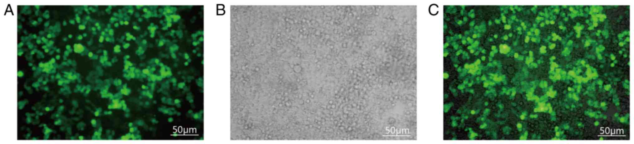
microscopy, and the proportion of fluorescent cells was recorded.
Transfection was defined as successful when the proportion of cells
with fluorescence exceeded 80% (Fig.
1).
Western blot analysis
To extract total protein, digested cells with
appropriate density were prepared from a single cell suspension and
centrifuged at 330 × g for 5 min at 37°C. A small volume of
ice-cold PBS solution was subsequently added into the
centrifugation bottle to resuspend and centrifuge the cells. This
step was repeated twice. Following the final wash, the supernatant
was removed and the cell pellet was resuspended in 200 µl RIPA
pyrolysis working solution (Biological Industries) and transferred
to an Eppendorf tube. The preparation was ultrasonicated for 10 sec
to destroy the cells. After shaking 10 times, the cells were
pipetted evenly on ice for 30 min, and centrifuged at 4°C for 15
min at 4,000 × g. The supernatant was stored at −20°C.
Protein was quantified using a bicinchoninic acid
protein quantification kit, according to the manufacturer's
protocols. The same volume of 2X protein loading buffer was added,
followed by denaturation at 100°C for 5 min and preservation at
−80°C for later use.
A volume containing 30 µg protein was used per well
for SDS-PAGE with a 5% concentration gel and 10% separation gel.
The previously prepared protein samples and markers were added
sequentially to the wells for electrophoresis at 80 and 120 V,
respectively. Following electrophoresis, the separation gel region,
in which the proteins with different molecular weights became
uniformly distributed, was removed and a PVDF membrane was soaked
in methanol for about 5 sec, followed by rinsing with transmembrane
solution. The transmembrane solution-soaked membrane was placed in
a wet transmembrane apparatus in which the membrane was sandwiched
between filter paper. Proteins were transferred from the gel to the
membrane for 1 h at 90 V. The transmembrane solution on the
membrane surface was washed away with Tris buffered saline-Tween 20
(TBST). The membrane was blocked using a solution of 10% skimmed
milk for 2 h at 37°C.
The membrane surface was flushed with TBST, followed
by shaking, and exposed to a primary antibody kit solution
(Invitrogen; Thermo Fisher Scientific, Inc.) (FOXm1; cat. no.
sc-271746; 1:500; Thermo Fisher Scientific, Inc.) at 4°C for 12 h.
The membrane was rinsed two to three times (10 min each) with TBST,
prior to shaking in the presence of pre-configured horseradish
peroxidase-IgG secondary antibody kit (1:1,000; Invitrogen; Thermo
Fisher Scientific, Inc.; cat. no. 31430) at 37°C for 2 h. The
membrane was rinsed two to three times (10 min each) with TBST. A
BeyoECL Star super-sensitized electrochemiluminescence kit, was
used to detect proteins according to the manufacturer's protocols.
ImageJ software (version 1.8.0; National Institutes of Health) was
used to analyze the gray scale values of the immunofluorescent
bands. The expression (%) of associated protein was calculated as
(gray value of target protein/gray value of internal reference)
×100.
Cell viability based on MTT assay
DMSO was purchased from Sigma-Aldrich; Merck KGaA.
MTT was purchased from Amresco, LLC. Cells in each group were
digested and single cell suspensions were prepared and evenly
seeded in 96-well plates with appropriate cell densities (70%). An
equal volume of PBS was added to the well to prevent a detrimental
effect on cell growth, due to water evaporation. Each group
comprised six wells, which were divided into 24, 48 and 72 h
samples (two wells for each time). Each well received 20 µl MTT
solution agent 4 h prior to each time-point to allow the formation
of blue formazan. At 24, 48 or 72 h, the liquid in each well was
removed and 150 µl DMSO was added, followed by shaking for 10 min.
The optical density (OD) value of each well was measured at a
wavelength of 490 mm, and blank wells were used for normalizing the
spectrophotometer. The maximum and minimum values of each well were
eliminated from the data, and the remaining data were averaged,
with the experiment repeated three times. The inhibition rate (%)
of FoxM1-siRNA on cell growth was calculated as (OD490Group
CON-OD490 Group T)/OD490 Group CON
×100.
Migration scratch assay
The cell groups were cultured in 6-well plates in
pairs (70%). When the cells had formed a monolayer covering the
bottom of the well, a 200-µl pipette tip was used to make a scratch
in each monolayer, with the width of the scratch ~0.6 mm. Each well
was washed two to three times using PBS to remove non-adherent
cells, cell debris and residual medium. RPMI-1640 medium was added
(0 h). The wells were imaged at 0, 24, 48 and 72 h with a
fluorescence microscope to observe the changes in the scratch area
in the images.
Invasion assay
Prior to the experiment, the prepared Matrigel (BD
Biosciences) was placed at 4°C for 12 h. The Matrigel and blank
RPMI-1640 medium were dispensed at a ratio of 1:9, and 20 µl
prepared Matrigel was evenly spread on each Transwell chamber. Care
was taken not to produce bubbles when adding the Matrigel. The
Transwell chambers were subsequently placed into one incubator for
12 h to allow coagulation of the Matrigel. The cell groups were
cultured for 48 h, digested, and subsequently seeded into the upper
chamber with a cell density of 2×10⁵ cells/100 µl of
serum-free, penicillin/streptomycin-free medium. A total of 600 µl
RPMI-1640 medium containing only 10% serum was added to the lower
chamber, which was subsequently transferred to an incubator for 18
h. The chamber was gently washed three times with PBS to remove the
cells on the surface of the upper chamber. The chamber was soaked
with 4% formaldehyde for 30 min at 37°C. The solution was removed
and the chamber was rinsed with PBS and allowed to air-dry. The
cells were then stained with 80% crystal violet for 15–20 min at
37°C and subsequently rinsed with distilled water. The
polycarbonate membrane was cut from the chamber and placed on a
glass slide. Subsequent to the membrane being air-dried, it was
covered using neutral gum and with a cover glass to further to
air-dry. Cells permeating the membrane were then observed by
optical microscopy (magnification, ×200). Randomly selected visual
fields (n=15) were examined to record the number of cells and the
mean value was calculated. The experiment was repeated three
times.
Statistical analyses
The data were processed using SPSS 17.0 software
(SPSS Inc.). The results are expressed as mean ± standard
deviation. The results of cell migration and invasion tests were
analyzed by one-way ANOVA. The results of MTT were analyzed using
the ANOVA of repeated measurements. P<0.05 considered to
indicate a statistical significant difference.
Results
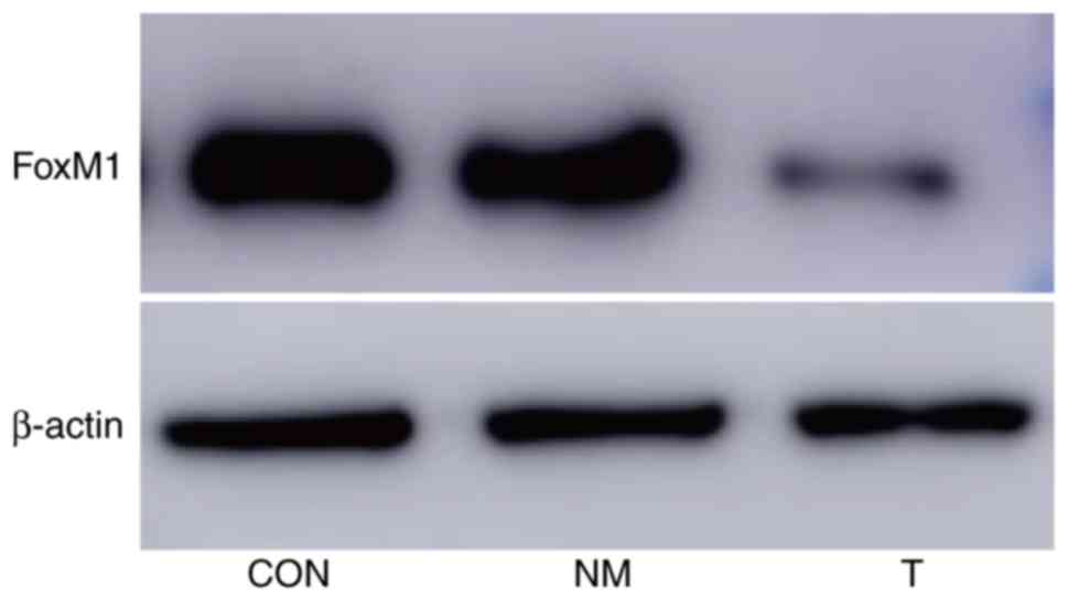
Expression of FoxM1 protein
The expression of FoxM1 protein in group T (cells
transfected with siRNA targeting FoxM1) was lower compared with the
other two groups (P<0.05); however, there was no statistical
significance between the CON and NM groups (P>0.05; Fig. 2; Table
I).
 | Table I.Effect of FoxM1 silencing on protein
expression (n=5). |
Table I.
Effect of FoxM1 silencing on protein
expression (n=5).
| Group | FoxM1 | F | P-value |
|---|
| CON |
0.96±0.01a | 1818.57 | <0.001 |
| NM |
0.95±0.01a |
|
|
| T | 0.27±0.03 |
|
|
Cell proliferation
Cell growth in group T was significantly inhibited
compared with the other two groups (P<0.05). There was no
significant difference between group NM and group CON (P>0.05;
Table II).
| Table II.Effect of FoxM1 silencing on K1 cell
growth (n=6). |
Table II.
Effect of FoxM1 silencing on K1 cell
growth (n=6).
|
| Group |
|
|
|---|
|
|
|
|
|
|---|
| Time-point (h) | CON | NM | T | F | P-value |
|---|
| 24 |
0.48±0.01a |
0.46±0.02a | 0.36±0.02 | 73.29 | <0.001 |
| 48 |
0.66±0.04a |
0.64±0.02a | 0.48±0.01 | 72.50 | <0.001 |
| 72 |
0.72±0.03a |
0.71±0.01a | 0.57±0.02 | 99.11 | <0.001 |
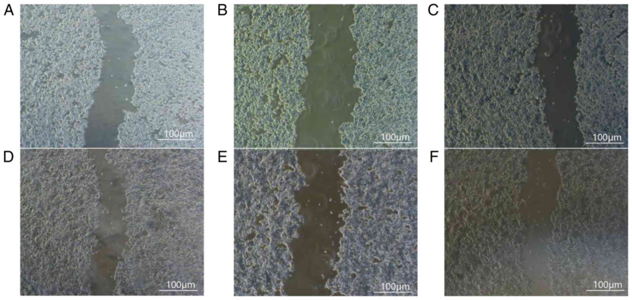
Cell migration
After 24 h, the relative distance between the
scratches in group CON (0.83±0.01) was significantly shorter than
in group T (0.93±0.01; P<0.05), indicating that the migration
speed of the K1 cells was significantly slower following FoxM1
silencing compared with the CON and NM groups. Comparison of cell
migration ability between the CON and NM groups indicated no
statistically significant difference (P>0.05; Fig. 3; Table
III).
| Table III.Effect of FoxM1 silencing on cell
migration (n=9). |
Table III.
Effect of FoxM1 silencing on cell
migration (n=9).
| Group | Scratch area at 24
h | F | P-value |
|---|
| CON |
0.83±0.01a | 137.71 | <0.001 |
| NM |
0.82±0.01a |
|
|
| T | 0.93±0.01 |
|
|
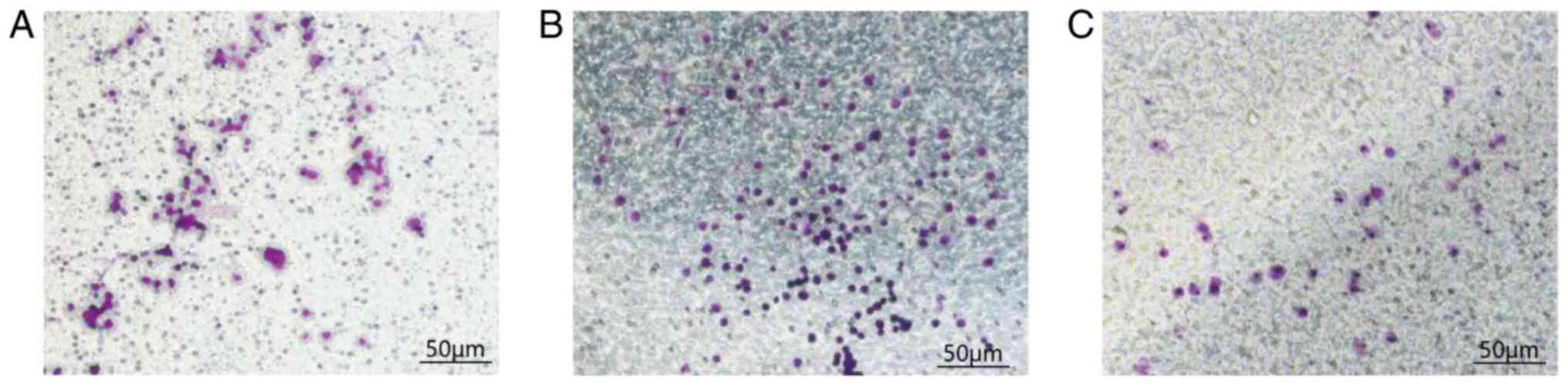
Detection of in vitro invasive ability
of tumor cells
There was no significant difference in the number of
cells penetrating the matrix membrane in the NM group compared with
the meaningless sequence group. This experiment confirmed that
silencing FoxM1 significantly reduced the invasive ability of K1
human thyroid cancer cells. The mean numbers of the cells that
penetrated the upper chamber, and reached the lower chamber were
37.20±3.96, 85.40±5.13 and 92.40±3.05/field in the T, NM and CON
groups, respectively. There was a statistical significance between
the CON and T groups (P<0.001), indicating that compared with
group CON, the number of cells penetrating the matrix membrane in
group T was significantly reduced. There was no statistical
significance between the CON and NM groups (P=0.20; Fig. 4 and Table
IV).
| Table IV.Effect of FoxM1 silencing on invasion
in a Transwell chamber assay (n=20). |
Table IV.
Effect of FoxM1 silencing on invasion
in a Transwell chamber assay (n=20).
| Group | Amount of cells
(cells/field) | F | P-value |
|---|
| CON |
92.40±3.05a | 264.09 | <0.001 |
| NM |
85.40±5.13a |
|
|
| T | 37.20±3.96 |
|
|
Discussion
The results of the present study indicated that the
proliferation, migration, and invasion of PTC cells are suppressed
following FoxM1-silencing. The results are consistent with prior
observations that inhibition of FoxM1 expression can alter the
biological changes in tumor cell proliferation, migration,
invasion, and other biological changes (16). Therefore, FoxM1 appears to promote
several cancer-associated functions of PTC cells. The incidence
rate of thyroid cancers in developing countries are high, as it
accounts for only 1% of all malignant tumors (17). They remain however, the most common
endocrine tumor, as thyroid cancers currently rank as the 10th most
frequent tumor disease in China (17), among which PTC is the most common,
accounting for ~70% of all types of thyroid cancers. Furthermore,
the incidence of PTC is increasing (18). Malignancy of PTC is less common and
its growth is relatively slow, making it prone to lymph node
metastasis (19). Surgery is the
most effective treatment method for thyroid cancers. However, the
complex anatomy, rich blood supply and the endocrine effects of PTC
can result in a number of postoperative complications (20). The postoperative 10-year survival
rate is high; however, the recurrence rate is also high, which
results in an increased mortality rate over time (21). Therefore, the need for novel targeted
therapeutic drugs has become urgent. Physiological processes,
including proliferation and apoptosis, are abnormal in tumor cells
(22). FoxM1 is a member of the
Forkhead transcription factor family (23). FoxM1 can regulate a number of
metabolic-associated processes to maintain the balance of tumor
cell proliferation and energy metabolism. Furthermore, FoxM1 is
also involved in the regulation of tumor cell apoptosis,
metastasis, and other related processes, and is associated with the
metastasis, angiogenesis and epithelial-mesenchymal transition of
tumor cells (24,25). Abnormal expression of FoxM1 is
associated with poor clinical classification and poor prognosis in
patients with cancer (26).
Based on the aforementioned characteristics, a
quantitative index diagnosis system of malignant tumors based on
the FoxM1 gene was previously established (27). Subsequent studies have documented an
accuracy of 94% against early oral, skin and neck cancers.
Therefore, FoxM1 gene expression can be suggested as a reliable
method for the early diagnosis of associated tumors and has great
practical potential in the clinical diagnosis and treatment of
tumors. FoxM1 has the same effect on other thyroid cancer cell
lines (28) as its role in TPC-1
cell line has been demonstrated. Alvarez-Fernández and Medema
(16) examined the underlying
molecular mechanism of FoxM1, therefore this was not the focus of
the present study; however, to the best of our knowledge, cell
scratch test data have not been provided in earlier studies. The
cell scratch test gauges the ability of cells, including cancer
cells, to migrate. Metastasis of cancer often results in a poor
prognosis. Therefore, controlling the spread of cancer by blunting
metastasis is a prudent strategy for cancer control and
prevention.
In summary, FoxM1 is crucial in the occurrence and
growth of PTC, and may be a valuable target for treatment. This
study demonstrated the effect of FoxM1 on the proliferation,
migration and invasion ability of PTC cells, however it was not
able to demonstrate the role of FoxM1 in PTC cells. Therefore,
further examination of the other biological effects of FoxM1 on PTC
cells is required, in order to verify the results of the present
study.
Acknowledgements
Not applicable.
Funding
This study was funded by the Hebei Institute of
Science and Technology Information (grant no. 20150523).
Availability of data and materials
All the data generated or analyzed in this study are
included in this published article.
Authors' contributions
GZ and XW made substantial contributions to
conception and design; GZ, YZ and MC acquired data; XW, JC and WC
analysed and interpreted data; GZ, GY, YZ, WC, MC and QL were
involved in drafting the manuscript and revising it critically for
important intellectual content; GY and YZ were responsible for the
preparation of experimental materials and equipment, WC and MC were
responsible for the final data statistics and collation, QL, GY and
YZ were responsible for performing the experiment, WC and YZ were
responsible for reviewing the relevant literature. GZ gave final
approval of the version to be published.
Ethics approval and consent to
participate
Not applicable.
Patient consent for publication
Not applicable.
Competing interests
The authors declare that they have no competing
interests.
References
|
1
|
Hedinger C, Williams ED and Sobin LH: The
WHO histological classification of thyroid tumors: A commentary on
the second edition. Cancer. 63:908–911. 1989. View Article : Google Scholar : PubMed/NCBI
|
|
2
|
Zhu T, Xu J, Tian T, Niu F and An CM:
Interpretation of the 2017 US Preventive Services Working Group
Recommendations for Thyroid Cancer Screening. Cancer Res.
45:710–714. 2018.
|
|
3
|
Stewart B and Wild C: World cancer report
2014. International Agency for Research on Cancer. 2014.
|
|
4
|
Kalinichenko VV, Major ML, Wang X,
Petrovic V, Kuechle J, Yoder HM, Dennewitz MB, Shin B, Datta A,
Raychaudhuri P and Costa RH: Foxm1b transcription factor is
essential for development of hepatocellular carcinomas and is
negatively regulated by the p19ARF tumor suppressor. Genes Dev.
18:830–850. 2004. View Article : Google Scholar : PubMed/NCBI
|
|
5
|
Lee S, Park YY, Kim SH, Nguyen OT, Yoo YS,
Chan GK, Sun X and Cho H: Human mitochondrial Fis1 links to cell
cycle regulators at G2/M transition. Cell Mol Life Sci. 71:711–725.
2014. View Article : Google Scholar : PubMed/NCBI
|
|
6
|
Zhang J, Yuan C, Wu J, Elsayed Z and Fu Z:
Polo-like kinase 1-mediated phosphorylation of Forkhead box protein
M1b antagonizes Its SUMOylation and facilitates its mitotic
function. J Biol Chem. 290:3708–3719. 2015. View Article : Google Scholar : PubMed/NCBI
|
|
7
|
Liu M, Dai B, Kang SH, Ban K, Huang FJ,
Lang FF, Aldape KD, Xie TX, Pelloski CE, Xie K, et al: FoxM1B is
overexpressed in human glioblastomas and critically regulates the
tumorigenicity of glioma cells. Cancer Res. 66:3593–3602. 2006.
View Article : Google Scholar : PubMed/NCBI
|
|
8
|
Cui J, Shi M, Xie D, Wei D, Jia Z, Zheng
S, Gao Y, Huang S and Xie K: FOXM1 promotes the Warburg effect and
pancreatic cancer progression via transactivation of LDHA
expression. Clin Cancer Res. 20:2595–2606. 2014. View Article : Google Scholar : PubMed/NCBI
|
|
9
|
Yung MM, Chan DW, Liu VW, Yao KM and Ngan
HY: Activation of AMPK inhibits cervical cancer cell growth through
AKT/FOXO3a/FOXM1 signaling cascade. BMC Cancer. 13:3272013.
View Article : Google Scholar : PubMed/NCBI
|
|
10
|
Khongkow P, Gomes AR, Gong C, Man EP,
Tsang JW, Zhao F, Monteiro LJ, Coombes RC, Medema RH, Khoo US and
Lam EW: Paclitaxel targets FOXM1 to regulate KIF20A in mitotic
catastrophe and breast cancer paclitaxel resistance. Oncogene.
35:990–1002. 2016. View Article : Google Scholar : PubMed/NCBI
|
|
11
|
Song IS, Jeong YJ and Han J: Mitochondrial
metabolism in cancer stem cells: A therapeutic target for colon
cancer. BMB Rep. 48:539–540. 2015. View Article : Google Scholar : PubMed/NCBI
|
|
12
|
Jin H, Park MH and Kim SM:
3,3′-Diindolylmethane potentiates paclitaxel-induced antitumor
effects on gastric cancer cells through the Akt/FOXM1 signaling
cascade. Oncol Rep. 33:2031–2036. 2015. View Article : Google Scholar : PubMed/NCBI
|
|
13
|
Huang X, Qin J and Lu S: Up-regulation of
miR-877 induced by paclitaxel inhibits hepatocellular carcinoma
cell proliferation though targeting FOXM1. Int J Clin Exp Pathol.
8:1515–1524. 2015.PubMed/NCBI
|
|
14
|
Khongkow P, Gomes AR, Gong C, Man EP,
Tsang JW, Zhao F, Monteiro LJ, Coombes RC, Medema RH, Khoo US and
Lam EW: Paclitaxel targets FOXM1 to regulate KIF20A in mitotic
catastrophe and breast cancer paclitaxel resistance. Oncogene.
35:990–1002. 2015. View Article : Google Scholar : PubMed/NCBI
|
|
15
|
Jiang Y, Liao Y, He H, Xin Q, Tu Z, Kong
S, Cui T, Wang B, Quan S, Li B, et al: FoxM1 Directs STAT3
expression essential for human endometrial stromal decidualization.
Sci Rep. 5:137352015. View Article : Google Scholar : PubMed/NCBI
|
|
16
|
Alvarez-Fernández M and Medema RH: Novel
functions of FoxM1: From molecular mechanisms to cancer therapy.
Front Oncol. 3:302013. View Article : Google Scholar : PubMed/NCBI
|
|
17
|
Chen WQ, Zheng RS, Zhang SW, Li N, Zhao P,
Li GL, Wu LY and He J: Report of incidence and mortality in china
cancer registries, 2008. Chin J Cancer Res. 24:171–180. 2012.
View Article : Google Scholar : PubMed/NCBI
|
|
18
|
McLeod DS, Sawka AM and Cooper DS:
Controversies in primary treatment of low-risk papillary thyroid
cancer. Lancet. 381:1046–1057. 2013. View Article : Google Scholar : PubMed/NCBI
|
|
19
|
Xue S, Wang P, Liu J, Li R, Zhang L and
Chen G: Prophylactic central lymph node dissection in cN0 patients
with papillary thyroid carcinoma: A retrospective study in China.
Asian J Surg. 39:131–136. 2016. View Article : Google Scholar : PubMed/NCBI
|
|
20
|
Yun JS, Lee YS, Jung JJ, Nam KH, Chung WY,
Chang HS and Park CS: The Zuckerkandl's tubercle: A useful
anatomical landmark for detecting both the recurrent laryngeal
nerve and the superior parathyroid during thyroid surgery. Endocr
J. 55:925–930. 2008. View Article : Google Scholar : PubMed/NCBI
|
|
21
|
Tuttle RM, Ball DW, Byrd D, Dilawari RA,
Doherty GM, Duh QY, Ehya H, Farrar WB, Haddad RI, Kandeel F, et al:
Thyroid carcinoma. J Natl Compr Canc Netw. 8:1228–1274. 2010.
View Article : Google Scholar : PubMed/NCBI
|
|
22
|
Dias RC, Marangoni DV, Riley LW and
Moreira BM: Identification of uropathogenic Escherichia coli clonal
group A (CgA) in hospitalised patients. Mem Inst Oswaldo Cruz.
104:787–789. 2009. View Article : Google Scholar : PubMed/NCBI
|
|
23
|
Laoukili J, Kooistra MR, Brás A, Kauw J,
Kerkhoven RM, Morrison A, Clevers H and Medema RH: FoxM1 is
required for execution of the mitotic programme and chromosome
stability. Nat Cell Biol. 7:126–136. 2005. View Article : Google Scholar : PubMed/NCBI
|
|
24
|
Chen H, Zou Y, Yang H, Wang J and Pan H:
Downregulation of FoxM1 inhibits proliferation, invasion and
angiogenesis of HeLa cells in vitro and in vivo. Int
J Oncol. 45:2355–2364. 2014. View Article : Google Scholar : PubMed/NCBI
|
|
25
|
Ahmad A, Wang Z, Kong D, Ali S, Li Y,
Banerjee S, Ali R and Sarkar FH: FoxM1 down-regulation leads to
inhibition of proliferation, migration and invasion of breast
cancer cells through the modulation of extra-cellular matrix
degrading factors. Breast Cancer Res Treat. 158:6072016. View Article : Google Scholar : PubMed/NCBI
|
|
26
|
Millour J, Constantinidou D, Stavropoulou
AV, Wilson MS, Myatt SS, Kwok JM, Sivanandan K, Coombes RC, Medema
RH, Hartman J, et al: FOXM1 is a transcriptional target of ERalpha
and has a critical role in breast cancer endocrine sensitivity and
resistance. Oncogene. 29:2983–2995. 2010. View Article : Google Scholar : PubMed/NCBI
|
|
27
|
The MT, Hutchison IL, Costea DE,
Neppelberg E, Liavaag PG, Purdie K, Harwood C, Wan H, Odell EW,
Hackshaw A and Waseem A: Exploiting FOXM1-orchestrated molecular
network for early squamous cell carcinoma diagnosis and prognosis.
Int J Cancer. 132:2095–2106. 2013. View Article : Google Scholar : PubMed/NCBI
|
|
28
|
Yuan H, Min Z and Chen J: Study on the
relationship between the expression of FoxM1 gene and the activity
and invasion of TPC-1 cells in papillary thyroid carcinoma. Chin J
Endocrine Surg. 8:33–37. 2014.
|