Introduction
Lung cancer is associated with high mortality and
its prevalence has increased worldwide in the past ten years
(1,2). Moreover, the incidence and mortality
rates of lung cancer have increased significantly in China
(3,4). Histologically, non-small cell lung
cancer (NSCLC) consists of several subtypes, including lung
adenocarcinoma (LUAD), lung squamous cell carcinoma (LUSC), large
cell lung cancer and other rarer types, of which LUAD and LUSC are
the two main histological types (5).
Although advances have been made in early diagnosis and systematic
therapy, the 5-year survival rate for patients with NSCLC remains
unsatisfactory (6). Therefore,
investigating the pathogenesis of NSCLC may aid the identification
of more sensitive and specific biomarkers as well as therapeutic
targets for patients with NSCLC.
With the development of abundant open data
resources, it is convenient and effective for researchers to screen
cancer-related therapeutic targets (7). Through an analysis of The Cancer Genome
Atlas (TCGA) database, the present study identified tripartite
motif-containing protein 59 (TRIM59) as a novel lung cancer-related
candidate target. The tripartite motif (TRIM) family of proteins
comprises >70 members that are evolutionarily conserved and
share RING, B-Box and coiled-coil (RBCC) multiple domains. TRIM
proteins consist of a common N-terminal Really Interesting New Gene
(RING) finger domain, one or two B-box motifs and a coiled-coil
region (8,9). Due to the highly conserved RING domain,
the majority of the proteins are E3 ubiquitin ligases that promote
post-translational modifications of various substrates (10) and affect a range of cellular
processes, including cell growth, development, differentiation,
apoptosis, inflammation and immunity (11). TRIM59 not only participates in
regulating protein expression, but also in the malignant behavior
of tumor cells (12).
A number of studies have reported that TRIM59 acts
as an oncogene in various types of cancer. Lin et al
(13) reported that knockdown of
TRIM59 inhibited tumor growth in prostate cancer. It was previously
demonstrated that downregulation of TRIM59 inhibited proliferation,
migration and invasion of breast cancer cells (14). Additionally, TRIM59 induced
epithelial-to-mesenchymal transition, and promoted migration and
invasion of medulloblastoma cells through the phosphoinositide
3-kinase (PI3K)/protein kinase B (AKT) signaling pathway (15). However, to the best of our knowledge,
there is little information available on the clinical significance
and prognostic value of TRIM59 expression in NSCLC. The present
study determined the expression of TRIM59 in NSCLC, and
investigated its association with the occurrence, development and
prognosis of NSCLC.
Materials and methods
TRIM59 expression in published TCGA
databases
TCGA (https://www.cancer.gov/) is a collaboration between
The National Cancer Institute (NCI) and The National Human Genome
Research Institute, which rigorously control the quality of the
datasets. Therefore, TCGA is a reliable source of information for
disease analysis (16). To evaluate
and analyze the TRIM59 expression level, three datasets, named
LUNG_exp_HiSeq-V2-2015-02-24, LUAD_exp_HiSeq-V2-2015-02-24 and
LUSC_exp_HiSeq-V2-2015-02-24, were downloaded from the University
of California, Santa Cruz Cancer Browser (https://genome-cancer.ucsc.edu). These datasets
contain a list of cancer-related characteristic information of
1,013 NSCLC tissue samples, which include 108 paired NSCLC tissue
samples, 57 pairs of LUAD tissues and 51 pairs of LUSC tissues. By
analyzing the files named ‘genomic Matrix’ in these datasets, the
mRNA expression levels of TRIM59 were obtained.
Patients and construction of a tissue
microarray (TMA)
In total, 140 NSCLC tissue and 10 normal adjacent
tissue samples were obtained from patients who had undergone
surgical resection at The Department of Thoracic Surgery of
Zhongshan Hospital, Fudan University between January 2005 and
December 2005. Complete clinical information was available for all
patients (112 male and 28 female) and the mean age of the patients
with NSCLC was 60.1 years (range, 26–79 years). Patients were
classified according to the TNM classification system, formulated
jointly by the American Joint Committee on Cancer and the Union for
International Cancer Control (17).
The clinical follow-up was recorded until July 2013. As previously
described by Gao et al (18),
the tissue samples from the 140 primary NSCLC cases and 10 normal
adjacent lung tissues were arranged in rows and columns to
construct a TMA. Patients involved in the present study had not
received chemotherapy, radiotherapy or biotherapy before
surgery.
Immunohistochemical staining and
quantification analysis
The standard indirect immunoperoxidase procedures
(Envision Plus; Dako; Agilent Technologies, Inc.) were adopted for
immunohistochemistry to detect the expression of TRIM59 in NSCLC.
Paraffin specimens were cut into slices (4-µm thick), which were
mounted on slides, baked, deparaffinized and hydrated following
conventional methods. Then, endogenous peroxidase activity was
quenched by incubating the sections with 3%
H2O2 for 20 min at room temperature. Sections
were then subjected to heat-induced antigen retrieval in 10 mM
citrate buffer (pH 6.0) for 10 min at 100°C. Slides were incubated
in 10 mM TBS with 4% normal goat serum (Abcam) for 1 h at room
temperature and incubated with the primary antibody against TRIM59
(1:500; cat. no. YT4737; ImmunoWay Biotechnology Company) at 4°C
overnight. Following primary antibody incubation, the sections were
washed with TBS and incubated with an HRP-conjugated goat anti
rabbit secondary antibody (1:500; cat. no. RS0002; ImmunoWay
Biotechnology Company) for 1 h at 37°C. The sections were stained
with DAB (3,3′-diaminobenzidine) (Dako; Agilent Technologies, Inc.)
at 37°C for 10 min and weakly counterstained with hematoxylin at
37°C for 1 min, dehydrated and covered with a coverslip. A light
microscope (Nikon Corporation; magnification, ×200) was used to
acquire the images. Breast cancer tissue was used as a strong
positive control for TRIM59 (19).
For the negative control, PBS was added to the slides instead of
primary antibody.
To quantify the expression of TRIM59 protein in
NSCLC tissues, four fields in each section were selected for
immunohistochemical scoring (magnification, ×200). Two experienced
pathologists assessed the immunohistochemical score independently.
Based on a protocol developed by Xu et al (20), the intensity and proportion of
positive tumor cells in the sections were used to calculate scores.
The cytoplasmic staining intensity was scored as follows: i) 0 (no
staining); ii) 1 (weak staining, light yellow); iii) 2 (moderate
staining, yellow brown); and iv) 3 (strong staining, brown). The
proportion of cytoplasmic positive cells was defined as 0–100% and
was classified as follows: i) 1 (0–25% positive cells); ii) 2
(2–50% positive cells); iii) 3 (51–75% positive cells); and iv) 4
(76–100% positive cells). Finally, the staining intensity was
multiplied by the proportion of positive cells to calculate the
scores that were used to represent the expression levels of TRIM59.
The higher the immunohistochemical scoring, the greater the
expression of TRIM59. According to the scores, the 140 patients
with NSCLC were classified into high expression (score ≥4) and low
expression (score <4) groups.
Statistical analysis
The χ2 test was used to investigate the
association between TRIM59 expression and various
clinicopathological parameters. The period from primary surgery
until the death of the patient or the latest follow-up was defined
as the overall survival (OS) time. Survival analysis was performed
using the Kaplan-Meier method and differences were tested using a
log-rank test. Univariate and Multivariate Wald test and cox
proportional hazard regression models were constructed to identify
the independent factors with a significant impact on patient OS
time. A paired t-test was used to analyze TRIM59 mRNA expression in
tumor and adjacent lung tissues. Statistical analyses were
conducted using SPSS software (version 19.0; IBM, Corp.) and
GraphPad Prism software (version 6.0; GraphPad Software, Inc.).
P<0.05 was considered to indicate a statistically significant
difference. All results are expressed as the mean ± standard
deviation.
Results
Characteristics of the patients
The clinicopathological characteristics of the 140
patients with NSCLC in the present study are presented in Table I. The mean age of the patients was
60.1±6.4 years (range, 26–79 years). The majority of the patients
with NSCLC were male and >50% of the tumors were LUSC. In total,
there were 80 LUSC (grade 1–3), 46 LUAD (grade 1–3), five
adenosquamous cell carcinoma, three large-cell neuroendocrine
carcinoma, three bronchiolo-alveolar carcinoma, two mucoepidermoid
carcinoma, one sarcomatoid carcinoma and 10 normal lung tissues.
The clinical follow-up was recorded until July 2013. There were 84
local recurrences or distant metastases and 91 deaths by the end of
the follow-up period.
 | Table I.Clinical and histological features of
the 140 patients with NSCLC in the present study. |
Table I.
Clinical and histological features of
the 140 patients with NSCLC in the present study.
| Characteristic | Number of
patients | % |
|---|
| Age
(years)a | 60 (26–79) | – |
| Sex |
|
Male | 112 | 80 |
|
Female | 28 | 20 |
| Location |
|
|
|
Left | 64 | 46 |
|
Right | 76 | 54 |
| Pathological
type |
|
|
|
Squamous cell carcinoma | 80 | 57 |
|
Adenocarcinoma | 46 | 33 |
|
Adenosquamous cell
carcinoma | 5 | 4 |
|
Bronchiolo-alveolar
carcinoma | 3 | 2 |
|
Sarcomatoid carcinoma | 1 | 1 |
|
Neuroendocrine carcinoma | 3 | 2 |
|
Mucoepidermoid carcinoma | 2 | 1 |
|
Differentiation |
|
|
|
Poorly | 59 | 42 |
|
Moderate | 49 | 35 |
|
Well | 32 | 23 |
| T stage |
|
|
| 1 | 15 | 11 |
| 2 | 105 | 75 |
| 3 | 20 | 14 |
| N stage |
|
|
| 0 | 82 | 59 |
| 1 | 31 | 22 |
| 2 | 27 | 19 |
| NSCLC stage |
|
|
| I | 67 | 48 |
| II | 44 | 31 |
|
III | 29 | 21 |
| Follow-up period
(months)a | 45 (3–101) | – |
TRIM59 expression in NSCLC
tissues
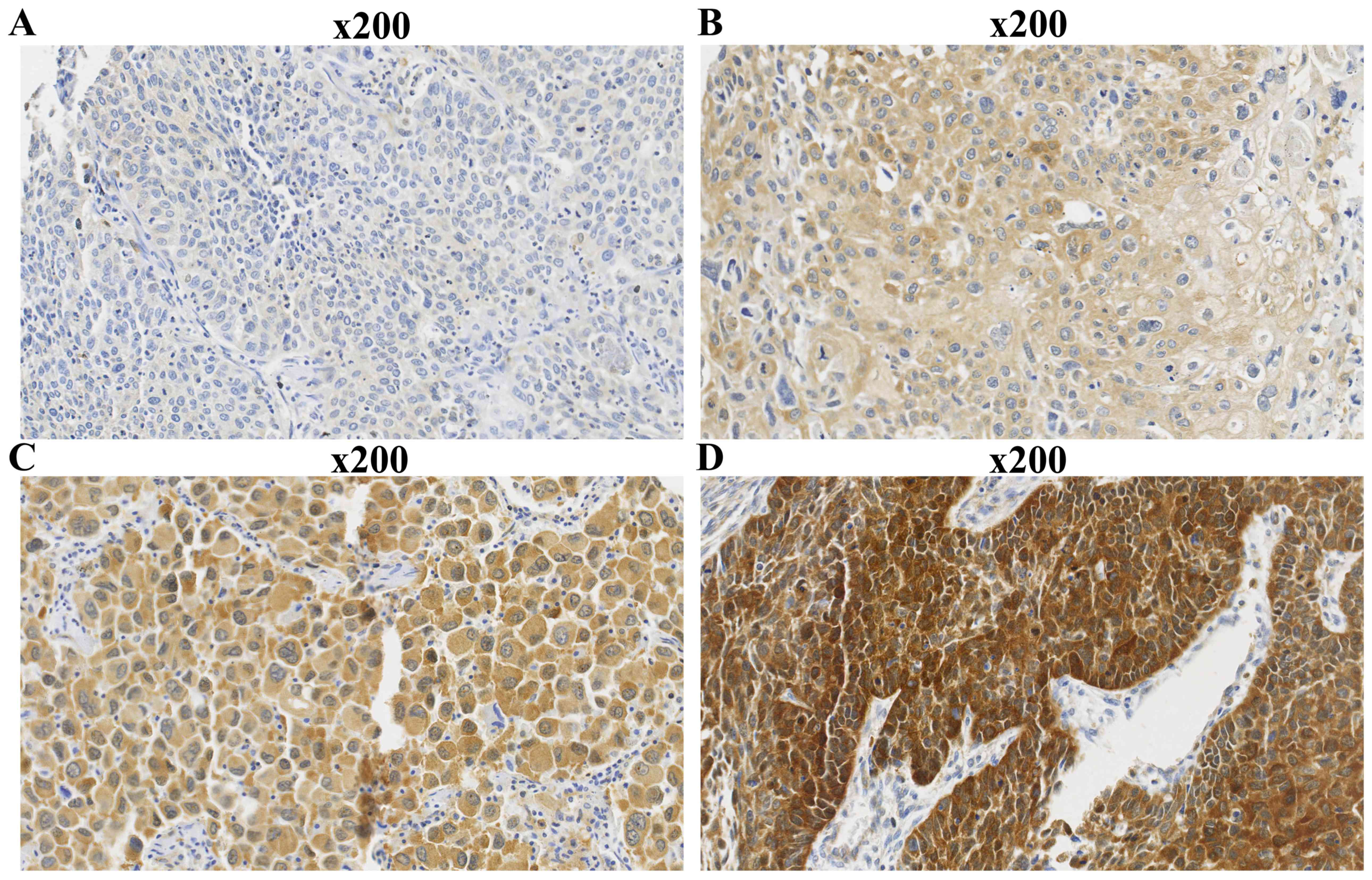
The immunohistochemical results showed that TRIM59
was mainly located in the cytoplasm of tumor cells, with low
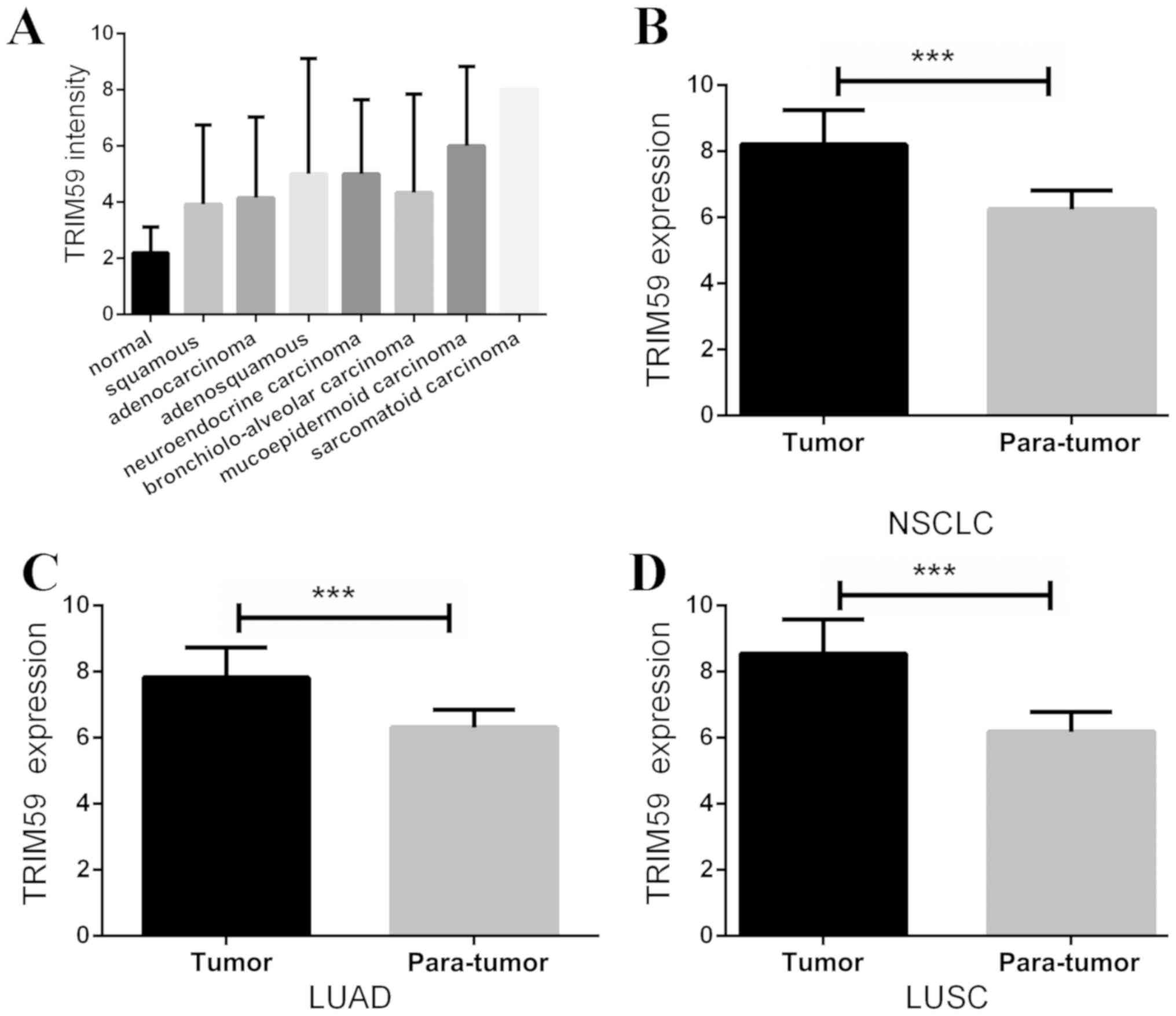
expression in normal lung tissue cells (Figs. 1 and 2). The average scores for all types of lung
cancer tissues were 2–3-fold higher than normal lung tissue
(Fig. 3A). The average scores for
LUSC and LUAD were 3.93±2.82 and 4.15±2.88, respectively, while the
normal lung score was 2.20±0.92. Among the 140 NSCLC tissue
samples, the high-TRIM59 expression group comprised 83 samples,
which was significantly higher than that found in normal tissues
(59.3 vs. 10.0%; χ2 value, 9.201; P=0.0024; Fig. 3). To further verify this conclusion,
TCGA datasets were analyzed. It was identified that TRIM59 was on
average 1.32-fold more highly expressed in NSCLC tissue samples
compared with paratumour samples (P<0.0001; Fig. 3B). Similarly, TRIM59 was on average
1.24-fold more highly expressed in LUAD (P<0.0001; Fig. 3C) and 1.40-fold more highly expressed
in LUSC, compared with paratumor samples (P<0.0001; Fig. 3D).
TRIM59 expression and
clinicopathological characteristics
The 140 patients with NSCLC were classified into low
and high TRIM59 expression groups and the clinicopathological
characteristics of the patients were compared. As shown in Table II, a significant association between
TRIM59 expression and tumor differentiation was identified
(P=0.012). However, no statistically significant associations were
found between TRIM59 and tumor size (P=0.781), lymph node status
(P=0.684), tumor stage (P=0.457) or any other clinicopathological
characteristics.
| Table II.Association between TRIM59 expression
and clinicopathological factors. |
Table II.
Association between TRIM59 expression
and clinicopathological factors.
| Clinicopathological
factor | Low TRIM59
expression | High TRIM59
expression | P-value |
|---|
| Age (years) | 59.8±10.4 | 60.3±9.9 | 0.265 |
| Sex |
|
|
|
|
Male | 46 | 66 | 0.863 |
|
Female | 11 | 17 |
|
| Location |
|
|
|
|
Left | 23 | 41 | 0.291 |
|
Right | 34 | 42 |
|
| Histological
typea |
|
|
|
|
Squamous | 34 | 46 | 0.896 |
|
Adenocarcinoma | 19 | 27 |
|
|
Differentiation |
|
|
|
|
Poorly | 26 | 33 |
|
|
Moderate | 25 | 24 | 0.012 |
|
Well | 6 | 26 |
|
| pT |
|
|
|
| 1 | 7 | 8 |
|
| 2 | 41 | 64 | 0.781 |
| 3 | 9 | 11 |
|
| pN |
|
|
|
| 0 | 35 | 47 |
|
| 1 | 13 | 18 | 0.684 |
| 2 | 9 | 18 |
|
| TNM stage |
|
|
|
| I | 28 | 39 |
|
| II | 20 | 24 | 0.457 |
|
III | 9 | 20 |
|
Prognostic value of TRIM59 in
NSCLC
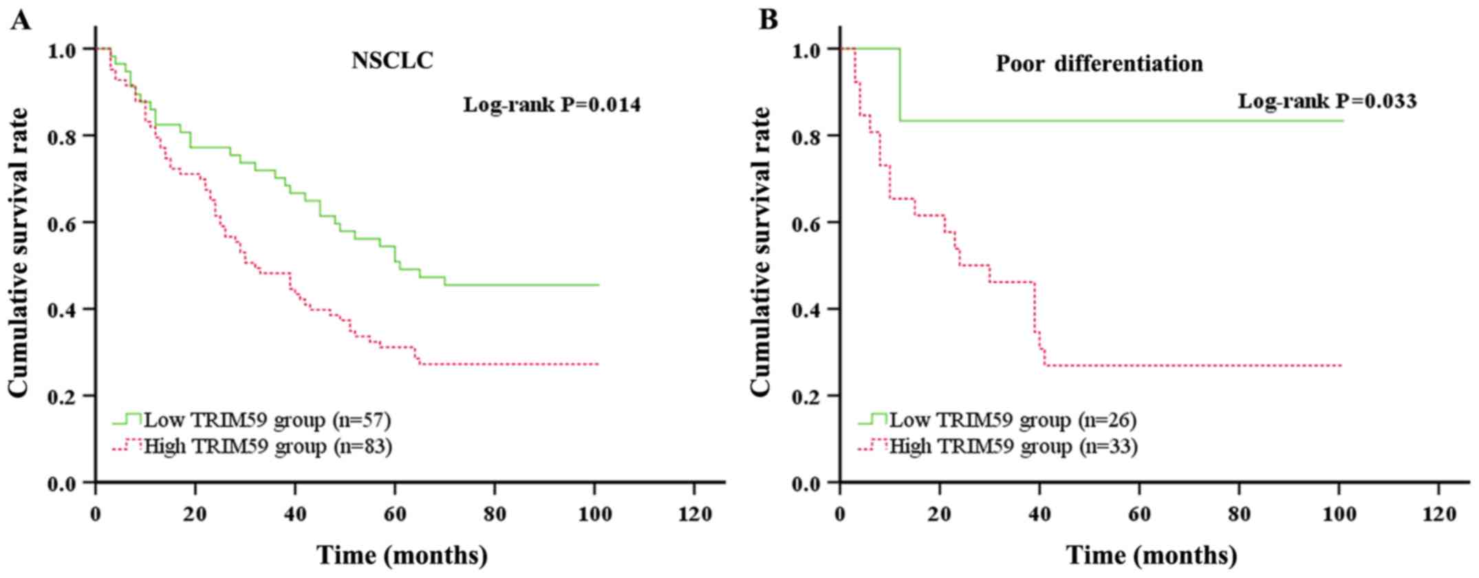
A long-term follow-up was conducted to examine the
association between TRIM59 expression and prognosis for patients
with NSCLC. The average OS time of patients in the high and low
TRIM59 expression groups were 46.586±4.011 and 63.378±4.994,
respectively (P=0.014; Fig. 4A).
Moreover, patients with poor differentiation had worse prognosis
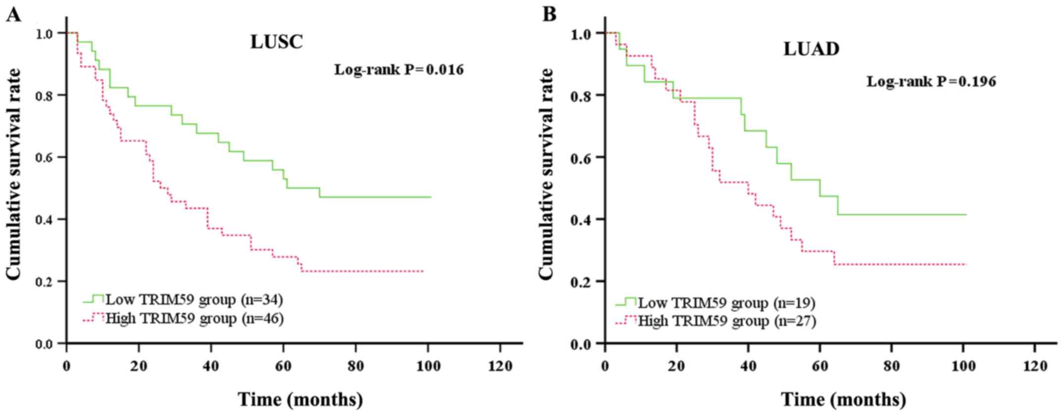
(P=0.033; Fig. 4B). Furthermore,
Kaplan-Meier survival analyses of LUSC and LUAD were conducted. The
results revealed that high expression of TRIM59 was significantly
associated with worse OS time in patients with LUSC (P=0.016;
Fig. 5A), whereas no association was
detected in patients with LUAD (P=0.196; Fig. 5B). Finally, univariate and
multivariate analyses were performed. As shown in Table III, high TRIM59 expression and
lymph node metastasis had independent prognostic values (P=0.018
and P=0.038, respectively).
| Table III.Multivariate Cox regression analysis
for potential factors influencing overall survival. |
Table III.
Multivariate Cox regression analysis
for potential factors influencing overall survival.
| A, Univariate
analysis |
|---|
|
|---|
|
|
|
|
|
|
| 95% CI for OR |
|---|
|
|
|
|
|
|
|
|
|---|
| Parameter | B | SE | Wald | P-value | OR | Lower | Upper |
|---|
| Age | 0.025 | 0.011 | 5.274 | 0.022 | 1.026 | 1.004 | 1.048 |
| Sex | −0.240 | 0.275 | 0.762 | 0.383 | 0.786 | 0.458 | 1.349 |
| Location | −0.216 | 0.210 | 1.059 | 0.303 | 0.806 | 0.534 | 1.216 |
| Histological
type | −0.123 | 0.213 | 0.337 | 0.562 | 0.884 | 0.583 | 1.341 |
|
Differentiation | −0.014 | 0.139 | 0.010 | 0.922 | 0.987 | 0.752 | 1.295 |
| pT | 0.839 | 0.423 | 3.940 | 0.047 | 2.314 | 1.011 | 5.298 |
| pN | 0.825 | 0.212 | 15.166 | 0.000 | 2.281 | 1.506 | 3.454 |
| Stage | 0.655 | 0.215 | 9.275 | 0.002 | 1.926 | 1.263 | 2.937 |
| TRIM59 | 0.535 | 0.222 | 5.799 | 0.016 | 1.708 | 1.105 | 2.640 |
|
| B, Multivariate
analysis |
|
|
|
|
|
|
|
| 95% CI for
OR |
|
|
|
|
|
|
|
|
|
Parameter | B | SE | Wald | P-value | OR | Lower | Upper |
|
| Age | 0.021 | 0.011 | 3.928 | 0.047 | 1.022 | 1.000 | 1.043 |
| pT | 0.682 | 0.430 | 2.520 | 0.122 | 1.978 | 0.852 | 4.591 |
| pN | 0.802 | 0.386 | 4.304 | 0.038 | 2.229 | 1.045 | 4.754 |
| Stage | −0.040 | 0.395 | 0.010 | 0.919 | 0.961 | 0.443 | 2.083 |
| TRIM59 | 0.528 | 0.223 | 5.597 | 0.018 | 1.696 | 1.095 | 2.626 |
Discussion
The TRIM family consists of a series of highly
conserved motif proteins with three domains that include a RING
finger, one or two B-box motifs and a coiled-coil region (21). Due to the variability of the
C-terminal domain of TRIM, the TRIM family proteins are further
divided into 11 subfamilies (22,23). In
the past decade, the role of TRIM proteins in the innate immunity
response to viral infection has attracted increasing attention
(24–26). Previous studies have demonstrated
that TRIM proteins are involved in several cell functions and
participate in the process of ubiquitination as E3 ubiquitin
ligases (27,28). For example, TRIM59 was revealed to
regulate autophagy by regulating the transcription and
ubiquitination of beclin 1, which in turn affected the progression
of NSCLC (29). Additionally, Zhou
et al (30) reported that
TRIM59 is upregulated in gastric cancer, and promotes the
ubiquitination and degradation of p53 that affects tumor growth.
Ubiquitination, as a post-translational modification, is involved
in many cellular processes, including signal transduction, protein
quality control, transcription, cell cycle, apoptosis and
development (31).
Certain TRIM proteins play important roles in the
occurrence and development of NSCLC. For example, higher expression
of TRIM29 was associated with worse prognosis in NSCLC (32). Dai et al (33) reported that knockdown of TRIM66
inhibited malignant behavior and epithelial-mesenchymal transition
in NSCLC cells. Additionally, Luo et al (34) reported that TRIM44 promoted NSCLC
development through activation of NF-κB signaling by upregulating
C-X-C motif chemokine 16 and matrix metalloproteinase 9 expression.
Furthermore, overexpression of TRIM44 enhanced the migratory and
invasive abilities of the lung cancer cell lines A549 and H441
(34). Previous studies reported
that TRIM59 was associated with the genesis, development and
prognosis of tumors (12,15,35). For
example, TRIM59 was upregulated and promoted cell proliferation,
migration and invasion in human osteosarcoma (36). Furthermore, it was found that TRIM59
facilitated the proliferation of colorectal cancer and promoted
metastasis via the PI3K/AKT signaling pathway (37). A previous study revealed that
upregulated TRIM59 served as a proto-oncogene and induced the
progression of prostate cancer in transgenic mice (38). The upregulation of TRIM59 not only
enhanced the expression of cyclin A, cyclin E, Bcl-xl, Bcl-2 and
phosphorylated-AKT, and downregulated the expression of p21, p27
and p53, resulting in poor prognosis in breast cancer, but also
affect breast cancer progression via the AKT signaling pathway
(12). The aforementioned studies
suggested that TRIM59 regulates a diverse range of cellular
functions during tumor progression. However, its role in
determining prognosis in NSCLC has not been fully established.
A previous study reported that TRIM59 expression was
significantly increased in various NSCLC cell lines in
vitro; however, data on the expression of TRIM59 in vivo
was lacking. The present study investigated the expression profile
and the prognostic value of TRIM59 in NSCLC. The expression of
TRIM59 in patients with NSCLC was examined using an
immunohistochemical method. Similar to a previous study by
Khatamianfar et al (39), it
was identified that TRIM59 may be a novel multiple cancer biomarker
for the immunohistochemical detection of tumorigenesis. In the
present study, TRIM59 was highly expressed in the majority of NSCLC
tissues and was mainly expressed in the cytoplasm of tumor cells.
TCGA datasets were used to further verify this result. Similar
results were obtained in TCGA datasets, in which the expression of
TRIM59 was significantly different between normal and tumor
tissues. Based on this differential expression, it was hypothesized
that there are functional roles associated with TRIM59. The
patients in the present study were divided into high and low groups
based on TRIM59 expression. It was observed that the TRIM59
expression level was associated with tumor differentiation, while
no association was observed between TRIM59 expression and any other
clinicopathological characteristics. Furthermore, it was identified
that patients with high expression of TRIM59 had a worse prognosis
than those with low TRIM59 expression, especially in patients with
LUSC and patients with poor differentiation. Consistent with the
results of a study performed by Zhan et al (40), TRIM59 promoted the proliferation and
migration of NSCLC cells by upregulating cell cycle-related
proteins, which may affect the prognosis of the patients with
NSCLC. Additionally, the multivariate analysis performed in the
present study indicated that high TRIM59 expression is an
independent prognostic factor for patients with NSCLC.
Collectively, the results suggested that TRIM59 functions as an
oncogene in NSCLC and is associated with the genesis and
development of NSCLC.
In conclusion, the present study revealed that high
TRIM59 expression was associated with worse prognosis in patients
with NSCLC and that TRIM59 may serve as an important prognostic
biomarker in patients with NSCLC. However, the present study had
some limitations. A limited number of normal tissue samples were
analyzed and the research was designed as a clinical retrospective
study without investigating a specific mechanism. It was
hypothesized that a TRIM59-related signaling pathway may
downregulate TRIM59 expression and delay tumor progression.
However, the exact mechanism of TRIM59 in NSCLC remains unclear and
further research is required.
Acknowledgements
Not applicable.
Funding
The present study was supported by The Medical
Scientific Research Foundation of Jiangsu Commission of Health
(grant no. H2018083),The High-Level Medical Talents Training
Project (grant no. 2016CZBJ042) and The Jiangsu Provincial Medical
Youth Talent [Jiangsu Health Scientific Education (2017; grant no.
3)].
Availability of data and materials
The datasets used or analyzed during the present
study are available from the corresponding author on reasonable
request. Moreover, datasets generated and/or analyzed during the
current study are available in the TCGA repository (https://genome-cancer.ucsc.edu).
Authors' contributions
ML, KY and JT conceived and designed the
experiments. ML and ZG performed the experiments. TZ and XM
acquired data and contributed to reagents, materials and analysis
tools. JT and YW analyzed the data. ML wrote the manuscript. KY and
JT supervised the study. All authors read and approved the final
manuscript.
Ethics approval and consent to
participate
The present study was approved by The Research
Ethics Committee of Zhongshan Hospital, Fudan University. All
patients or their family members provided written informed
consent.
Patient consent for publication
Not applicable.
Competing interests
The authors declare that they have no competing
interests.
References
|
1
|
Siegel RL, Miller KD and Jemal A: Cancer
statistics, 2017. CA Cancer J Clin. 67:7–30. 2017. View Article : Google Scholar : PubMed/NCBI
|
|
2
|
Hoffman RM and Sanchez R: Lung cancer
screening. Med Clin North Am. 101:769–785. 2017. View Article : Google Scholar : PubMed/NCBI
|
|
3
|
Chen W, Zheng R, Zeng H and Zhang S:
Epidemiology of lung cancer in China. Thorac Cancer. 6:209–215.
2015. View Article : Google Scholar : PubMed/NCBI
|
|
4
|
Sun KX, Zheng RS, Zeng HM, Zhang SW, Zou
XN, Gu XY, Xia CF, Yang ZX, Li H, Chen WQ and He J: The incidence
and mortality of lung cancer in China, 2014. Zhonghua Zhong Liu Za
Zhi. 40:805–811. 2018.(In Chinese). PubMed/NCBI
|
|
5
|
Mengoli MC, Longo FR, Fraggetta F, Cavazza
A, Dubini A, Alì G, Guddo F, Gilioli E, Bogina G, Nannini N, et al:
The 2015 world health organization classification of lung tumors:
New entities since the 2004 classification. Pathologica. 110:39–67.
2018.PubMed/NCBI
|
|
6
|
Hirsch FR, Suda K, Wiens J and Bunn PA Jr:
New and emerging targeted treatments in advanced non-small-cell
lung cancer. Lancet. 388:1012–1024. 2016. View Article : Google Scholar : PubMed/NCBI
|
|
7
|
Li CY, Xiong DD, Huang CQ, He RQ, Liang
HW, Pan DH, Wang HL, Wang YW, Zhu HW and Chen G: Clinical value of
miR-101-3p and biological analysis of its prospective targets in
breast cancer: A study based on the cancer genome atlas (TCGA) and
bioinformatics. Med Sci Monit. 23:1857–1871. 2017. View Article : Google Scholar : PubMed/NCBI
|
|
8
|
Ikeda K and Inoue S: TRIM proteins as RING
finger E3 ubiquitin ligases. Adv Exp Med Biol. 770:27–37. 2012.
View Article : Google Scholar : PubMed/NCBI
|
|
9
|
Hatakeyama S: TRIM family proteins: Roles
in autophagy, immunity, and carcinogenesis. Trends Biochem Sci.
42:297–311. 2017. View Article : Google Scholar : PubMed/NCBI
|
|
10
|
Rajsbaum R, Garcia-Sastre A and Versteeg
GA: TRIMmunity: The roles of the TRIM E3-ubiquitin ligase family in
innate antiviral immunity. J Mol Biol. 426:1265–1284. 2014.
View Article : Google Scholar : PubMed/NCBI
|
|
11
|
Chen W, Zhao K, Miao C, Xu A, Zhang J, Zhu
J, Su S and Wang Z: Silencing Trim59 inhibits invasion/migration
and epithelial-to-mesenchymal transition via TGF-β/Smad2/3
signaling pathway in bladder cancer cells. Onco Targets Ther.
10:1503–1512. 2017. View Article : Google Scholar : PubMed/NCBI
|
|
12
|
Liu Y, Dong Y, Zhao L, Su L, Diao K and Mi
X: TRIM59 overexpression correlates with poor prognosis and
contributes to breast cancer progression through AKT signaling
pathway. Mol Carcinog. 57:1792–1802. 2018. View Article : Google Scholar : PubMed/NCBI
|
|
13
|
Lin WY, Wang H, Song X, Zhang SX, Zhou PS,
Sun JM and Li JS: Knockdown of tripartite motif 59 (TRIM59)
inhibits tumor growth in prostate cancer. Eur Rev Med Pharmacol
Sci. 20:4864–4873. 2016.PubMed/NCBI
|
|
14
|
Zhang Y and Yang WB: Down-regulation of
tripartite motif protein 59 inhibits proliferation, migration and
invasion in breast cancer cells. Biomed Pharmacother. 89:462–467.
2017. View Article : Google Scholar : PubMed/NCBI
|
|
15
|
Gao R, Lv G, Zhang C, Wang X and Chen L:
TRIM59 induces epithelial-to-mesenchymal transition and promotes
migration and invasion by PI3K/AKT signaling pathway in
medulloblastoma. Oncol Lett. 15:8253–8260. 2018.PubMed/NCBI
|
|
16
|
Yuan Y, Van Allen EM, Omberg L, Wagle N,
Amin-Mansour A, Sokolov A, Byers LA, Xu Y, Hess KR, Diao L, et al:
Assessing the clinical utility of cancer genomic and proteomic data
across tumor types. Nat Biotechnol. 32:644–652. 2014. View Article : Google Scholar : PubMed/NCBI
|
|
17
|
Jin Y, Chen M and Yu X: Comparison of the
7(th) and proposed 8(th) editions of the AJCC/UICC TNM staging
system for non-small cell lung cancer undergoing radical surgery.
Sci Rep. 6:335872016. View Article : Google Scholar : PubMed/NCBI
|
|
18
|
Gao ZJ, Wang Y, Yuan WD, Yuan JQ and Yuan
K: HIF-2α not HIF-1α overexpression confers poor prognosis in
non-small cell lung cancer. Tumour Biol. 39:10104283177096372017.
View Article : Google Scholar : PubMed/NCBI
|
|
19
|
Li XR, Ji F, Ouyang J, Wu W, Qian LY and
Yang KY: Overexpression of RhoA is associated with poor prognosis
in hepatocellular carcinoma. Eur J Surg Oncol. 32:1130–1134. 2006.
View Article : Google Scholar : PubMed/NCBI
|
|
20
|
Xu H, Yu S, Yuan X, Xiong J, Kuang D,
Pestell RG and Wu K: DACH1 suppresses breast cancer as a negative
regulator of CD44. Sci Rep. 7:43612017. View Article : Google Scholar : PubMed/NCBI
|
|
21
|
Esposito D, Koliopoulos MG and Rittinger
K: Structural determinants of TRIM protein function. Biochem Soc
Trans. 45:183–191. 2017. View Article : Google Scholar : PubMed/NCBI
|
|
22
|
Micale L, Chaignat E, Fusco C, Reymond A
and Merla G: The tripartite motif: Structure and function. Adv Exp
Med Biol. 770:11–25. 2012. View Article : Google Scholar : PubMed/NCBI
|
|
23
|
Streich FC Jr, Ronchi VP, Connick JP and
Haas AL: Tripartite motif ligases catalyze polyubiquitin chain
formation through a cooperative allosteric mechanism. J Biol Chem.
288:8209–8221. 2013. View Article : Google Scholar : PubMed/NCBI
|
|
24
|
Ozato K, Shin DM, Chang TH and Morse HC
III: TRIM family proteins and their emerging roles in innate
immunity. Nat Rev Immunol. 8:849–860. 2008. View Article : Google Scholar : PubMed/NCBI
|
|
25
|
McNab FW, Rajsbaum R, Stoye JP and O'Garra
A: Tripartite- motif proteins and innate immune regulation. Curr
Opin Immunol. 23:46–56. 2011. View Article : Google Scholar : PubMed/NCBI
|
|
26
|
Chan E, Towers GJ and Qasim W: Gene
therapy strategies to exploit TRIM derived restriction factors
against HIV-1. Viruses. 6:243–263. 2014. View Article : Google Scholar : PubMed/NCBI
|
|
27
|
Tomar D and Singh R: TRIM family proteins:
Emerging class of RING E3 ligases as regulator of NF-κB pathway.
Biol Cell. 107:22–40. 2015. View Article : Google Scholar : PubMed/NCBI
|
|
28
|
Gushchina LV, Kwiatkowski TA, Bhattacharya
S and Weisleder NL: Conserved structural and functional aspects of
the tripartite motif gene family point towards therapeutic
applications in multiple diseases. Pharmacol Ther. 185:12–25. 2018.
View Article : Google Scholar : PubMed/NCBI
|
|
29
|
Han T, Guo M, Gan M, Yu B, Tian X and Wang
JB: TRIM59 regulates autophagy through modulating both the
transcription and the ubiquitination of BECN1. Autophagy.
14:2035–2048. 2018. View Article : Google Scholar : PubMed/NCBI
|
|
30
|
Zhou Z, Ji Z, Wang Y, Li J, Cao H, Zhu HH
and Gao WQ: TRIM59 is up-regulated in gastric tumors, promoting
ubiquitination and degradation of p53. Gastroenterology.
147:1043–1054. 2014. View Article : Google Scholar : PubMed/NCBI
|
|
31
|
Watanabe M and Hatakeyama S: TRIM proteins
and diseases. J Biochem. 161:135–144. 2017.PubMed/NCBI
|
|
32
|
Song X, Fu C, Yang X, Sun D, Zhang X and
Zhang J: Tripartite motif-containing 29 as a novel biomarker in
non-small cell lung cancer. Oncol Lett. 10:2283–2288. 2015.
View Article : Google Scholar : PubMed/NCBI
|
|
33
|
Dai HY, Ma Y, Da Z and Hou XM: Knockdown
of TRIM66 inhibits malignant behavior and epithelial-mesenchymal
transition in non-small cell lung cancer. Pathol Res Pract.
214:1130–1135. 2018. View Article : Google Scholar : PubMed/NCBI
|
|
34
|
Luo Q, Lin H, Ye X, Huang J, Lu S and Xu
L: Trim44 facilitates the migration and invasion of human lung
cancer cells via the NF-κB signaling pathway. Int J Clin Oncol.
20:508–517. 2015. View Article : Google Scholar : PubMed/NCBI
|
|
35
|
Chen G, Chen W, Ye M, Tan W and Jia B:
TRIM59 knockdown inhibits cell proliferation by down-regulating the
Wnt/β-catenin signaling pathway in neuroblastoma. Biosci Rep.
39(pii): BSR201812772019. View Article : Google Scholar : PubMed/NCBI
|
|
36
|
Liang J, Xing D, Li Z, Shen J, Zhao H and
Li S: TRIM59 is upregulated and promotes cell proliferation and
migration in human osteosarcoma. Mol Med Rep. 13:5200–5206. 2016.
View Article : Google Scholar : PubMed/NCBI
|
|
37
|
Sun Y, Ji B, Feng Y, Zhang Y, Ji D, Zhu C,
Wang S, Zhang C, Zhang D and Sun Y: TRIM59 facilitates the
proliferation of colorectal cancer and promotes metastasis via the
PI3K/AKT pathway. Oncol Rep. 38:43–52. 2017. View Article : Google Scholar : PubMed/NCBI
|
|
38
|
Valiyeva F, Jiang F, Elmaadawi A, Moussa
M, Yee SP, Raptis L, Izawa JI, Yang BB, Greenberg NM, Wang F and
Xuan JW: Characterization of the oncogenic activity of the novel
TRIM59 gene in mouse cancer models. Mol Cancer Ther. 10:1229–1240.
2011. View Article : Google Scholar : PubMed/NCBI
|
|
39
|
Khatamianfar V, Valiyeva F, Rennie PS, Lu
WY, Yang BB, Bauman GS, Moussa M and Xuan JW: TRIM59, a novel
multiple cancer biomarker for immunohistochemical detection of
tumorigenesis. BMJ Open. 2(pii): e0014102012. View Article : Google Scholar : PubMed/NCBI
|
|
40
|
Zhan W, Han T, Zhang C, Xie C, Gan M, Deng
K, Fu M and Wang JB: TRIM59 promotes the proliferation and
migration of non-small cell lung cancer cells by upregulating cell
cycle related proteins. PLoS One. 10:e01425962015. View Article : Google Scholar : PubMed/NCBI
|