Introduction
Ovarian cancer is the gynaecological malignancy with
the highest mortality rate, with >120,000 mortalities worldwide
annually (1,2). The 5-year survival rate for ovarian
cancer is only 30–40% (3).
Approximately 70% of patients with ovarian cancer are diagnosed in
the later stages of the disease (4).
Chemotherapy is a commonly used method for the treatment of ovarian
cancer; however, patients with ovarian cancer are likely to develop
resistance to chemotherapy drugs owing to continuous use (5). Therefore, development of low toxicity
and efficient novel drugs for the treatment of ovarian cancer is
urgently required.
Naphthalimide has been a focus of research in the
development of antitumour drugs (6,7).
Naphthalimides, a class of compounds that bind to DNA by
intercalation, exhibit relatively marked anticancer activity
against various human cancer cell lines (7,8). Since it
has toxic side effects, the primary amine group of naphthalimide
has been optimized, and one of the derivatives is UNBS3157
(9). UNBS3157 was designed to avoid
the blood toxicity of the clinical metabolism amonafide (10). UNBS3157 is rapidly hydrolysed in
physiological saline to produce UNBS5162 which exhibits anticancer
activity (11). UNBS5162, a novel
type of naphthalimide, was used to avoid the specific effects of
the metabolite amonafide, including haemotoxicity (12). UNBS5162 significantly attenuates cell
cycle progression of human cancer cells by significantly prolonging
the duration of G2 phase (13). UNBS5162 has been identified to
decrease levels of CXC chemokine ligand (CXCL) chemokines,
including CXCL1, CXCL5 and CXCL8, in experimental prostate cancer
and has a good inhibitory effect on the proliferation of tumour
cells in vitro and in vivo (14). In summary, UNBS5162 has anticancer
effects, and can decrease certain side effects. He et al
(12) identified that UNBS5162
inhibits the proliferation of oesophageal cancer squamous cells
through the phosphoinositide 3-kinase (PI3K)/protein kinase B (AKT)
signalling pathway. Liu et al (11) identified that UNBS5162 inhibits the
proliferation of human lung cancer cells by promoting cell
apoptosis. It has also been identified that UNBS5162 has a
therapeutic effect in the treatment of human retinoblastoma
(13). However, to the best of our
knowledge, UNBS5162 has not been used in the treatment of ovarian
cancer.
In the present study, we investigated the effect of
UNBS5162 treatment on the proliferation, migration and invasion of
ovarian cancer SKOV3 cells, which may be regulated by the
enhancement of apoptosis and inhibition of PI3K/AKT.
Materials and methods
Cell lines and cell culture
The ovarian cancer cell line SKOV3 was obtained from
the American Type Culture Collection (Manassas, VA, USA). Cells
were cultured in Dulbecco's modified Eagle's medium (DMEM; HyClone;
GE Healthcare Life Sciences, Logan, UT, USA) supplemented with 10%
fetal bovine serum (FBS; Gibco; Thermo Fisher Scientific, Inc.,
Waltham, MA, USA), 100 U/ml penicillin (Sigma-Aldrich; Merck KGaA,
Darmstadt, Germany) and 0.1 mg/ml streptomycin (Sigma-Aldrich;
Merck KGaA) at 37°C in an incubator containing 5% CO2.
When the cells entered the exponential growth phase, they were
washed three times with PBS and digested with trypsin (Beijing
Solarbio Science & Technology, Co., Ltd., Beijing, China).
Following rounding of the cells, DMEM was added to end the
digestion, and the cells were treated with DMEM containing 10% FBS
repeatedly to obtain a single cell suspension and, finally, the
cells were maintained in 6-well plates prior to use in subsequent
experiments. A total of 0.1% dimethylsulfoxide (DMSO) was the
negative control (NC) group.
Cell Counting Kit-8 (CCK-8) viability
assay
SKOV3 cells were digested in 0.25% trypsin solution
at room temperature for 2 min and counted to prepare a cell
suspension. Subsequently, 96-well plates were seeded at 1,000
cells/well with 100 µl cell suspension, whereas 0.2, 2, 20 or 200
µM UNBS5162 was added to the experimental groups. The cells were
cultured in a CO2 incubator as aforementioned, and cell
viability was determined every 24 h using the CCK-8 assay (Beijing
Solarbio Science & Technology, Co., Ltd.). To each well, 10 µl
CCK-8 reagent was added and cells were incubated at 37°C for 1.5 h.
The optical density (OD) values were determined using a microplate
reader at 450 nm. Each assay was performed in triplicate.
Cell invasion and migration
assays
Matrigel (BD Biosciences, San Jose, CA, USA) was
dissolved overnight in serum-free DMEM (diluted 1:6), and 100 µl
was added to the upper chamber of 24-well Transwell plates (EMD
Millipore, Billerica, MA, USA). Following shaking evenly, the
Matrigel was placed in a CO2 incubator for 4–6 h at 37°C
until gel formation. The culture medium was dried, and 500 µl
serum-free medium was added to the bottom of wells to hydrate the
basement membrane for 30 min. The cell suspensions, treated with 20
µM UNBS5162 for 24 h, were prepared using serum-free DMEM. A 100 µl
volume of the cell suspension (1×10⁵ cells) was loaded
into the upper chamber of the Transwell, and 500 µl DMEM containing
10% FBS was added to the bottom of Transwell for incubation
overnight. The next day, the Transwell was removed, and cells
remaining on the upper chamber were removed with a cotton swab.
Following washing with PBS, the cells adhering to the membrane were
fixed in 4% paraformaldehyde for 30 min at room temperature. Cells
were stained with 0.1% crystal violet for 20 min at room
temperature. Following washing with PBS, five fields of view were
selected randomly under an light and inverted microscope, and
images were captured for enumeration of the cells. For the
migration assay, the experimental procedure was similar, except
that Matrigel was not added to the Transwell chamber, and 5,000
cells were used. Each assay was performed three times
independently.
Cell apoptosis assay
Following treatment of SKOV3 cells with 20 µM
UNBS5162 for 24 h, cells were harvested, digested with trypsin
without EDTA, centrifuged at 200 × g for 5 min at room temperature
and resuspended in pre-cooled PBS at 4°C. The cells were
centrifuged once again as mentioned above, and the supernatant was
carefully aspirated. Subsequently, 1X binding buffer (Biomiga Inc.,
San Diego, CA, USA) was added to resuspend the cells, and cells
were used at a density of 1–5×10⁶ cells/ml. A 100-µl
aliquot of the cell suspension was transferred into a 5 ml flow
tube, stained with 5 µl annexin V/fluorescein isothiocyanate (FITC)
(Beijing 4A Biotech Co., Ltd., Beijing, China) for 5 min at room
temperature and kept in a dark place. The samples were stained with
10 µl propidium iodide (PI) and 400 µl PBS, and cells were
collected and detected using a flow cytometry. The results were
analysed by FlowJo software (version 7.6.3; FlowJo, LLC, Ashland,
OR, USA). The assay was performed in triplicate.
Western blot assay
Following treatment of SKOV3 cells with 0.1% DMSO
and 20 µM UNBS5162 for 24 h, proteins were extracted with
radioimmunoprecipitation lysis buffer (including protease
inhibitor) (Beijing ComWin Biotech Co., Ltd., Beijing, China).
Protein concentration was determined using the bicinchoninic acid
(Beijing ComWin Biotech Co., Ltd., Beijing, China) method. Proteins
were heated at 95°C for 5 min, and ~20 µg protein/group was added
to each well in a vertical electrophoresis tank for SDS-PAGE (10%
gel) prior to transfer onto a polyvinylidene difluoride membrane.
Following blocking with 5% non-fat milk in Tris-buffered saline
containing 0.1% Tween-20 (TBST; Beijing ComWin Biotech Co., Ltd.,
Beijing, China) at room temperature for 1 h, the membrane was
incubated with primary antibodies at 4°C overnight. Primary
antibodies were against the following: Rabbit anti-human antibodies
against AKT (dilution, 1:1,000; cat. no. 4691; Cell Signalling
Technology, Inc., Danvers, MA, USA), phospho (p)-AKT (dilution,
1:1,000; cat. no. 4060; Cell Signalling Technology, Inc.),
mammalian target of rapamycin (mTOR; dilution, 1:1,000; cat. no.
2983; Cell Signalling Technology, Inc.), p-mTOR (dilution, 1:1,000;
cat. no. 5536; Cell Signalling Technology, Inc.), p70 S6 kinase
(p70S6K; dilution, 1:1,000; cat. no. 14485-1-AP; ProteinTech Group,
Inc., Chicago, IL, USA), p-p70S6K (dilution, 1:1,000; cat. no.
9204; Cell Signalling Technology, Inc.), cyclin D1 (dilution,
1:1,000; cat. no. 60186-1-Ig; ProteinTech Group, Inc.), B-cell
lymphoma 2 (Bcl-2; dilution, 1:1,000; cat. no. 12789-1-AP;
ProteinTech Group, Inc.), Bcl-2-associted X protein (Bax; dilution,
1:1,000; cat. no. 23931-1-AP; ProteinTech Group, Inc.),
pro-caspase-3 (dilution, 1:1,000; cat. no. 12742; Cell Signalling
Technology, Inc.), active caspase-3 (dilution, 1:1,000; cat. no.
25546-1-AP; ProteinTech Group, Inc.) and α-tubulin (dilution,
1:5,000; cat. no. 11224-1-AP; ProteinTech Group, Inc.). Following
incubation, the membrane was washed three times in Tris-buffered
saline containing 0.1% Tween-20 for 5 min each, and then incubated
with horseradish peroxidase-labelled goat anti-rabbit antibody
(dilution, 1:5,000; cat. no. SA00001-2; ProteinTech Group, Inc.)
secondary antibodies at room temperature for 1 h. Following
washing, an enhanced chemiluminescence (ProteinTech Group, Inc.)
chromogenic substrate was added to visualize the bands. The grey
value was determined using Quantity One software (version 4.62;
Bio-Rad Laboratories, Inc., Hercules, CA, USA). The expression of
each protein was calculated relative to tubulin. Each blot was
performed three times.
Statistical analysis
Results were analysed using Student's t-test or
analysis of variance with Fisher's least significant difference
post hoc test with SPSS statistical analysis software (version
18.0; SPSS, Inc., Chicago, IL, USA). Results are expressed as the
mean ± standard deviation. P<0.05 was considered to indicate a
statistically significant difference.
Results
UNBS5162 inhibits SKOV3 ovarian cancer
cell proliferation
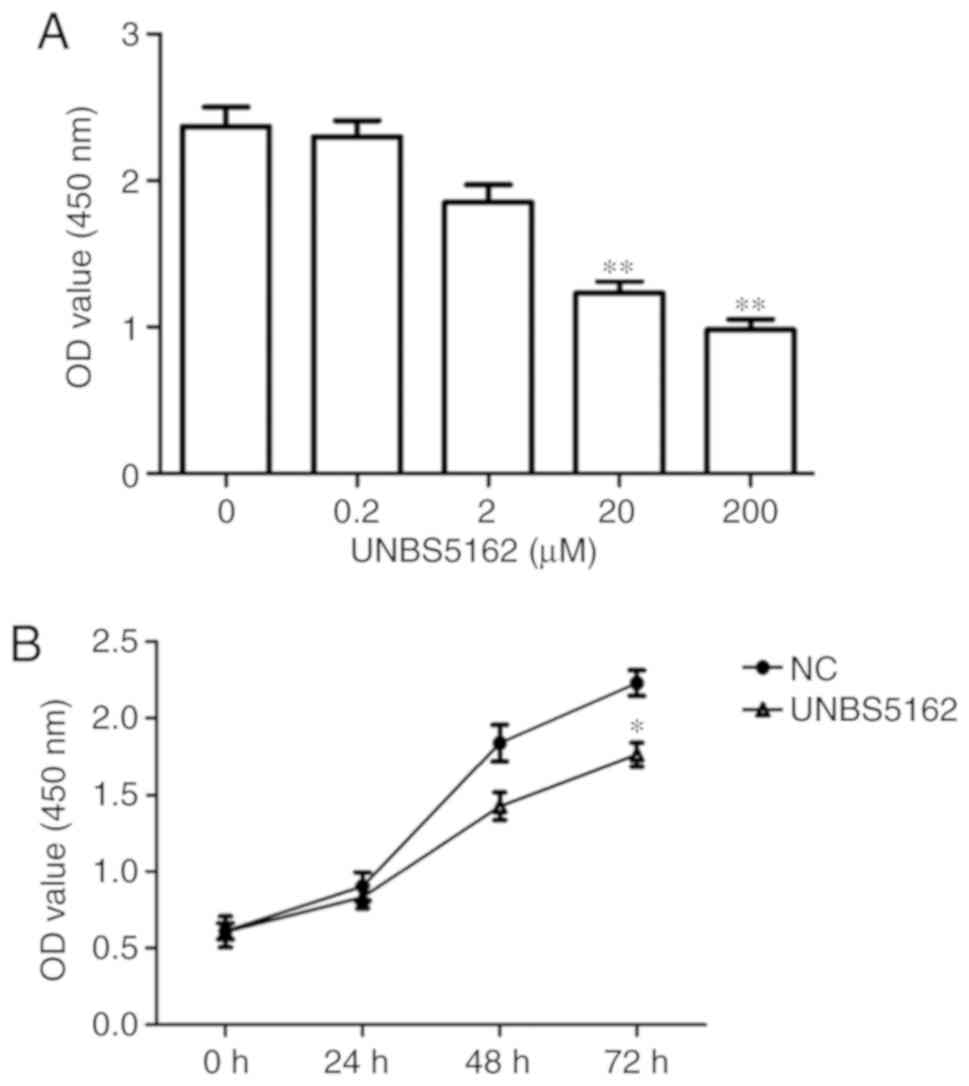
A CCK-8 assay was used to determine the effect of
UNBS5162 on SKOV3 ovarian cancer cell viability. The viability of
SKOV3 cells significantly decreased following 20 and 200 µM
UNBS5162 treatment in a dose-dependent manner (P<0.01 vs. NC;
Fig. 1A). Therefore, in subsequent
experiments, 20 µM UNBS5162 was used, as it had a significant and
similar inhibitory effect compared with 200 µM UNBS5162 and lower
general toxicity. UNBS5162 decreased the number of SKOV3 cells in a
time-dependent manner, with a significantly decreased viability
compared with the NC at 72 h (P<0.05; Fig. 1B). These results suggested that
UNBS5162 could effectively inhibit the viability of the ovarian
cancer cell line SKOV3.
UNBS5162 inhibits SKOV3 ovarian cancer
cell invasion and migration
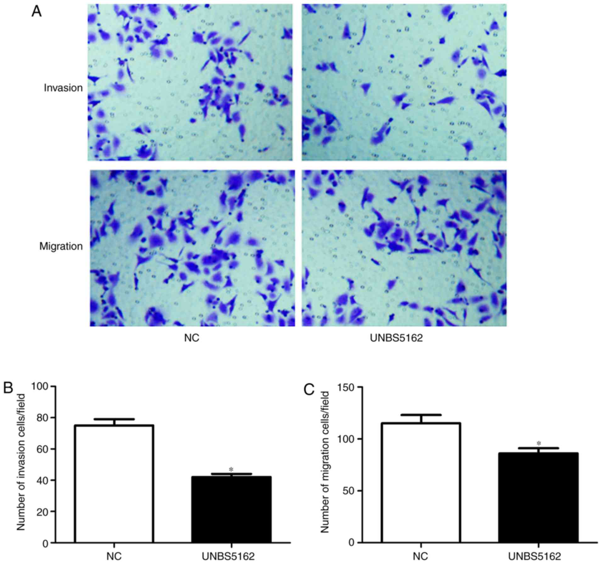
The effects of UNBS5162 on the invasion and
migration of the ovarian cancer cell line SKOV3 were investigated
using Transwell assays. The number of crystal violet-stained cells
in the invasion and migration assays was decreased by UNBS5162
(Fig. 2A). These results were
revealed to be significant (P<0.05; Fig. 2B and C), suggesting that UNBS5162
inhibits migration and invasion of ovarian cancer cells.
UNBS5162 promotes SKOV3 cell
apoptosis
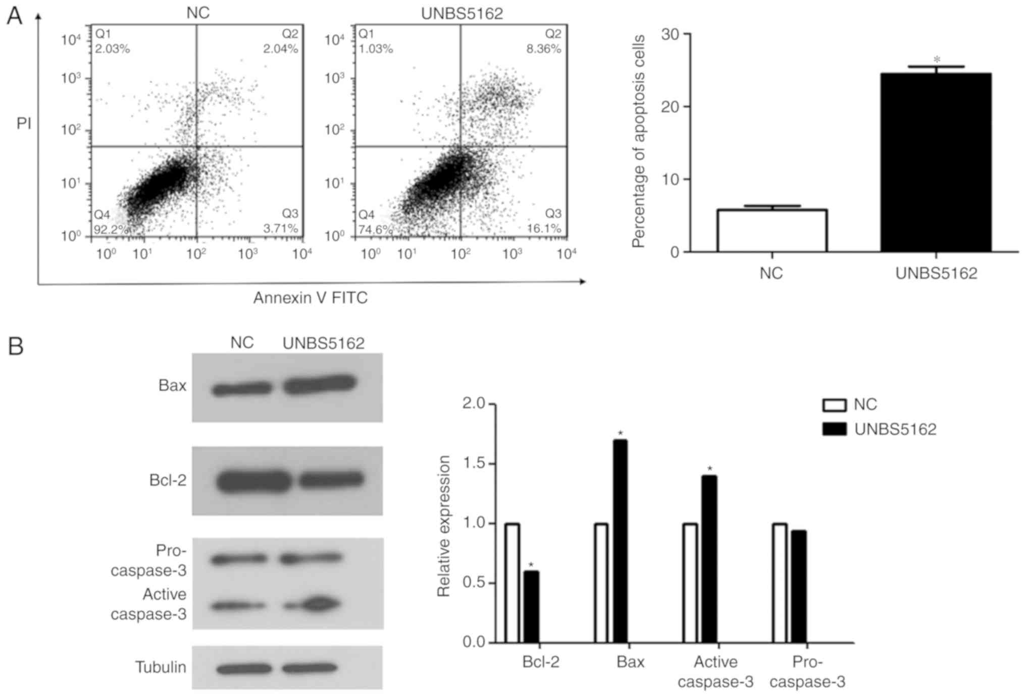
The effect of UNBS5162 on SKOV3 cell apoptosis was
determined using an annexin V/FITC and PI double-staining assay.
The apoptosis rate in the UNBS5162-treated group was significantly
higher compared with the NC group (24.46% vs. 5.75%; P<0.05;
Fig. 3A). Furthermore, apoptosis
regulators, such as the anti-apoptotic protein Bcl-2, the
pro-apoptotic protein active caspase-3 and Bax, were analysed by
western blotting (Fig. 3B).
Consistent with the flow cytometric results, the expression of the
anti-apoptotic protein Bcl-2 decreased, and the expression of the
pro-apoptotic protein active caspase-3 and Bax increased in the
UNBS5162-treated group compared with the NC group (P<0.05,
Fig. 3D). These results indicated
that UNBS5162 promotes SKOV3 cell apoptosis.
UNBS5162 suppresses the PI3K/AKT
pathway in SKOV3 ovarian cancer cells
The PI3K/AKT signalling pathway is an important
signalling pathway in tumours. The mTOR, p70S6K and cyclin D1
proteins were selected as indicators to determine the activity of
the PI3K/AKT signalling pathway following UNBS5162 treatment. The
western blot results indicated that the p-AKT/total AKT and
p-mTOR/total mTOR expression ratios were decreased significantly in
UNBS5162-treated SKOV3 cells compared with the NC (P<0.05;
Fig. 4). Similarly, the
p-p70S6K/total p70S6K expression ratio and cyclin D1 expression
level decreased following UNBS5162 treatment (P<0.05; Fig. 4). These results suggested that the
proliferation of UNBS5162-treated SKOV3 cells is suppressed via the
PI3K/AKT signalling pathway.
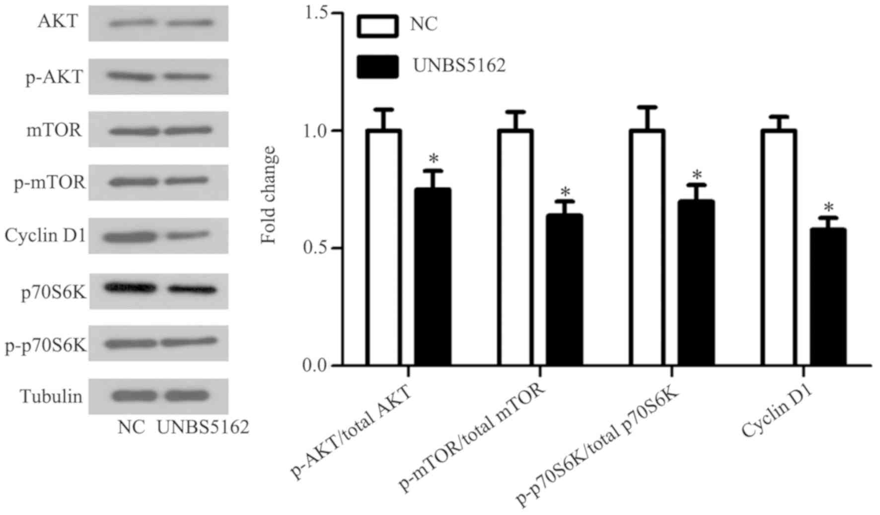 | Figure 4.Effects of UNBS5162 on the
phosphoinositide 3-kinase/AKT signalling pathway in SKOV3 cells.
Expression levels of AKT, p-AKT, mTOR, p-mTOR, p70S6K, p-p70S6K and
cyclin D1 were determined in the UNBS5162-treated and untreated
SKOV3 cells using western blot analysis. The relative protein
levels of p-AKT/total AKT, p-mTOR/total mTOR, p-p70S6K/total p70S6K
and cyclin D1 were quantified. *P<0.05 vs. the NC group. AKT,
protein kinase B; p-, phospho-; mTOR, mammalian target of
rapamycin; p70S6K, p70 S6 kinase; NC, negative control. |
Discussion
In the present study, UNBS5162, a derivative of
naphthalimide, was investigated to determine its antitumour effect
and the underlying molecular mechanism. Specifically, it was
identified that UNBS5162 inhibited SKOV3 cell proliferation,
invasion and migration. Furthermore, it was identified that cell
apoptosis was promoted by UNBS5162. Finally, the effects of
UNBS5162 on SKOV3 were identified to be associated with the
inhibition of the PI3K/AKT signalling pathway.
Owing to the difference in structure of DNA in
normal cells and tumour cells, DNA is an ideal target for
anticancer drugs (15). DNA
intercalators serve an important function in the treatment of
tumours. Naphthalimide compounds are DNA intercalators that can
insert into the middle of a base pair, causing changes in the
topology of the DNA, and thus affecting topoisomerase function and
recognition of DNA (8,16). The naphthalimide derivatives,
including amonafide (17), mitonafide
(18) and UNBS5162 (19), remain in clinical research. However,
in clinical trials, mitonafide and amonafide exhibited marked side
effects on the central nervous system, leading to thrombocytopenia,
anaemia and leukopenia, and thus these two drugs failed to enter
Phase III clinical trials (20). In
addition, the clinical effects of these two drugs on solid cancers
are limited. To decrease further the toxic side effects by removing
the primary amine groups, a novel naphthalimide derivative,
UNBS3157, was developed which did not exhibit the blood toxicity of
amonafide (9,10). In a previous study, UNBS3157 was
hydrolysed in physiological saline to produce a novel compound that
accounts for its anti-cancer activity, and the product was named
UNBS5162 (21). However, limited
research has been performed in using UNBS5162 on human ovarian
cancer.
The results of the present study indicated that
UNBS5162 affects the expression of apoptosis-associated proteins.
The results of western blot analysis indicated that the expression
of Bcl-2 decreased, and the expression of Bax and active caspase-3
increased in the UNBS5162-treated group. These three proteins are
well-characterized regulators of apoptosis (22). Bcl-2 is an anti-apoptotic protein,
whereas Bax and active caspase-3 are pro-apoptotic proteins
(23). Apoptosis is an important
antitumour mechanism, and a number of antineoplastic agents exert
their function in cancer via apoptosis (24). Studies have indicated that
oestrogen-induced apoptosis can be used to treat and prevent breast
cancer (25). Shukla et al
(26) reported a number of
applications of apoptosis in prostate cancer. Similarly, UNBS5162
could inhibit A549 non-small cell lung cancer cell proliferation by
promoting apoptosis (11). In the
present study, it was identified that 20 µM UNBS5162 exhibited a
marked and similar inhibitory effect on, and low toxicity towards,
SKOV3 ovarian cancer cells. The activation of apoptosis is
regulated by a number of signalling pathways, among which the
PI3K/AKT signalling pathway is important (27). PI3K, as a member of the lipid kinase
family, serves a major function in the regulation of apoptosis,
angiogenesis, cellular metabolism, senescence and other processes
(28). The PI3K/AKT signalling
pathway was identified to be the most frequently affected cancer
pathway at the genetic level (29).
The PI3K/AKT signalling pathway has a function in ovarian cancer
(30). The key protein mTOR, which is
a downstream kinase of the PI3K/AKT signalling pathway, has an
important function in the proliferation, migration and invasion of
cancer cells (31). Previous studies
also have indicated that p70S6K and cyclin D1 are located
downstream of the PI3K/AKT signalling pathway, which is associated
with cell proliferation (32,33). The results of the present study are
consistent with those of these previous studies, indicating that
UNBS5162 targets key components of the PI3K/AKT signalling pathway,
which may be important in the successful treatment of ovarian
cancer.
The present study was performed to elucidate the
underlying molecular mechanism of the effect of UNBS5162 in ovarian
cancer; however, it may have certain limitations. For instance, the
use of other cell lines, inhibition such as using short interfering
RNA to interfere with the PI3K/AKT signalling pathway and the
impact of other signalling pathways are being assessed in our
laboratory. The lack of data on non-cancerous cells used as a
negative control is also a limitation of the present study. In
addition, in vivo experimental verification is required.
In summary, the results of the present study
indicate that UNBS5162 could inhibit ovarian cancer cell
proliferation, invasion and migration, and promote cell apoptosis,
potentially through the PI3K/AKT signalling pathway. The results of
the present study suggest the possibility of further clinical
applications of UNBS5162 in the treatment of ovarian cancer.
Acknowledgements
Not applicable.
Funding
No funding was received.
Availability of data and materials
The datasets used and/or analysed during the current
study are available from the corresponding author on reasonable
request.
Authors' contributions
QW and WS conceived and designed the study,
performed the study, analysed and interpreted the data, and wrote
the paper.
Ethics approval and consent to
participate
Not applicable.
Patient consent for publication
Not applicable.
Competing interests
The authors declare that they have no competing
interests.
Glossary
Abbreviations
Abbreviations:
|
CCK-8
|
Cell Counting Kit-8
|
|
PI3K
|
phosphoinositide 3-kinase
|
|
DMEM
|
Dulbecco's modified Eagle's medium
|
References
|
1
|
Worzfeld T, Pogge von Strandmann E, Huber
M, Adhikary T, Wagner U, Reinartz S and Müller R: The unique
molecular and cellular microenvironment of ovarian cancer. Front
Oncol. 7:242017. View Article : Google Scholar : PubMed/NCBI
|
|
2
|
Yan B, Yin F, Wang QI, Zhang W and Li LI:
Integration and bioinformatics analysis of DNA-methylated genes
associated with drug resistance in ovarian cancer. Oncol Lett.
12:157–166. 2016. View Article : Google Scholar : PubMed/NCBI
|
|
3
|
Faber MT, Kjaer SK, Dehlendorff C,
Chang-Claude J, Andersen KK, Høgdall E, Webb PM, Jordan SJ;
Australian Cancer Study (Ovarian Cancer); Australian Ovarian Cancer
Study Group, ; et al: Cigarette smoking and risk of ovarian cancer:
A pooled analysis of 21 case-control studies. Cancer Causes
Control. 24:989–1004. 2013. View Article : Google Scholar : PubMed/NCBI
|
|
4
|
Weidle UH, Birzele F, Kollmorgen G and
Rueger R: Mechanisms and targets involved in dissemination of
ovarian cancer. Cancer Genomics Proteomics. 13:407–423. 2016.
View Article : Google Scholar : PubMed/NCBI
|
|
5
|
Correa RJ, Valdes YR, Peart TM, Fazio EN,
Bertrand M, McGee J, Préfontaine M, Sugimoto A, DiMattia GE and
Shepherd TG: Combination of AKT inhibition with autophagy blockade
effectively reduces ascites-derived ovarian cancer cell viability.
Carcinogenesis. 35:1951–1961. 2014. View Article : Google Scholar : PubMed/NCBI
|
|
6
|
Li S, Xu S, Tang Y, Ding S, Zhang J, Wang
S, Zhou G, Zhou C and Li X: Synthesis, anticancer activity and
DNA-binding properties of novel 4-pyrazolyl-1,8-naphthalimide
derivatives. Bioorg Med Chem Lett. 24:586–590. 2014. View Article : Google Scholar : PubMed/NCBI
|
|
7
|
Zhao L, Li J, Li Y, Liu J, Wirth T and Li
Z: Selenium-containing naphthalimides as anticancer agents: Design,
synthesis and bioactivity. Bioorg Med Chem. 20:2558–2563. 2012.
View Article : Google Scholar : PubMed/NCBI
|
|
8
|
Ingrassia L, Lefranc F, Kiss R and
Mijatovic T: Naphthalimides and azonafides as promising anti-cancer
agents. Curr Med Chem. 16:1192–1213. 2009. View Article : Google Scholar : PubMed/NCBI
|
|
9
|
Dumont P, Ribaucour F, Quaquebeke EV,
Darro F and Kiss R: UNBS3157, a new amonafide derivative with
improved in vivo efficacy and decreased toxicity. Cancer Res. 66
Supp:11052006.PubMed/NCBI
|
|
10
|
Van Quaquebeke E, Mahieu T, Dumont P,
Dewelle J, Ribaucour F, Simon G, Sauvage S, Gaussin JF, Tuti J, El
Yazidi M, et al:
2,2,2-Trichloro-N-({2-[2-(dimethylamino)ethyl]-1,3-dioxo-2,3-
dihydro-1H-benzo[de]isoquinolin-5-yl}carbamoyl)acetamide
(UNBS3157), a novel nonhematotoxic naphthalimide derivative with
potent antitumor activity. J Med Chem. 50:4122–4134. 2007.
View Article : Google Scholar : PubMed/NCBI
|
|
11
|
Liu C, Xing J and Gao Y: UNBS5162 inhibits
the proliferation of human A549 non-small-cell lung cancer cells by
promoting apoptosis. Thorac Cancer. 9:105–111. 2018. View Article : Google Scholar : PubMed/NCBI
|
|
12
|
He D and Zhang S: UNBS5162 inhibits the
proliferation of esophageal cancer squamous cells via the PI3K/AKT
signalling pathway. Mol Med Rep. 17:549–555. 2018.PubMed/NCBI
|
|
13
|
Wang B, Shen J and Wang J: UNBS5162
inhibits proliferation of human retinoblastoma cells by promoting
cell apoptosis. Onco Targets Ther. 10:5303–5309. 2017. View Article : Google Scholar : PubMed/NCBI
|
|
14
|
Mijatovic T, Mahieu T, Bruyere C, De Nève
N, Dewelle J, Simon G, Dehoux MJ, van der Aar E, Haibe-Kains B,
Bontempi G, et al: UNBS5162, a novel naphthalimide that decreases
CXCL chemokine expression in experimental prostate cancers.
Neoplasia. 10:573–586. 2008. View Article : Google Scholar : PubMed/NCBI
|
|
15
|
Agudelo D, Bourassa P, Bérubé G and
Tajmir-Riahi HA: Intercalation of antitumor drug doxorubicin and
its analogue by DNA duplex: Structural features and biological
implications. Int J Biol Macromol. 66:144–150. 2014. View Article : Google Scholar : PubMed/NCBI
|
|
16
|
Gellerman G: Recent developments in the
synthesis and applications of anticancer amonafide derivatives. A
mini review. Letters Drug Design Discov. 13:47–63. 2016. View Article : Google Scholar
|
|
17
|
Zhao J, Lu M, Lai H, Lu H, Lalevée J,
Barner-Kowollik C, Stenzel MH and Xiao P: Delivery of amonafide
from fructose-coated nanodiamonds by oxime ligation for treatment
of human breast cancer. Biomacromolecules. 19:481–489. 2018.
View Article : Google Scholar : PubMed/NCBI
|
|
18
|
Ge C, Chang L, Zhao Y, Chang C, Xu X, He
H, Wang Y, Dai F, Xie S and Wang C: Design, synthesis and
evaluation of naphthalimide derivatives as potential anticancer
agents for hepatocellular carcinoma. Molecules. 22:E3422017.
View Article : Google Scholar : PubMed/NCBI
|
|
19
|
Mahieu T, Mijatovic T, Quaquebeke EV,
Lefranc F, Vynckt FV, Darro F and Kiss R: UNBS5162 is a novel
naphthalimide derivative that induces autophagy and senescence in
human prostate cancer cells. Mol Cancer Ther. 6:3373S2007.
|
|
20
|
Antonini I, Volpini R, Dal Ben D,
Lambertucci C and Cristalli G: Design, synthesis, and biological
evaluation of new mitonafide derivatives as potential antitumor
drugs. Bioorg Med Chem. 16:8440–8446. 2008. View Article : Google Scholar : PubMed/NCBI
|
|
21
|
Mahadevan D, Northfelt DW, Chalasani P,
Rensvold D, Kurtin S, Von Hoff DD, Borad MJ and Tibes R: Phase I
trial of UNBS5162, a novel naphthalimide in patients with advanced
solid tumors or lymphoma. Int J Clin Oncol. 18:934–941. 2013.
View Article : Google Scholar : PubMed/NCBI
|
|
22
|
Mei JM and Niu CS: Effects of CDNF on
6-OHDA-induced apoptosis in PC12 cells via modulation of Bcl-2/Bax
and caspase-3 activation. Neurol Sci. 35:1275–1280. 2014.
View Article : Google Scholar : PubMed/NCBI
|
|
23
|
Zhuang RJ, Ma J, Shi X, Ju F, Ma SP, Wang
L, Cheng BF, Ma YW, Wang M, Li T, et al: Cold-inducible protein
RBM3 protects UV irradiation-induced apoptosis in neuroblastoma
cells by affecting p38 and JNK pathways and Bcl2 family proteins. J
Mol Neurosci. 63:142–151. 2017. View Article : Google Scholar : PubMed/NCBI
|
|
24
|
Nakamura Y, Ise K, Yamazaki Y, Fujishima
F, McNamara KM and Sasano H: Serotonin receptor 4
(5-hydroxytryptamine receptor Type 4) regulates expression of
estrogen receptor beta and cell migration in hormone-naive prostate
cancer. Indian J Pathol Microbiol. 60:33–37. 2017.PubMed/NCBI
|
|
25
|
Jordan VC: The new biology of
estrogen-induced apoptosis applied to treat and prevent breast
cancer. Endocr Relat Cancer. 22:R1–R31. 2015. View Article : Google Scholar : PubMed/NCBI
|
|
26
|
Shukla S, Fu P and Gupta S: Apigenin
induces apoptosis by targeting inhibitor of apoptosis proteins and
Ku70-Bax interaction in prostate cancer. Apoptosis. 19:883–894.
2014. View Article : Google Scholar : PubMed/NCBI
|
|
27
|
Zhou ZW, Li XX, He ZX, Pan ST, Yang Y,
Zhang X, Chow K, Yang T, Qiu JX, Zhou Q, et al: Induction of
apoptosis and autophagy via sirtuin1- and PI3K/Akt/mTOR-mediated
pathways by plumbagin in human prostate cancer cells. Drug Des
Devel Ther. 9:1511–1554. 2015. View Article : Google Scholar : PubMed/NCBI
|
|
28
|
Akinleye A, Avvaru P, Furqan M, Song Y and
Liu D: Phosphatidylinositol 3-kinase (PI3K) inhibitors as cancer
therapeutics. J Hematol Oncol. 6:882013. View Article : Google Scholar : PubMed/NCBI
|
|
29
|
Fang WL, Huang KH, Lan YT, Lin CH, Chang
SC, Chen MH, Chao Y, Lin WC, Lo SS, Li AF, et al: Mutations in
PI3K/AKT pathway genes and amplifications of PIK3CA are associated
with patterns of recurrence in gastric cancers. Oncotarget.
7:6201–6220. 2016. View Article : Google Scholar : PubMed/NCBI
|
|
30
|
Huang J, Zhang L, Greshock J, Colligon TA,
Wang Y, Ward R, Katsaros D, Lassus H, Butzow R, Godwin AK, et al:
Frequent genetic abnormalities of the PI3K/AKT pathway in primary
ovarian cancer predict patient outcome. Genes Chromosomes Cancer.
50:606–618. 2011. View Article : Google Scholar : PubMed/NCBI
|
|
31
|
Zhang L, Huo X, Liao Y, Yang F, Gao L and
Cao L: Zeylenone, a naturally occurring cyclohexene oxide, inhibits
proliferation and induces apoptosis in cervical carcinoma cells via
PI3K/AKT/mTOR and MAPK/ERK pathways. Sci Rep. 7:16692017.
View Article : Google Scholar : PubMed/NCBI
|
|
32
|
Halacli SO and Dogan AL: FOXP1 regulation
via the PI3K/Akt/p70S6K signalling pathway in breast cancer cells.
Oncol Lett. 9:1482–1488. 2015. View Article : Google Scholar : PubMed/NCBI
|
|
33
|
Wang HY, Yang SL, Liang HF and Li CH: HBx
protein promotes oval cell proliferation by up-regulation of cyclin
D1 via activation of the MEK/ERK and PI3K/Akt pathways. Int J Mol
Sci. 15:3507–3518. 2014. View Article : Google Scholar : PubMed/NCBI
|