Introduction
Cervical cancer has the second highest incidence of
female common tumors, with the highest mortality rate among female
cancers. Approximately 300,000 people die of it every year around
the world (1). In recent years, the
incidence of cervical cancer has increased and tends to be younger
in age, posing a serious threat to the health of females (2). Without obvious symptoms in early onset,
once found, cervical cancer is in the middle and late stage, with a
poor prognosis. Therefore, its early diagnosis is of great
significance for its treatment and prognosis (3).
At present, the pathogenesis of cervical cancer is
still not fully clarified. Studies have shown that the occurrence
and development of cervical cancer is an extremely complex
multi-stage process regulated by many factors. The regulation of
nucleic acid level is closely related to the occurrence of cervical
cancer. In particular, the expression of target genes of non-coding
microRNA (miRNA) is influenced by its target regulation, thereby
affecting the occurrence and development of cervical cancer
(4,5).
For example, miR-31 (6) has an impact
on the biological function of cervical cancer cells by the target
regulation of gene ARID1A.
In recent years, related studies have shown that
miR-1202 can function in the occurrence and development of multiple
tumors by inhibiting the proliferation of tumor cells. Among them,
miR-1202 has been studied in breast cancer (7) and endometrial cancer (8). However, there is no related report on
the expression and diagnostic value of it in cervical cancer.
miR-195 is an important cancer suppressor gene (9). Studies have shown that it inhibits tumor
cells in many tumors such as hepatocellular carcinoma (10), breast cancer (11) and non-small cell lung cancer (12). Yet, its expression and diagnostic
value in cervical cancer is still not very clear.
Therefore, in this study, in order to provide a
better plan for the early diagnosis of cervical cancer, the
expression of miR-1202 and miR-195 in cervical cancer was explored.
Thus, the clinical value of the combined detection of miR-1202 and
miR-195 in the early diagnosis was investigated.
Patients and methods
General information
A retrospective analysis of 70 cervical cancer
patients treated in The Third Affiliated Hospital of Kunming
Medical University and Yunnan Cancer Hospital (Kunming, China) from
October 2015 to December 2017 was performed, lesion tissues were
used as the experimental group. The average age of the patients was
40.2±3.7 years. According to the FIGO staging criteria, there were:
stage I with 26 cases, stage II with 29 cases, stage III with 9
cases and stage IV with 6 cases. There were 47 cases of cervical
squamous cell carcinoma and 23 cases of cervical adenocarcinoma
according to the pathological type. Normal cervical tissues from
another 67 healthy females confirmed by physical examination at the
same period were selected as the control group. The average age was
41.1±2.8 years. All the tissues were taken out and quickly stored
at −80°C. There was no significant difference in age and BMI
between the two groups of patients (P>0.05; Table I).
 | Table I.Comparison of general information
between the two groups of patients [n (%)]. |
Table I.
Comparison of general information
between the two groups of patients [n (%)].
| Factors | Experimental group
(n=70) | Control group
(n=67) | χ2 | P-value |
|---|
| Age |
|
| 0.002 | 0.966 |
| ≤40 | 41 (58.57) | 39 (58.21) |
|
|
|
>40 | 29 (41.43) | 28 (41.79) |
|
|
| BMI |
|
| 0.005 | 0.942 |
| ≤22 | 37 (52.86) | 35 (52.24) |
|
|
|
>22 | 33 (47.14) | 32 (47.76) |
|
|
| Married or not |
|
| 0.055 | 0.814 |
| Yes | 51 (72.86) | 50 (74.63) |
|
|
| No | 19 (27.14) | 17 (25.37) |
|
|
| Fertile or not |
|
| 0.165 | 0.685 |
| Yes | 49 (70.00) | 49 (73.13) |
|
|
| No | 21 (30.00) | 18 (26.87) |
|
|
| Family history |
|
| – | – |
| Yes | 23 (32.86) | – |
|
|
| No | 47 (67.14) | – |
|
|
| Staging |
|
| – | – |
| Stage
I | 26 (37.14) | – |
|
|
| Stage
II | 29 (41.43) | – |
|
|
| Stage
III | 9 (12.86) | – |
|
|
| Stage
IV | 6 (8.57) | – |
|
|
| Pathological
types |
|
| – | – |
| Cervical
squamous cell carcinoma | 47 (67.14) | – |
|
|
| Cervical
adenocarcinoma | 23 (32.86) | – |
|
|
Inclusion and exclusion criteria
Inclusion criteria were: i) the experimental group
included patients confirmed with cervical cancer by pathology, and
ii) the control group included females confirmed as healthy by
physical examination. Exclusion criteria were: i) patients having
undergone radiotherapy and chemotherapy prior to taking specimens;
ii) patients with severe other organ diseases; iii) patients who
did not cooperate with the examination. and iv) patients with
cognitive and communication impairment. The participants and their
family members signed an informed consent form to cooperate with
the medical staff to complete relevant medical treatment and the
study was approved by the Ethics Committee of The Third Affiliated
Hospital of Kunming Medical University and Yunnan Cancer
Hospital.
Experimental instruments and
materials
Refrigerator at −80°C (Sanyo, Tokyo, Japan); reverse
transcription-quantitative polymerase chain reaction (RT-qPCR) (iQ5
Multicolor; Bio-Rad Laboratories, Inc., Hercules, CA, USA); TRIzol
reagent (Invitrogen; Thermo Fisher Scientific, Inc., Waltham, MA,
USA); RT-qPCR kit and minScript reverse transcription kit (Takara
Biotechnology Co., Ltd., Dalian, China).
Methods
The lesion tissues and normal cervical tissues were
taken out from the refrigerator at −80°C. Trizol reagent was added,
tissue RNA was extracted, and 1 µl of total RNA was obtained. The
specific stem ring primers were added, respectively. Reverse
transcription cDNA was performed according to the manufacturers
instructions. Reaction temperature: at 42°C for 15 min and at 85°C
for 5 sec. Primers for miR-1202 and miR-195 were amplified
according to sequence design, with U6 as an internal reference
control (Table II). Amplification
conditions: denaturation at 95°C for 30 sec, annealing at 60°C for
10 sec, extension at 72°C for 20 sec, for a total of 40 cycles, and
extension at 72°C for 5 min to stop the reaction. The expression of
miR-1202 and miR-195, resspectively, was calculated and analyzed
using the 2−ΔCq method (13).
| Table II.RT-qPCR reaction miRNA-related
primers. |
Table II.
RT-qPCR reaction miRNA-related
primers.
| Factors | Upstream primer
sequences | Downstream primer
sequences |
|---|
| miR-1202 |
5′-ATCCAGTGCGTGTCGTG-3′ |
5′-TGCTGTGCCAGCTGCAGT-3′ |
| miR-195 |
5′-TAGCAGCA-CAGAAATATTGGC-3′ |
5′-TGCTGTGCCAGCTGCAGT-3′ |
| U6 |
5′-GCTTCGGCAGCACATATACTA-AAAT-3′ |
5′-CGCTTCACGAATTTGCGTGTCAT-3′ |
Statistical analysis
Chi-square test was used for enumeration data.
Measurement data were expressed as mean ± standard deviation, using
the t-test. SPSS19.0 (Shanghai Yuchuang Network Technology Co.,
Ltd., Shanghai, China) statistical software was used for analysis.
P<0.05 was considered to indicate a statistically significant
difference.
Results
Comparison of relative expression of
miR-1202 and miR-195 between the two groups
The relative expression of miR-1202 in the
experimental group was 0.45±0.13, lower than that in the normal
control group 0.97±0.32; P<0.05, and that of miR-195 in the
experimental group was 0.53±0.15, lower than that in the normal
control group 0.96±0.25 (P<0.05; Table III).
| Table III.Comparison of relative expression of
miR-1202 and miR-195 in the two groups. |
Table III.
Comparison of relative expression of
miR-1202 and miR-195 in the two groups.
| Factors | Experimental group
(n=70) | Control group
(n=67) | t | P-value |
|---|
| miR-1202 | 0.45±0.13 | 0.97±0.32 | 12.56 | <0.001 |
| miR-195 | 0.53±0.15 | 0.96±0.25 | 12.27 | <0.001 |
Comparison of relative expression of
miR-1202 and miR-195 in different stages
In the cervical cancer patients of early stage
(I–II) and middle-late stage (III–IV), the expression levels of
miR-1202 in the experimental group were 0.65±0.22 and 0.31±0.10,
respectively, and those of miR-195 were 0.78±0.18 and 0.29±0.16,
respectively. Based on statistical analysis, there was a
significant difference in the overall level (P<0.001),
suggesting that the later the clinical staging is, the lower the
expression levels of miR-1202 and miR-195 are (Table IV).
| Table IV.Comparison of relative expression of
miR-1202 and miR-195 in different stages. |
Table IV.
Comparison of relative expression of
miR-1202 and miR-195 in different stages.
| Staging | miR-1202 | miR-195 |
|---|
| Stage I–II
(n=55) | 0.65±0.22 | 0.78±0.18 |
| Stage III–IV
(n=15) | 0.31±0.10 | 0.29±0.16 |
| t | 5.800 | 9.554 |
| P-value | <0.001 | <0.001 |
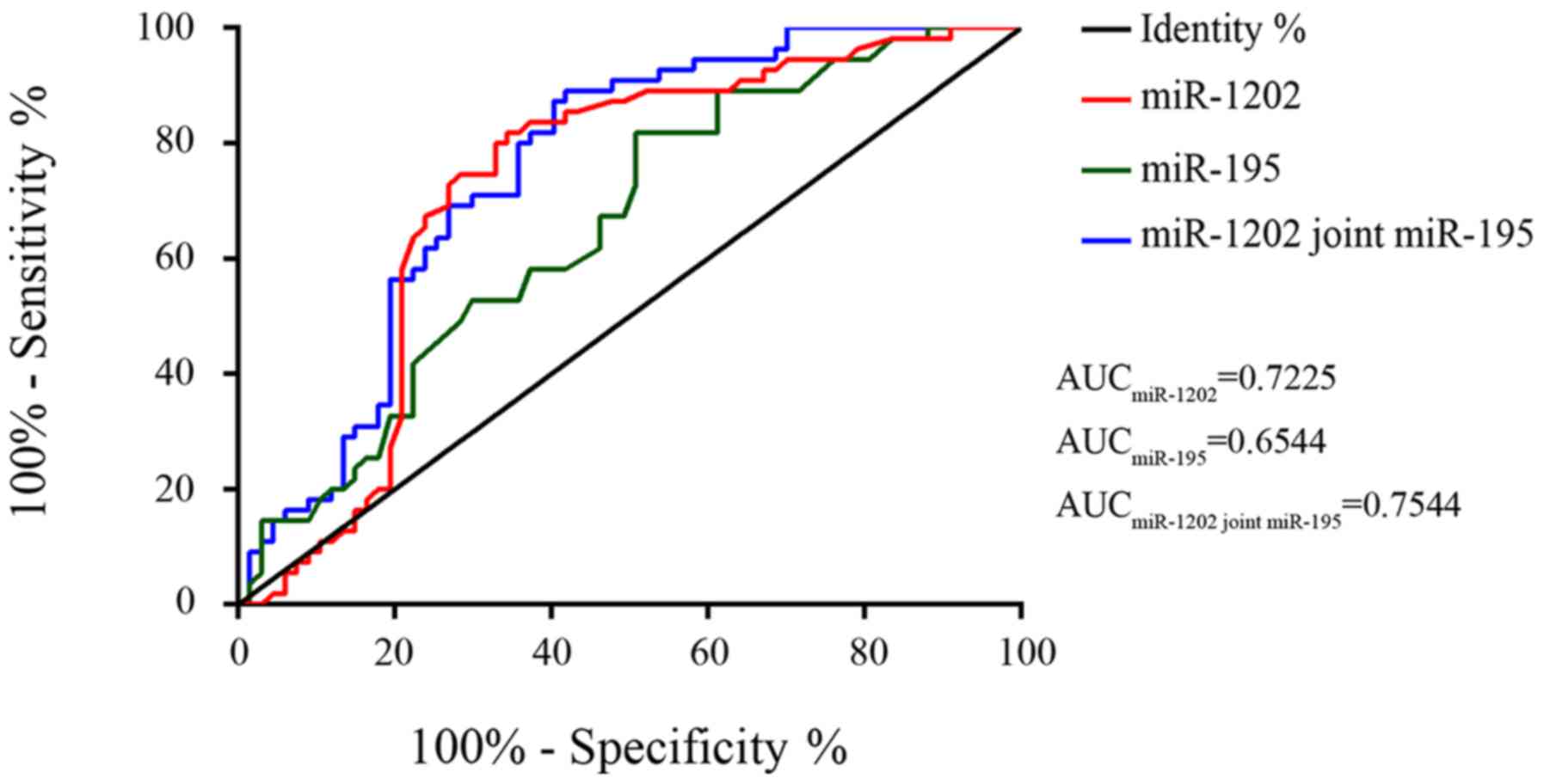
Diagnostic values of miR-1202 and
miR-195 detection alone and their combined detection in early stage
(I–II) of cervical cancer
Calculated with early patients and healthy
population, in the diagnosis of early cervical cancer patients, the
diagnosis standard of miR-1202 alone was <0.845. The
sensitivity, specificity and area under the curve (AUC) value was
81.82%, 65.67% and 0.7225, respectively. That of miR-195 diagnosis
alone was <0.950. The sensitivity, specificity and AUC value was
80.00%, 49.25% and 0.6544, respectively. The combined diagnosis of
miR-1202 and miR-195 was 89.09%, 58.21% and 0.7544, respectively.
Among them, the sensitivity and AUC value of the combined diagnosis
were significantly higher than those of miR-1202 and miR-195
diagnosis alone (Table V and Fig. 1).
| Table V.Diagnostic values of miR-1202 and
miR-195 detection alone and their combined detection in early
cervical cancer. |
Table V.
Diagnostic values of miR-1202 and
miR-195 detection alone and their combined detection in early
cervical cancer.
| Diagnostic
methods | Sensitivity
(%) | Specificity
(%) | AUC |
|---|
| miR-1202 | 81.82 | 65.67 | 0.7225 |
| miR-195 | 80.00 | 49.25 | 0.6544 |
| miR-1202 combined
with miR-195 | 89.09 | 58.21 | 0.7544 |
Discussion
Cervical cancer is one of the most common malignant
tumors of the reproductive system in females, with the highest
incidence that is still rising (14).
Studies have reported (15) that
early cervical cancer is not easy to be detected, but once found,
it is often in the middle and late stage, with a higher recurrence,
invasion and metastasis rate, leading to poor treatment and
prognosis of cervical cancer patients. The pathogenesis of cervical
cancer is a very complicated process, involving the abnormalities
of the structures and expression of many coding and non-coding
genes, from normal cervical epithelial cells to infiltration of
cancer cells (16). As a
single-stranded non-coding regulatory RNA, by participating in the
regulation of target gene expression, miRNA plays a role in various
biological processes such as cell proliferation, apoptosis,
differentiation and migration (17).
The biological function of miR-1202 is mainly related to cell
proliferation and apoptosis. Studies of Du et al (18) showed that miR-1202 induced endoplasmic
reticulum stress and apoptosis through the target regulation of
Rab1A in glioma. It also inhibited the proliferation of glioma
cells. Studies have shown (19) that
as a cancer suppressor gene, miR-195 is closely related to the
occurrence of breast cancer, bladder cancer, colorectal cancer and
other tumors, through the regulation of target genes. In addition,
related studies have reported that the expression of miR-195 is
downregulated in the lesion tissues of cervical cancer. The
downregulation is associated with deep interstitial infiltration
and lymph node metastasis in cervical cancer patients (20).
In order to clarify the diagnostic values of
miR-1202 and miR-195 in cervical cancer, in this study, their
expression in lesion and normal cervical tissues was compared. It
was found that the expression in the experimental group was lower
than that in the control group (P<0.05). Then, the relative
expression of miR-1202 and miR-195 in different stages was
compared. The results showed that the later the clinical staging
was, the lower the expression levels were (P<0.05). The study of
Wipperman et al (21) showed
that miR-1202 played a role as a tumor suppressor gene in cervical
cancer, so its expression in cervical cancer was downregulated. In
the study of Philp et al (22), it was found that miR-195 could act as
a cancer suppressor gene in cervical cancer, so its expression in
cervical cancer tissues was downregulated compared to normal
tissues. The results are consistent with our findings, confirming
the role of miR-1202 and miR-195 as cancer suppressor genes in
cervical cancer. The diagnostic values of miR-1202 and miR-195
detection alone and their combined detection in early cervical
cancer were evaluated. The results showed that the sensitivity,
specificity and AUC value of miR-1020 diagnosis alone was 81.82%,
65.67% and 0.7225, respectively and in miR-195 diagnosis alone was
80.00%, 49.25% and 0.6544, respectively, and in the combined
diagnosis of miR-1202 and miR-195 was 89.09%, 58.21% and 0.7544,
respectively. The sensitivity and AUC value of the combined
diagnosis were significantly higher than those of miR-1202 and
miR-195 diagnosis alone, showing that the value of the combined
diagnosis of miR-1202 and miR-195 in early cervical cancer is
higher than that of their diagnosis alone.
In conclusion, the expression of miR-1202 and
miR-195 in cervical cancer tissues is significantly downregulated.
The value of the combined detection is higher than that of the
detection alone in early cervical cancer, which can be used as a
preferred solution for the clinical diagnosis of early cervical
cancer. However, there are few research studies on miR-1202 and
miR-195 in cervical cancer, and the sample size is small.
Therefore, more investigations are required to carry out extensive
research.
Acknowledgements
Not applicable.
Funding
This study was supported by Health Science and
Technology Project of Yunnan Province (2017NS189).
Availability of data and materials
The datasets used and/or analyzed during the present
study are available from the corresponding author on reasonable
request.
Authors' contributions
XY conceived the study and drafted the manuscript.
HY directed the project and was also involved in the conception and
design of the study. ZY, LZ and HN acquired the data. LZ and YW
analyzed the data and revised the manuscript. All authors read and
approved the final manuscript.
Ethics approval and consent to
participate
The study was approved by the Ethics Committee of
The Third Affiliated Hospital of Kunming Medical University and
Yunnan Cancer Hospital (Kumming, China) and the participants and
their family members signed an informed consent form.
Patient consent for publication
Not applicable.
Competing interests
The authors declare that they have no competing
interests.
References
|
1
|
Turkistanli EC, Sogukpinar N, Saydam BK
and Aydemir G: Cervical cancer prevention and early detection-the
role of nurses and midwives. Asian Pac J Cancer Prev. 4:15–21.
2003.PubMed/NCBI
|
|
2
|
Shi YR, Liu J, He W and Yang Y: Expression
of Micro-RNA 218 in cervical cancer and its effect on
proliferation, apoptosis and invasion of HeLa cells. Sichuan Da Xue
Xue Bao Yi Xue Ban. 47:697–702. 2016.(In Chinese). PubMed/NCBI
|
|
3
|
Jung S, Yi L, Kim J, Jeong D, Oh T, Kim
CH, Kim CJ, Shin J, An S and Lee MS: The role of vimentin as a
methylation biomarker for early diagnosis of cervical cancer. Mol
Cells. 31:405–411. 2011. View Article : Google Scholar : PubMed/NCBI
|
|
4
|
Shen Y, Chen H, Gao L, Zhang W, He J, Yang
X, Qin L, Xue X and Guo Z: MiR-638 acts as a tumor suppressor gene
in gastric cancer. Oncotarget. 8:108170–108180. 2017. View Article : Google Scholar : PubMed/NCBI
|
|
5
|
Zhang WN, Li W, Wang XL, Hu Z, Zhu D, Ding
WC, Liu D, Li KZ, Ma D and Wang H: CLDN1 expression in cervical
cancer cells is related to tumor invasion and metastasis.
Oncotarget. 7:87449–87461. 2016. View Article : Google Scholar : PubMed/NCBI
|
|
6
|
Yang X, Da M, Zhang W, Qi Q, Zhang C and
Han S: Role of Lactobacillus in cervical cancer. Cancer Manag Res.
10:1219–1229. 2018. View Article : Google Scholar : PubMed/NCBI
|
|
7
|
Chen H, Fan Y, Xu W, Chen J, Meng Y, Fang
D and Wang J: Exploration of miR-1202 and miR-196a in human
endometrial cancer based on high throughout gene screening
analysis. Oncol Rep. 37:3493–3501. 2017. View Article : Google Scholar : PubMed/NCBI
|
|
8
|
McMullen JRW, Selleck M, Wall NR and
Senthil M: Peritoneal carcinomatosis: limits of diagnosis and the
case for liquid biopsy. Oncotarget. 8:43481–43490. 2017. View Article : Google Scholar : PubMed/NCBI
|
|
9
|
Zhou Q, Han LR, Zhou YX and Li Y: MiR-195
Suppresses cervical cancer migration and invasion through targeting
Smad3. Int J Gynecol Cancer. 26:817–824. 2016. View Article : Google Scholar : PubMed/NCBI
|
|
10
|
Yan JJ, Chang Y, Zhang YN, Lin JS, He XX
and Huang HJ: miR-195 inhibits cell proliferation via targeting
AEG-1 in hepatocellular carcinoma. Oncol Lett. 13:3118–3126. 2017.
View Article : Google Scholar : PubMed/NCBI
|
|
11
|
Nadeem F, Hanif M, Ahmed A, Jamal Q and
Khan A: Clinico-pathological features associated to MiRNA-195
expression in patients with breast cancer: evidence of a potential
biomarker. Pak J Med Sci. 33:1242–1247. 2017. View Article : Google Scholar : PubMed/NCBI
|
|
12
|
Feng C, Zhang L, Sun Y, Li X, Zhan L, Lou
Y, Wang Y, Liu L and Zhang Y: GDPD5, a target of miR-195-5p, is
associated with metastasis and chemoresistance in colorectal
cancer. Biomed Pharmacother. 101:945–952. 2018. View Article : Google Scholar : PubMed/NCBI
|
|
13
|
Livak KJ and Schmittgen TD: Analysis of
relative gene expression data using real-time quantitative PCR and
the 2(-Delta Delta C(T)) method. Methods. 25:402–408. 2001.
View Article : Google Scholar : PubMed/NCBI
|
|
14
|
Li H, Wu X and Cheng X: Advances in
diagnosis and treatment of metastatic cervical cancer. J Gynecol
Oncol. 27:e432016. View Article : Google Scholar : PubMed/NCBI
|
|
15
|
Zhou Z, Liu X, Hu K and Zhang F: The
clinical value of PET and PET/CT in the diagnosis and management of
suspected cervical cancer recurrence. Nucl Med Commun. 39:97–102.
2018.PubMed/NCBI
|
|
16
|
Lv KT, Liu Z, Feng J, Zhao W, Hao T, Ding
WY, Chu JP and Gao LJ: MiR-22-3p regulates cell proliferation and
inhibits cell apoptosis through targeting the eIF4EBP3 gene in
human cervical squamous carcinoma cells. Int J Med Sci. 15:142–152.
2018. View Article : Google Scholar : PubMed/NCBI
|
|
17
|
George OL and Ness SA: Situational
awareness: Regulation of the myb transcription factor in
differentiation, the cell cycle and oncogenesis. Cancers (Basel).
6:2049–2071. 2014. View Article : Google Scholar : PubMed/NCBI
|
|
18
|
Du B, Zhang P, Tan Z and Xu J: MiR-1202
suppresses hepatocellular carcinoma cells migration and invasion by
targeting cyclin dependent kinase 14. Biomed Pharmacother.
96:1246–1252. 2017. View Article : Google Scholar : PubMed/NCBI
|
|
19
|
Li Z, Wang H, Wang Z and Cai H: MiR-195
inhibits the proliferation of human cervical cancer cells by
directly targeting cyclin D1. Tumour Biol. 37:6457–6463. 2016.
View Article : Google Scholar : PubMed/NCBI
|
|
20
|
Mou Z, Xu X, Dong M and Xu J:
MicroRNA-148b acts as a tumor suppressor in cervical cancer by
inducing G1/S-phase cell cycle arrest and apoptosis in a
caspase-3-dependent manner. Med Sci Monit. 22:2809–2815. 2016.
View Article : Google Scholar : PubMed/NCBI
|
|
21
|
Wipperman J, Neil T and Williams T:
Cervical cancer: evaluation and management. Am Fam Physician.
97:449–454. 2018.PubMed/NCBI
|
|
22
|
Philp L, Jembere N, Wang L, Gao J, Maguire
B and Kupets R: Pap tests in the diagnosis of cervical cancer: help
or hinder? Gynecol Oncol. 150:61–66. 2018. View Article : Google Scholar : PubMed/NCBI
|