Introduction
Bladder cancer was diagnosed in ~74,000 patients in
the USA and in >430,000 patients worldwide, which makes it the
fourth most common cancer in males and the 11th most common cancer
in females in the USA during 2015 (1). Due to its invasive nature,
muscle-invasive bladder cancer (MIBC) has a high rate of mortality
with a poor quality of life and an overall 5-year survival rate of
35–60% (2). The burden of cancer in
non-MIBC (NMIBC) patients is primarily caused by subsequent
adjuvant therapy, including intravesical chemotherapy; and the
progression risk and high recurrence rate ranged from 50–90% with a
5-year survival rate of 70–80% worldwide in 2018 (3). Owing to the high recurrence and
heterogeneity of this disease, the characterization of the
molecular profile of tumors and investigating putative biomarkers
is imperative. A number of molecules, including microRNA (miRNA),
long chain non-coding RNAs and other RNAs, have been demonstrated
to be associated with the occurrence and development of bladder
cancer, and the underlying molecular mechanisms and prognostic
features have been analyzed extensively (4).
Circular RNAs (circRNAs) have been recently
determined as a novel type of non-coding RNAs (5), and the enigmatic structure, origin, and
classification have been elucidated (6). Previous studies also demonstrated that
circRNAs are involved in the development of a number of types of
diseases, including breast cancer and liver cancer (7,8). However,
only a few articles have been reported, particularly with respect
to bladder cancer (9,10).
In the present study, the differences in the
expression of a valuable circRNA, circ lysophosphatidic acid
receptor 1 (LPAR1) (hsa_circ_0087960), were detected, and its
prognosis value in 125 patients with MIBC was evaluated.
Additionally, its potential function and the underlying molecular
mechanism were investigated.
Materials and methods
Patients and clinical specimens
A total of 125 bladder cancer tissues and 68 paired
cancer tissue and adjacent non-tumorous tissues were obtained by
radical cystectomy at Fudan University Cancer Center (Shanghai,
China) between December 2006 and December 2015. Among them, the
patients corresponding to the tissues used for disease-specific
survival (DSS) analysis were treated with radical bladder radical
resection between January 2007 and June 2012. All tissue specimens
were preserved in RNA Later Stabilization Solution (Thermo Fisher
Scientific, Inc., Waltham, MA, USA) at −80°C following surgical
resection. Patients were excluded from the present study if they
had received prior radiotherapy, systemic venous chemotherapy or
immune therapy. The general clinical characteristics of the 125
participants were summarized in Table
I.
 | Table I.Comparison of patient
clinicopathological characteristic for 125 patients with
muscle-invasive bladder cancer. |
Table I.
Comparison of patient
clinicopathological characteristic for 125 patients with
muscle-invasive bladder cancer.
| Characteristic | Low circLPAR1
level | High circLPAR1
level |
P-valuea |
|---|
| Age, years |
|
| 0.126 |
| Median (range) | 62 (38–78) | 61 (25–74) |
|
| Sex, n (%) |
|
| 0.465 |
|
Male | 51 (48.1) | 55 (51.9) |
|
|
Female | 11 (57.9) | 8 (42.1) |
|
| Pathological T
stage, n (%) |
|
| 0.441 |
| T2 | 41 (47.1) | 46 (52.9) |
|
|
T3+T4 | 21 (55.3) | 17 (44.7) |
|
| Pathological N
stage, n (%) |
|
| 0.372 |
| N0 | 48 (47.5) | 53 (52.5) |
|
|
N1+N2 | 14 (58.3) | 10 (41.7) |
|
| Pathological Grade,
n (%) |
|
| 0.073 |
|
Low | 10 (35.7) | 18 (64.3) |
|
|
High | 52 (53.6) | 45 (46.4) |
|
| Maximum tumor
diameter, cm |
|
| 0.318 |
| Median
(range) | 4.0 (1.0–19.0) | 3.4 (0.2–18.0) |
|
| Tumor number,
n |
|
| 0.675 |
| Median
(range) | 1.7 (1.0–5.0) | 1.5 (1.0–6.0) |
|
| Recurrence after
transurethral resection of bladder tumor, n (%) |
|
| 0.561 |
|
Yes | 17 (44.7) | 21 (55.3) |
|
| No | 45 (51.7) | 42 (48.3) |
|
| Neoadjuvant
chemotherapy prior to radical cystectomy, n (%) |
|
| 0.039b |
|
Yes | 46 (56.8) | 35 (43.2) |
|
| No | 16 (36.4) | 28 (63.6) |
|
| Urinary diversion,
n (%) |
|
| 0.086 |
|
Continent reservoir | 40 (47.1) | 45 (52.9) |
|
|
Orthotopic neobladder | 15 (46.9) | 17 (53.1) |
|
|
Cutaneous ureterostomy | 7 (87.5) | 1 (12.5) |
|
The present study was conducted in accordance with
the ethical standards of the Helsinki Declaration II and approved
by the Ethics Committee of Fudan University Shanghai Cancer Center
(approval no. 050432-4-1212B). Written informed consent was
obtained from each patient prior to any study-specific experiments
being performed.
RNA preparation
Total tissue RNA was isolated from the bladder
cancer tissues and paired adjacent tissues using TRIzol®
reagent (Thermo Fisher Scientific, Inc.) and was quantified by a
NanoDrop ND-1000 spectrophotometer (NanoDrop Technologies; Thermo
Fisher Scientific, Inc.). The RNA integrity was assessed by 1.5%
agarose gel electrophoresis. cDNA was synthesized using the
PrimeScript RT Master mix (Takara Biotechnology Co., Ltd, Dalian,
China) from 500 ng total RNA.
Reverse transcription-quantitative
polymerase chain reaction (RT-qPCR)
RT-qPCR was conducted and analyzed on a Mx3000P
Quantitative PCR system Thermo Fisher Scientific, Inc.) using
SYBR® Premix Ex Taq II (Takara Biotechnology, Co.,
Ltd.), according to the manufacturer's protocol. The divergent
primers were designed to amplify specific circRNAs, based on
previous literature (11). β-actin
was used as an endogenous control. The primer sequences of the
candidate circRNAs were listed as follows in the 5′ to 3′
direction: circLPAR1, forward (F), CGTGTTCACCACCTACAACCA, and
reverse (R), ATGCTGTAGGTGTCAGTCCT; LPAR1 F, GCTGCCATCTCTACTTCCATC,
and R, AAGCGGCGGTTGACATAGATT; and β-actin F, CATGTACGTTGCTATCCAGGC,
and R, CTCCTTAATGTCACGCACGAT. The thermocycling conditions for
RT-qPCR was as follows: 95°C for 1 min, 95°C for 5 sec, 40 cycles
of 60°C for 30 sec, 95°C for 15 sec, 60°C for 1 min and 95°C 15
sec. Analysis of relative gene expression data was achieved using
the 2−ΔΔCq method (11).
Sanger sequencing and RNase R
digestion
The locations of the RT-qPCR amplified products were
identified by 1.5% agarose gel electrophoresis at 150 V for 20 min.
A total of 5 µl DNA sample was loaded per lane. Subsequently, these
fragments were recovered from the gel and inserted into a pMD18
vector (Takara Biotechnology, Co., Ltd.). Following transformation
and amplification, the plasmids were isolated and sequenced. Sanger
sequencing was used to show the back-spliced events of candidate
circRNAs. For RNase R digestion, 2 µg total RNA was incubated for
30 min at 37°C with 3 U/µg of RNase R (Epicenter Technologies,
Madison, WI, USA), and the resulting product was subsequently
purified using a RNeasy MinElute Cleaning kit (Qiagen GmbH, Hilden,
Germany), according to the manufacturer's protocol and previous
literature (11). The visualization
reagent gel loading dye purple was obtained from New England
BioLabs, Inc., Ipswich, MA, USA.
Bioinformatics analysis
The predicted target miRNAs, including the conserved
sites of the seed region binding to the circRNA, were analyzed by
TargetScan 5.2 (http://www.targetscan.org/vert_72/) and miRanda 2.4
(http://www.microrna.org/) based on the miRBase
(http:\www.mirbase.org) and The Cancer Genome
Atlas network data of bladder cancer (11–13).
Additionally, the Gene Ontology (GO; http:\www.geneontology.org) and Kyoto Encyclopedia of
Genes and Genomes (KEGG; http:\www.genome.jpkegg) pathway annotation were also
analyzed for circLPAR1 using the DAVID annotation tool (14), and these genes were categorized
hierarchically. The Fisher's exact test was used to classify the
enrichment of the pathway category. The false discovery rate
algorithm was applied to adjust the P-values to the threshold of
<0.05 (15). The National Centre
for Biotechnology Information database (www.ncbi.nlm.nih.gov) was used to perform the
literature review and TIGR (https://www.jcvi.org) was used for gene
annotation.
Cell culture and treatments
The 5637 and T24 cells (American Type Culture
Collection, Manassas, VA, USA)were cultured in RPMI-1640 (Hyclone;
GE Healthcare Life Sciences, Logan, UT, USA) supplemented with 10%
fetal bovine serum (FBS; Hyclone; GE Healthcare Life Sciences) and
1% penicillin/streptomycin (Hyclone; GE Healthcare Life Sciences)
at 37°C and 5% CO2. For wound healing, Matrigel and
Luciferase reporter assays 293 cells, obtained from the American
Type Culture Collection (Manassas, VA, USA) were cultured in
Dulbecco's modified Eagle's medium (Hyclone; GE Healthcare Life
Sciences, with 10% FBS and 1% antibiotics. Transcription was
blocked by the addition of 2 mg/ml Actinomycin D or dimethyl
sulfoxide (Sigma-Aldrich; Merck KGaA, Darmstadt, Germany), which
served as a control for the cell culture medium.
Vector construction
The PCR templates of circLPAR1 wide-type (WT) and
mutation-type (MT) of the binding site with miR-762 was constructed
by gene synthesis as follows: circLPAR1 WT (226 bp),
5′-CAGCAAACAAGAAAATTTGTCTCCCGTAGTTCTGGGGCGTGTTCACCACCTACAACCACAGAGCTGTCATGGCTGCCATCTCTACTTCCATCCCTGTAATTTCACAGCCCCAGGTGGACGTCTGATTTATGAAGCTCCCCATCCACCTATCTGAGTACCTGACTTCTCAGGACTGACACCTACAGCATCAGGTACACAGCTTCTCCTAGCATGACTTCGATCTGAT-3′;
and circLPAR1 MT of the binding site with miR-762 (226 bp),
5′-CAGCAAACAAGAAAATTTGTCTCCCGTAGTTCTGGGGCGTGTTCACCACCTACAACCACAGAGCTGTCATGGCTGCCATCTCTACTTCCATCCCTGTAATTTCAGTCGGGCAGGTGGACGTCTGATTTATGAAGCTCCCCATCCACCTATCTGAGTACCTGACTTCTCAGGACTGACACCTACAGCATCAGGTACACAGCTTCTCCTAGCATGACTTCGATCTGAT-3′.
The bold text represents the circLPAR1 mutation site, and the
underlined text represents the back splice site.
The genomic region of circLPAR1-WT and MT was
amplified by PrimerSTAR Max DNA Polymerase mix (Takara
Biotechnology Co., Ltd.) and subcloned into a pcDNA3.0 vector,
which was kindly provided by the Shenglin Huang lab (Fudan
University Shanghai Cancer Center, Shanghai, China).
Oligonucleotide transfection
Oligonucleotides (si-circLPAR1, relative miRNA
inhibitor and relative miRNA mimics) were synthesized by Guangzhou
RiboBio Co., Ltd (Guangzhou, China). Cells at 70–80% confluency in
6-well plates were transfected with 3 µl oligonucleotides using 5
µl Lipofectamine® RNAiMax (Thermo Fisher Scientific,
Inc.). The sequences of si-circLPAR1 are shown as follows (5′-3′):
si-circPVT1-1, CAGGTGGACGTCTGATTTA; si-circPVT1-2,
AGGTGGACGTCTGATTTAT; micrONTM hsa-miR-762 mimic, sense,
GGGGCUGGGGCCGGGGCCGAGC, and antisense, GCUCGGCCCCGGCCCCAGCCCC;
micrOFFTM hsa-miR-762 inhibitor, sense, GCUCGGCCCCGGCCCCAGCCCC;
miRNA mimic negative control, sense, UUUGUACUACACAAAAGUACUG, and
antisense, CAGUACUUUUGUGUAGUACAAA; miRNA inhibitor negative
control, sense, CAGUACUUUUGUGUAGUACAAA. Subsequent experiments were
performed 48 h after transfection.
Wound healing assay
The 5637 and T24 bladder cancer cells were seeded in
6-well culture plates at a density of 1×10⁶ cells/well
in RPMI-1640 (Hyclone; GE Healthcare Life Sciences) supplemented
with 10% FBS (Hyclone; GE Healthcare Life Sciences) and 1%
penicillin/streptomycin, and incubated for 12 h at 37°C. The cell
monolayer was wounded by scratching a line with sterile 200 µl
pipette tip. The detached cells were removed by washing the cell
monolayer with PBS for 5 sec using a sterile rubber dropper.
Subsequently, the mixtures of Lipofectamine RNAiMax and the
si-circLPAR1 or negative control were added. The images were
captured at 48 h using a digital camera.
Matrigel assay
si-circLPAR1-transfected 5637/T24 cells at a density
of 2 ×10⁵ cells/0.2 ml RPMI-1640 were seeded in the
upper wells of Matrigel-precoated Transwell plates (Corning Costar
Co., Lowell, CA, USA). Lower chambers comprised of 0.5 ml RPMI-1640
containing 10% FBS. At 48 h, the membranes were treated with 4%
formaldehyde for 30 min at room temperature, followed by 2% crystal
violet staining for 15 min at room temperature. Cells that invaded
across the Transwell membrane were counted using a light microscope
(magnification, ×40) in 10 randomly selected high-power fields.
Luciferase reporter assay
In the luciferase reporter assay, circLPAR1 was
inserted into the region directly downstream of a cytomegalovirus
promoter-driven firefly luciferase cassette in the pcDNA3.0 vector.
Mutations in the miR-762 binding sites in the circLPAR1 sequence
were inserted using a Mut Express II Fast Mutagenesis kit (Vazyme,
Piscataway, NJ, USA) as described previously. The primers were
listed as follows: Luc-cricLPAR1-F;
AACGGCCGCCAGTGTGCTGGCAGCAAACAAGAAAATTTGTCTCCC and Luc-cricLPAR1-R;
TAGAATAGGGCCCTCTAGAGATCAGATCGAAGTCATGCTAGGAG. 293 cells were seeded
into 96-well plates at a density of 1×10⁴ cells/well 24
h prior to transfection. The cells were then co-transfected using
Lipofectamine® RNAiMax (Thermo Fisher Scientific, Inc.)
with a mixture of 50 ng firefly luciferase reporter inserted in a
circLPAR1 wide-type/mutation fragment, 5 ng Renilla
luciferase reporter (from the Shenglin Huang lab, Fudan University
Shanghai Cancer Center, Shanghai, China) and miRNA mimics/negative
control (Guangzhou RiboBio Co., Ltd). At 48 h post-incubation, the
firefly and Renilla luciferase activities were quantified
using a dual-luciferase reporter assay (Promega Corporation,
Madison, WI, USA). The relative luciferase activity was calculated
for each miRNA relative to the negative control miRNA mimic.
Statistical analysis
In this retrospective study, Student's unpaired
t-test for two groups or one-way analysis of variance followed by
Tukey's post-hoc test for multiple comparisons were used to compare
continuous variables in different groups. Data are presented as the
mean ± standard error of the mean. Prognostic factors were assessed
using univariate and multivariate Cox regression. The overall
survival curve was plotted using the Kaplan-Meier method and a
log-rank test. P<0.05 was considered to indicate a statistically
significant difference. All statistical analyses were performed
using the SPSS software version 16.0 (SPSS, Inc., Chicago, IL,
USA). Mann-Whitney U test was used for continuous variables and
χ2 test was used for categorical variables.
Results
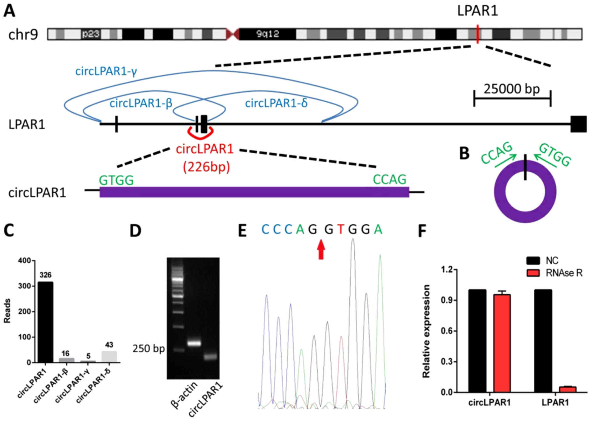
Identification and validation of
circLPAR1 in bladder cancer tissue
circLPAR1 (hsa_circ_0087960) is the product of gene
LPAR1 formed during the transcription process, 226 bp in length,
derived from exons 2 and 3 (Fig. 1A and
B). The transcriptome sequencing results indicated that the
LPAR1 gene encodes three other circRNAs, including LPAR1-β, LPAR1-γ
and LPAR1-δ (Fig. 1C). The
amplification products of circLAPR1 were assessed by RT-qPCR, and
the divergent primers certified that the cyclization site was
expressed in the bladder cancer samples (Fig. 1D). The Sanger sequencing results also
indicated the occurrence of cyclization (Fig. 1E). The RNase R exonuclease digestion
further confirmed that the RNA species was stable in circular form
and resistant to digestion by RNase R (Fig. 1F).
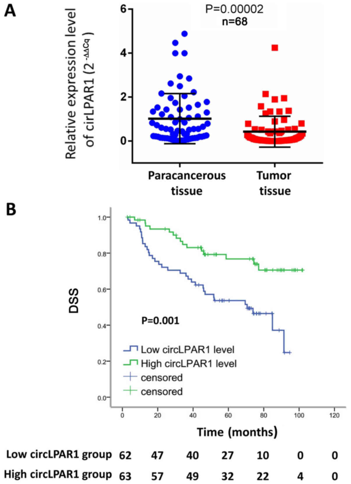
circLPAR1 as a potential predictor of
DSS for MIBC
Firstly, the level of circLPAR1 expression was
investigated by RT-qPCR in 68 MIBC tissues and paired adjacent
non-tumorous tissues. The results demonstrated that the level of
expression was significantly reduced in MIBC tissue, compared with
para-carcinoma tissue (P=0.00002; Fig.
2A). The present study subsequently evaluated the potential
prognostic value of the circRNA and identified that circLPAR1
expression was significantly associated with neoadjuvant
chemotherapy prior to radical cystectomy (P=0.039; Table I). The mean DSS was 54.8±2.6 months
(median, 53.2 months; 95% confidence interval CI, 49.7–60.0
months). In the univariate and multivariate analyses, a low circRNA
expression level (2−∆∆Cq <0.0023) was significantly
associated with poor DSS, compared with a high circRNA expression
level (P=0.001; Table II). The
median DSS was 52.4 months (95% CI, 42.9–57.3 months) and 56.0
months (95% CI, 52.0–66.6 months) for low and high circRNA
expression groups, respectively (P=0.001), and the survival curve
for DSS was illustrated in Fig.
2B.
| Table II.Univariate and multivariate Cox
regression analyses for disease-specific survival in 125 patients
with muscle-invasive bladder cancer. |
Table II.
Univariate and multivariate Cox
regression analyses for disease-specific survival in 125 patients
with muscle-invasive bladder cancer.
|
| Univariate
analysis | Multivariate
analysis |
|---|
|
|
|
|
|---|
| Variables | P-value | HR (95% CI) | P-value | HR (95% CI) |
|---|
| Age (years) |
|
|
|
|
| <61
vs. ≥61 | 0.910 | 0.968
(0.548–1.710) |
|
|
| Sex |
|
|
|
|
| Male
vs. female | 0.266 | 0.614
(0.260–1.451) |
|
|
| Pathological T
stagea |
|
|
|
|
| T2 vs.
T3+T4 | 0.008 | 2.176
(1.228–3.857) | 0.027 | 1.885
(1.040–3.416) |
| Pathological N
stagea |
|
|
|
|
| N0 vs.
N1+N2 | 0.070 | 1.780
(0.955–3.318) |
|
|
| Pathological
Grade |
|
|
|
|
| Low vs.
high | 0.021 | 2.598
(1.154–5.851) | 0.042 | 2.355
(1.031–5.380) |
| Maximum tumor
diameter (cm) |
|
| 0.089 | 1.682
(1.040–3.416) |
| <3.5
vs. ≥3.5 | 0.015 | 2.041
(1.149–3.626) |
|
|
| Tumor number
(n) |
|
|
|
|
| 1 vs.
≥2 | 0.403 | 0.762
(0.403–1.441) |
|
|
| circLPAR1
level |
|
|
|
|
| Low vs.
highb | 0.001 | 0.364
(0.197–0.673) | 0.011 | 0.444
(0.237–0.832) |
| Recurrence after
TURBT |
|
|
|
|
| Yes vs.
no | 0.356 | 1.351
(0.713–2.558) |
|
|
| Chemotherapy before
radical cystectomy |
|
|
|
|
| Yes vs.
no | 0.19 | 1.466
(0.828–2.596) |
|
|
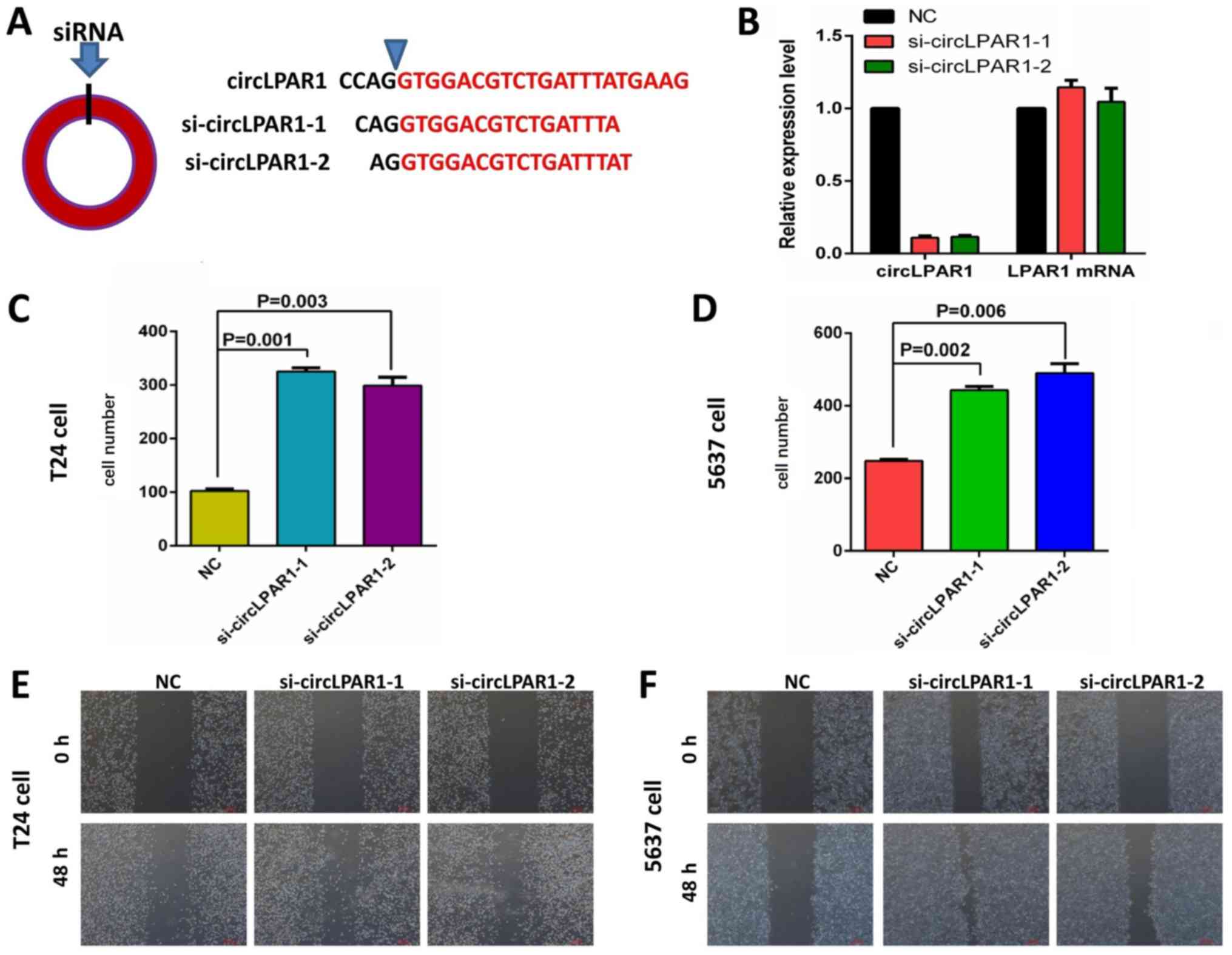
Altered invasion of bladder cancer
cells by si-circLPAR1
The present study designed and synthesized two
siRNAs for circLPAR1 (Fig. 3A). The
RT-qPCR results indicated that these siRNAs could successfully
knockdown the expression of circLPAR1 without affecting the
expression of the host gene, LPAR1 (Fig.
3B). Further cell function tests including Matrigel
demonstrated that in T24 bladder cancer cells, the invasion was
significantly enhanced following the knockdown of circLPAR1
(Fig. 3C). Similar results were also
obtained for the 5637 bladder cancer cells (Fig. 3D). In addition, wound healing assays
for these cell lines demonstrated similar results (Fig. 3E and F).
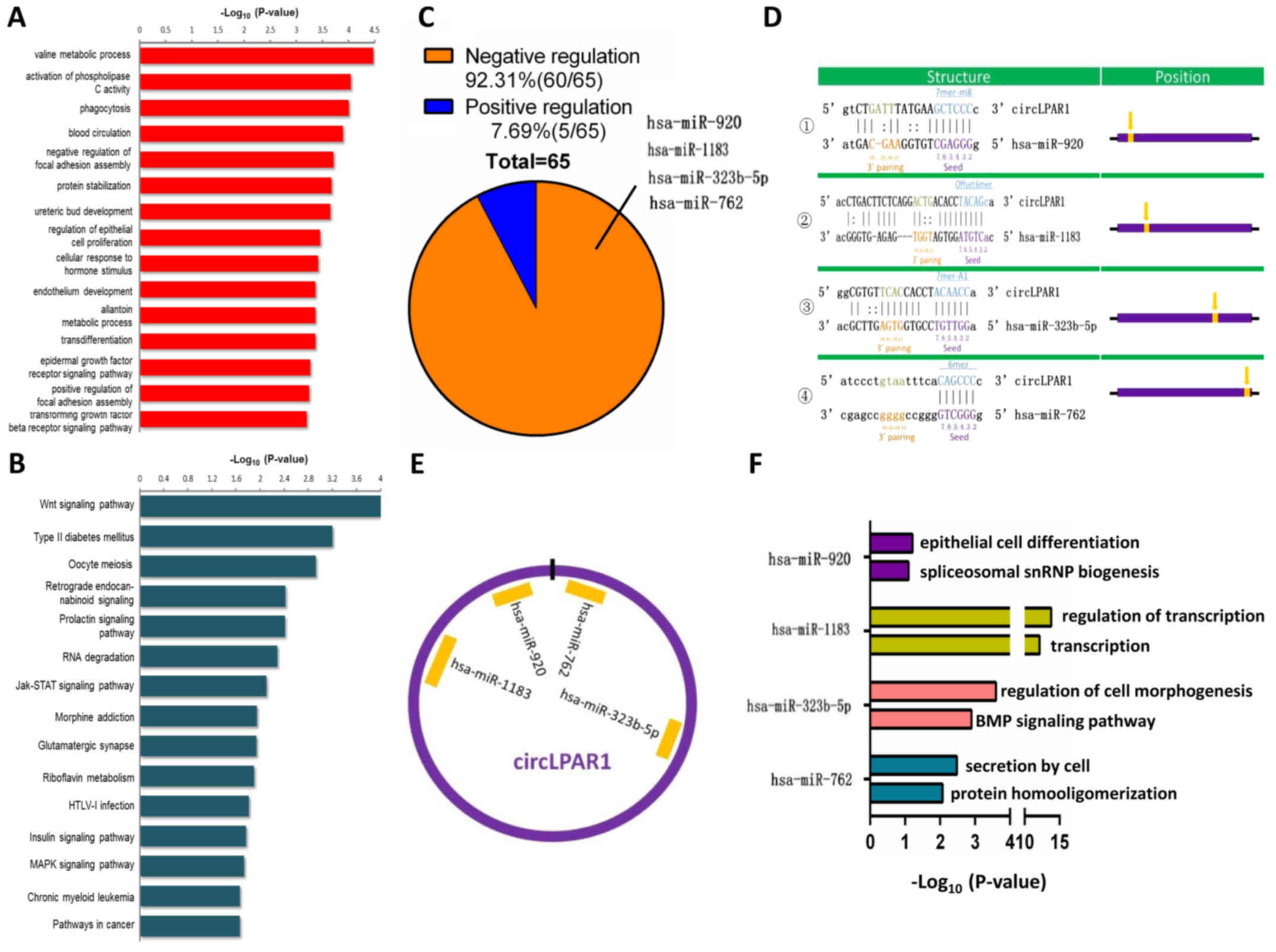
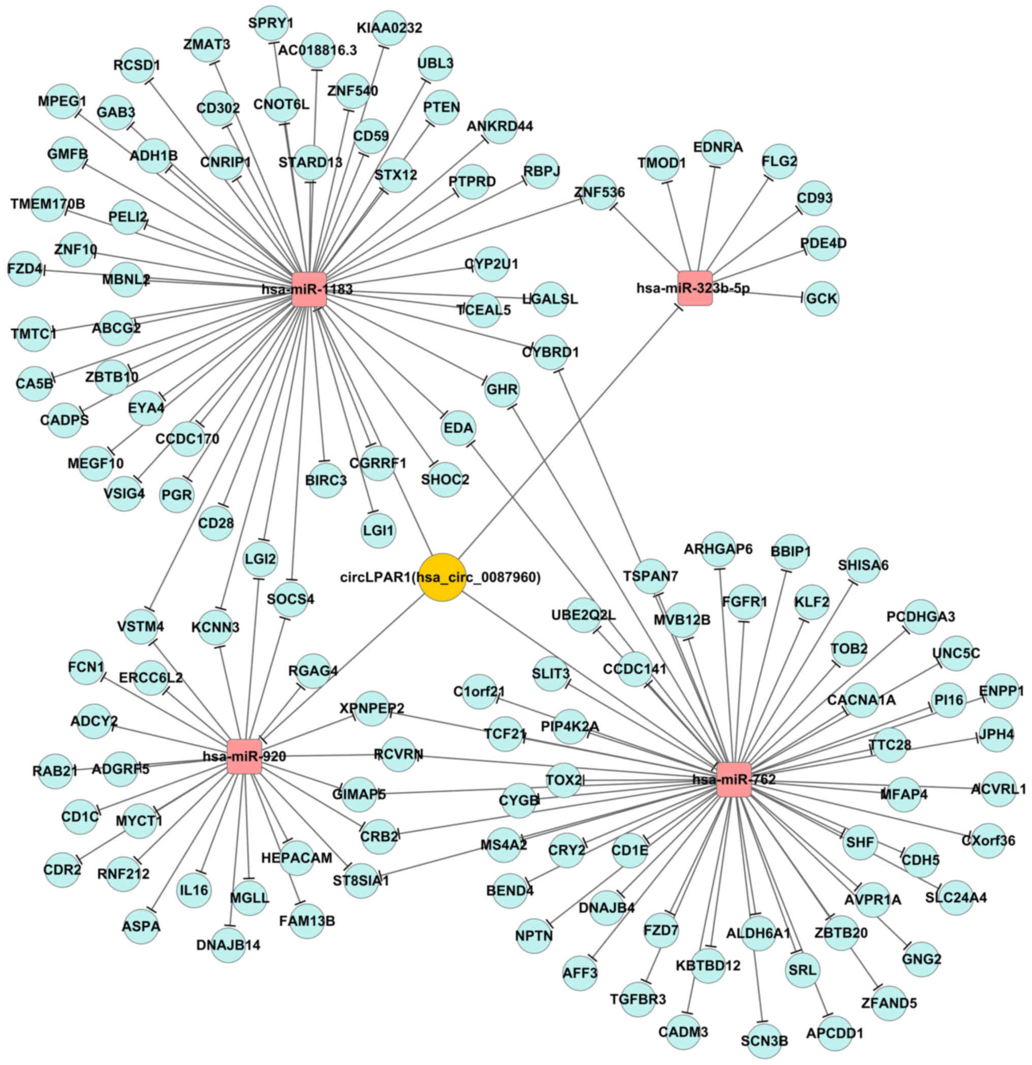
Bioinformatics analysis of circLPAR1
in bladder cancer
The present study identified that there were 56, 7,
52 and 25 associated mRNAs for these miRNAs (fold-change ≥2),
respectively(data not shown). The bioinformatics analysis revealed
that a total of 372 GO-Analysis-BP items were associated with
circLPAR1, and that 22 KEGG pathways were associated with circLPAR1
(Fig. 4A and B). Additionally, there
were 60 negatively regulated miRNAs and 5 positively regulated
miRNAs for circLPAR1 (Fig. 4C). The
data demonstrated that circLPAR1 harbors four miRNAs (hsa-miR-920,
hsa-miR-1183, hsa-miR-323b-5p and hsa-miR-762) according to the
matching seed sequences (Fig. 4D),
and a schematic representation illustrated the putative binding
sites of circLPAR1 with the four associated miRNAs (Fig. 4E). The present study also presented
the two most markedly varying molecular function and biological
process GO items for each of the four matched miRNAs, including
miR-920, miR-1183, miR-323b-5p and miR-762 (Fig. 4F).
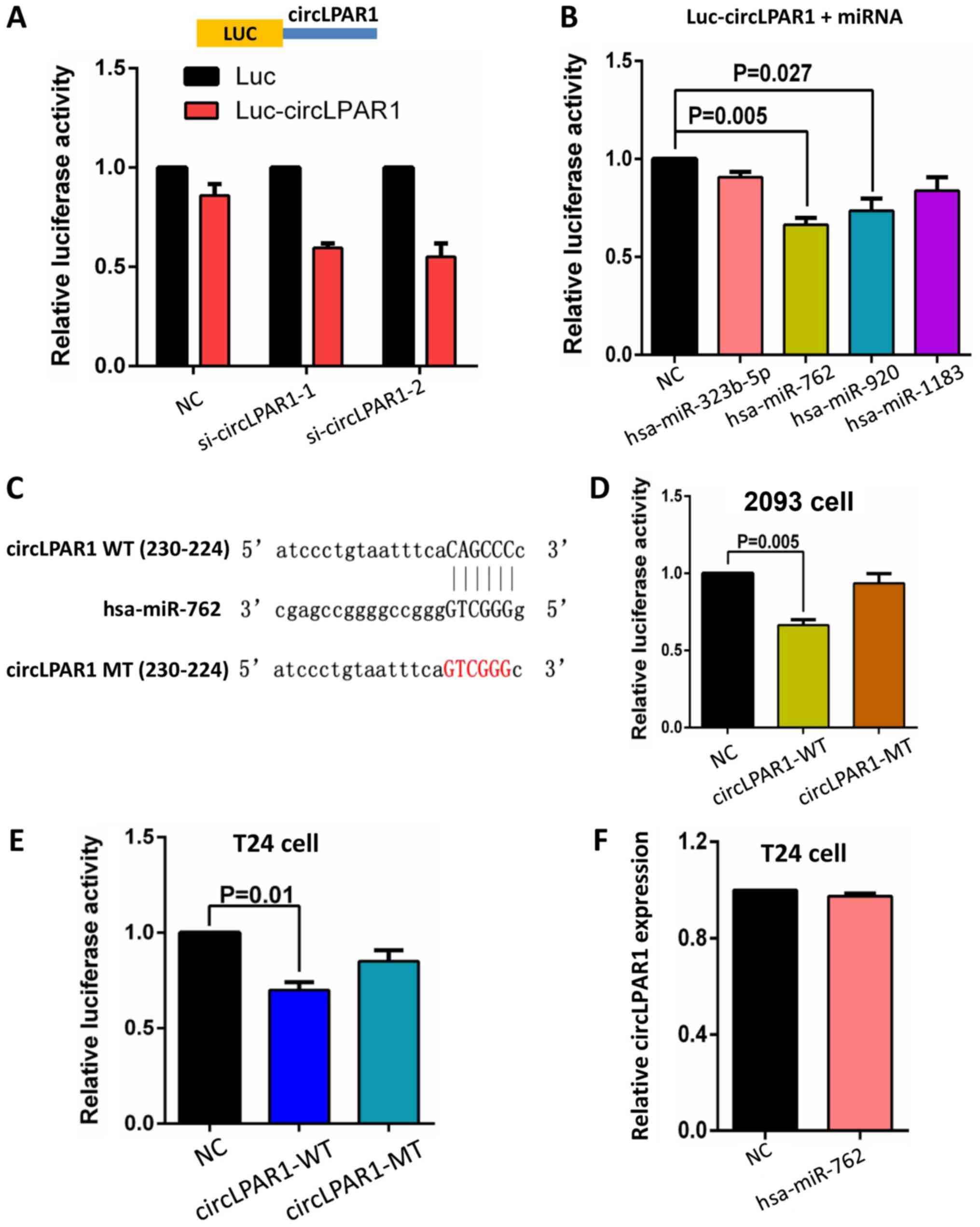
circLPAR1 binds to miR-762 and
inhibits its activity
A circLPAR1 fragment was constructed and inserted
downstream of the luciferase reporter gene. We hypothesized that
miRNAs associated with circLPAR1 may potentially inhibit the
luciferase activity by miRNA-mediated activation of deadenylation
and the subsequent exonucleolytic degradation. The inclusion of
circLPAR1 sequence in the 3′ untranslated region (3′UTR) was
observed to cause the downregulation of luciferase activity and
knockdown of the endogenous circLPAR1 further decreased the
luciferase activity (Fig. 5A).
Additionally, a luciferase reporter assay was performed using these
four miRNAs. Each miRNA mimic was co-transfected with the
luciferase reporters into 293 cells. Compared with the negative
control RNA, miR-762 and miR-920 significantly reduced the
luciferase reporter activity (P=0.005 and 0.027, respectively;
Fig. 5B).
The present study mutated the miR-762 target site by
including the circLPAR1 sequence in the 3′UTR (Fig. 5C). The transfection of miR-762 and the
luciferase reporter gene inserted circLPAR1 mutated fragment did
not significantly affect the luciferase activity in 293 cells
(Fig. 5D). Similar results were
obtained in T24 cells (Fig. 5E). The
transfection of miR-762 in T24 cells did not significantly decrease
the level of circLPAR1, compared with the negative control, as
assessed by RT-qPCR (Fig. 5F), which
indicated that circLPAR1 may not be digested or suppressed by
miR-762.
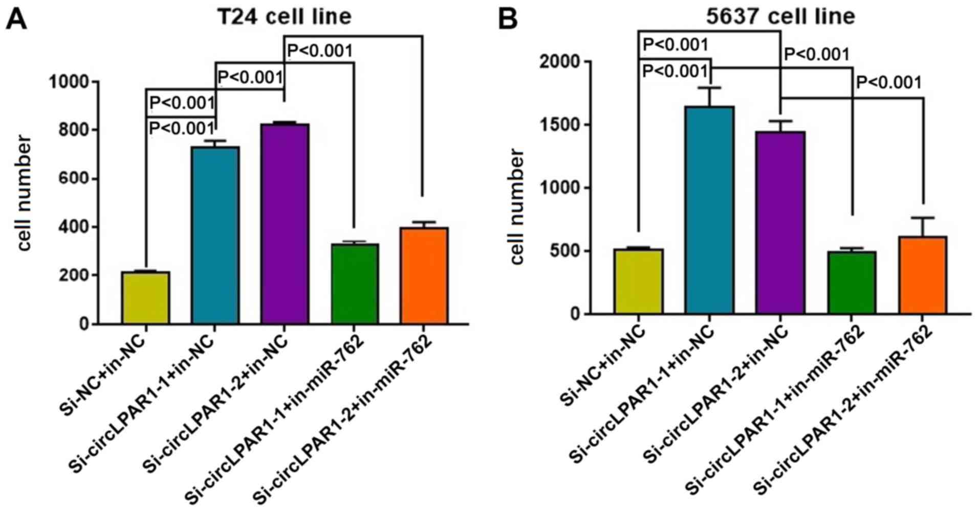
Matrigel assays demonstrated that in T24 bladder
cancer cells, the invasion was significantly enhanced following the
knockdown of circLPAR1 but suppressed by miR-762 (Fig. 6A). Similar results were also obtained
for the 5637 bladder cancer cells (Fig.
6B). Additionally, the circRNA-miRNA-mRNA interaction network
for circLPAR1 was also illustrated in Fig. 7.
Discussion
Urothelial carcinoma is the most common type of
bladder cancer (16). Certain drugs,
such as PD-L1 inhibitors, propose novel treatment options and an
improved patient prognosis (17);
however, the heterogeneity of tumors necessitates the investigation
of individualized treatments and prognostic biomarkers. Non-coding
RNA is an intensive research topic in molecular biology and has
been the focus of numerous studies (11–14);
however, the oncologic value of circRNA, as a novel non-coding RNA,
remains unclear in a clinical setting. Thus, the present study
focused on the clinical application of circRNAs in bladder cancer
and also investigated the underlying mechanism of action.
The majority of circRNAs are derived from exons and
frequently exist in eukaryotic cells (18). A series of endogenous RNA molecules
are speculated to serve a role in regulating gene and protein
expression, and exhibit functions in several pathophysiological
processes, including protein and miRNA binding (19–21). In
the present study a closed ring structure is one of the most vital
characteristics for circRNAs, and hence, circRNAs are evaluated for
the occurrence of circular molecules. In our previous study, the
parent gene LPAR1 in bladder cancer tissue could be formed from
four circRNAs, of which one circRNA derived from exons 2 and 3, was
highly abundant (11). In the present
study, this circRNA was termed as circLPAR1, which constituted of
226 bp. The results of the present study confirmed that circLPAR1
was resistant to RNase R digestion. Divergent primers were used for
amplification, and the RT-qPCR product also demonstrated that the
fragment originated from across the back-splice site. Furthermore,
the distinct band on agarose gel electrophoresis was confirmed as
the ring structure by Sanger sequencing. These studies
substantiated the circular structure of circLPAR1.
circRNAs are associated with numerous diseases, such
as nervous system dysplasia, atherosclerosis and Alzheimer's
disease (22–24). Additionally, circRNAs serve a major
role in the occurrence and development of human tumors (8,25).
Reportedly, the abundance of several circRNAs, including circHIPK3,
differed significantly in the tumor and normal tissues (11,26). Some
scholars indicated that the content of circular RNA
(hsa_circ_002059) in gastric cancer was significantly reduced,
compared with adjacent tissues, and may be a potential novel and
stable biomarker for the diagnosis of gastric carcinoma (11,27). An
additional study demonstrated that the level of the circular RNA
hsa_circ_0001649 was not only decreased in the liver cancer tissue
but was also associated with tumor size and lymph node metastasis
(28). Zhong et al (29) elaborated the molecular mechanism of
circTCF25 promoting the occurrence of bladder cancer by the
inhibition of miR-103a-3p/miR-107.
In the present study, the expression of circLPAR1
was observed to be decreased in 68 bladder cancer tissues, compared
with paired para-cancer tissues. The low level of circLPAR1
expression was defined the level less than the median value
(2C∆∆q=0.0023), and the high level was defined as the
level equal or more than the median value. Further survival
analysis indicated that the DSS from patients with low a expression
level of circLPAR1 was 4 months shorter than those with a high
expression, and the risk of mortality in the low expression group
was two times greater than that in the high expression group. A
potential hypothesis was that circLPAR1 may activate or inhibit
certain signaling pathways, thus resulting in disease progression
(30). In order to investigate the
effect of circLPAR1 on cell function, siRNA was employed to
knockdown the expression of circLPAR1, and the invasion and
metastasis ability of cells was observed to be significantly
decreased. Collectively, these results indicate that that circLPAR1
may be associated with invasion and metastasis.
The most common mechanism of circRNA is speculated
to involve the negative regulation of the expression and function
of miRNAs (14,31). In the present study, the
bioinformatics analysis indicated that four miRNAs may be the
potential targets of circLPAR1. Based on the luciferase reporter
assay, miR-762 and miR-920 were observed to reduce the luciferase
activity, and miR-762 varies significantly. Additionally, the
inhibition of luciferase activity could be alleviated in different
cells after mutating the circLPAR1 sites that could potentially
bind to miR-762. These results indicated that circLPAR1 could bind
to miR-762 and inhibit its activity as a miRNA sponge. Previous
studies also reported that miR-762 promoted the proliferation and
invasion of breast cancer and the development of oral squamous cell
carcinoma (32,33). Collectively, these results revealed a
plausible explanation that circLPAR1 may serve a crucial role in
the invasion and metastasis by miR-762.
However, the theory of circRNAs serving as the miRNA
sponge remains controversial. Although the sponge-like effect of
circRNAs has been proposed previously and confirmed experimentally
(31), recent literature does not
support this conclusion or indicate the presence of other
interacting isoforms (7,20). Notably, a host gene can produce
multiple circRNAs, one of which may exhibit abundant functions,
including in cell proliferation and migration (11). Simultaneously, focus on the existing
functional features of the host genes is required.
In conclusion, the present study demonstrated that
circLPAR1, may be a potential novel and stable biomarker for the
prognosis of bladder cancer that may associate with the invasion by
miR-762. It indicated that the invasion of group si-circLPAR1 and
inhibitor-NC is same as Fig. 3C and
D. But for the group si-circLPAR1 and inhibitor-miR-762, the
invasion returned to the level of si-NC and inhibitor-NC (Fig. 6).
circLPAR1 may be a potential novel and stable
biomarker for the prognosis of MIBC and may be associated with the
invasion and metastasis by miR-762.
Acknowledgements
The authors would like to thank Professor Sheng-Lin
Huang of Fudan University Cancer Institute (Shanghai, China), who
read this manuscript and provided some suggestions.
Funding
This study was supported by the Shanghai Health
Bureau of Research Fund (20144Y0180) and Natural Science Foundation
of China (grant nos. 81672544 and 81572531).
Availability of data and materials
The datasets used and/or analyzed during the current
study are available from the corresponding author on reasonable
request.
Authors' contributions
GWL and HYS improved and refined the project details
and performed the experiments; HYS and HYX provided important
recommendations and all tissue specimens; GHS and DWY designed and
supervised the study; QPZ made substantial contributions to
conception and design; GWL, HY and YJS made substantial
contributions to acquisition of clinicopathological data. All
authors reviewed and approved the manuscript.
Ethics approval and consent to
participate
The present study was conducted in accordance with
the ethical standards of the Helsinki Declaration II and approved
by the Ethics Committee of Fudan University Shanghai Cancer Center
(approval no. 050432-4-1212B). Written informed consent was
obtained from each patient prior to any study-specific experiments
being performed.
Patient consent for publication
Not applicable.
Competing interests
The authors declare that they have no competing
interests.
References
|
1
|
American Cancer Society: Cancer facts and
figures 2015. American Cancer Society; Atlanta, GA: 2015
|
|
2
|
Krogsbøll LT, Jørgensen KJ and Gøtzsche
PC: Screening with urinary dipsticks for reducing morbidity and
mortality. Cochrane Database Syst Rev. 1:CD0100072015.PubMed/NCBI
|
|
3
|
Singh R, Ansari JA, Maurya N, Mandhani A,
Agrawal V and Garg M: Epithelial-to-Mesenchymal transition and its
correlation with clinicopathologic features in patients with
urothelial carcinoma of the bladder. Clin Genitourin Cancer.
15:e187–e197. 2017. View Article : Google Scholar : PubMed/NCBI
|
|
4
|
Chen J, Miao Z, Xue B, Shan Y, Weng G and
Shen B: Long Non-coding RNAs in urologic malignancies: Functional
roles and clinical translation. J Cancer. 7:1842–1855. 2016.
View Article : Google Scholar : PubMed/NCBI
|
|
5
|
Qu S, Yang X, Li X, Wang J, Gao Y, Shang
R, Sun W, Dou K and Li H: Circular RNA: A new star of noncoding
RNAs. Cancer Lett. 365:141–148. 2015. View Article : Google Scholar : PubMed/NCBI
|
|
6
|
Li JH, Liu S, Zhou H, Qu LH and Yang JH:
starBase v2.0: Decoding miRNA-ceRNA, miRNA-ncRNA and protein-RNA
interaction networks from large-scale CLIP-Seq data. Nucleic Acids
Res. 42:(Database Issue). D92–D97. 2014. View Article : Google Scholar : PubMed/NCBI
|
|
7
|
You X, Vlatkovic I, Babic A, Will T,
Epstein I, Tushev G, Akbalik G, Wang M, Glock C, Quedenau C, et al:
Neural circular RNAs are derived from synaptic genes and regulated
by development and plasticity. Nat Neurosci. 18:603–610. 2015.
View Article : Google Scholar : PubMed/NCBI
|
|
8
|
Hansen TB, Kjems J and Damgaard CK:
Circular RNA and miR-7 in cancer. Cancer Res. 73:5609–5612. 2013.
View Article : Google Scholar : PubMed/NCBI
|
|
9
|
Chi BJ, Zhao DM, Liu L, Yin XZ, Wang FF,
Bi S, Gui SL, Zhou SB, Qin WB, Wu DM and Wang SQ: Downregulation of
hsa_circ_0000285 serves as a prognostic biomarker for bladder
cancer and is involved in cisplatin resistance. Neoplasma. Sep
16–2018.(Epub ahead of print). View Article : Google Scholar
|
|
10
|
Liu H, Bi J, Dong W, Yang M, Shi J, Jiang
N, Lin T and Huang J: Invasion-related circular RNA circFNDC3B
inhibits bladder cancer progression through the
miR-1178-3p/G3BP2/SRC/FAK axis. Mol Cancer. 17:1612018. View Article : Google Scholar : PubMed/NCBI
|
|
11
|
Zheng Q, Bao C, Guo W, Li S, Chen J, Chen
B, Luo Y, Lyu D, Li Y, Shi G, et al: Circular RNA profiling reveals
an abundant circHIPK3 that regulates cell growth by sponging
multiple miRNAs. Nat Commun. 7:112152016. View Article : Google Scholar : PubMed/NCBI
|
|
12
|
Enright AJ, John B, Gaul U, Tuschl T,
Sander C and Marks DS: MicroRNA targets in drosophila. Genome Biol.
5:R12003. View Article : Google Scholar : PubMed/NCBI
|
|
13
|
Pasquinelli AE: MicroRNAs and their
targets: Recognition, regulation and an emerging reciprocal
relationship. Nat Rev Genet. 13:271–282. 2012. View Article : Google Scholar : PubMed/NCBI
|
|
14
|
Dennis G Jr, Sherman BT, Hosack DA, Yang
J, Gao W, Lane HC and Lempicki RA: DAVID: Database for annotation,
visualization, and integrated discovery. Genome Biol. 4:P32003.
View Article : Google Scholar : PubMed/NCBI
|
|
15
|
Reiner A, Yekutieli D and Benjamini Y:
Identifying differentially expressed genes using false discovery
rate controlling procedures. Bioinformatics. 19:368–375. 2003.
View Article : Google Scholar : PubMed/NCBI
|
|
16
|
Grivas PD, Melas M and Papavassiliou AG:
The biological complexity of urothelial carcinoma: Insights into
carcinogenesis, targets and biomarkers of response to therapeutic
approaches. Semin Cancer Biol. 35:125–132. 2015. View Article : Google Scholar : PubMed/NCBI
|
|
17
|
Aoun F, Kourie HR, Sideris S, Roumeguere
T, van Velthoven R and Gil T: Checkpoint inhibitors in bladder and
renal cancers: Results and perspectives. Immunotherapy.
7:1259–1271. 2015. View Article : Google Scholar : PubMed/NCBI
|
|
18
|
Zhang Y, Zhang XO, Chen T, Xiang JF, Yin
QF, Xing YH, Zhu S, Yang L and Chen LL: Circular intronic long
noncoding RNAs. Mol Cell. 51:792–806. 2013. View Article : Google Scholar : PubMed/NCBI
|
|
19
|
Jeck WR and Sharpless NE: Detecting and
characterizing circular RNAs. Nat Biotechnol. 32:453–461. 2014.
View Article : Google Scholar : PubMed/NCBI
|
|
20
|
Memczak S, Jens M, Elefsinioti A, Torti F,
Krueger J, Rybak A, Maier L, Mackowiak SD, Gregersen LH, Munschauer
M, et al: Circular RNAs are a large class of animal RNAs with
regulatory potency. Nature. 495:333–338. 2013. View Article : Google Scholar : PubMed/NCBI
|
|
21
|
Conn SJ, Pillman KA, Toubia J, Conn VM,
Salmanidis M, Phillips CA, Roslan S, Schreiber AW, Gregory PA and
Goodall GJ: The RNA binding protein quaking regulates formation of
circRNAs. Cell. 160:1125–1134. 2015. View Article : Google Scholar : PubMed/NCBI
|
|
22
|
Floris G, Zhang L, Follesa P and Sun T:
Regulatory role of circular RNAs and neurological disorders. Mol
Neurobiol. 54:5156–5165. 2017. View Article : Google Scholar : PubMed/NCBI
|
|
23
|
Burd CE, Jeck WR, Liu Y, Sanoff HK, Wang Z
and Sharpless NE: Expression of linear and novel circular forms of
an INK4/ARF-associated non-coding RNA correlates with
atherosclerosis risk. PLoS Genet. 6:e10012332010. View Article : Google Scholar : PubMed/NCBI
|
|
24
|
Lukiw WJ: Circular RNA (circRNA) in
Alzheimer's disease (AD). Front Genet. 4:3072013. View Article : Google Scholar : PubMed/NCBI
|
|
25
|
Li F, Zhang L, Li W, Deng J, Zheng J, An
M, Lu J and Zhou Y: Circular RNA ITCH has inhibitory effect on ESCC
by suppressing the Wnt/β-catenin pathway. Oncotarget. 6:6001–6013.
2015.PubMed/NCBI
|
|
26
|
Li Y, Zheng Q, Bao C, Li S, Guo W, Zhao J,
Chen D, Gu J, He X and Huang S: Circular RNA is enriched and stable
in exosomes: A promising biomarker for cancer diagnosis. Cell Res.
25:981–984. 2015. View Article : Google Scholar : PubMed/NCBI
|
|
27
|
Li P, Chen S, Chen H, Mo X, Li T, Shao Y,
Xiao B and Guo J: Using circular RNA as a novel type of biomarker
in the screening of gastric cancer. Clin Chim Acta. 444:132–136.
2015. View Article : Google Scholar : PubMed/NCBI
|
|
28
|
Qin M, Liu G, Huo X, Tao X, Sun X, Ge Z,
Yang J, Fan J, Liu L and Qin W: Hsa_circ_0001649: A circular RNA
and potential novel biomarker for hepatocellular carcinoma. Cancer
Biomark. 16:161–169. 2016. View Article : Google Scholar : PubMed/NCBI
|
|
29
|
Zhong Z, Lv M and Chen J: Screening
differential circular RNA expression profiles reveals the
regulatory role of circTCF25-miR-103a-3p/miR-107-CDK6 pathway in
bladder carcinoma. Sci Rep. 6:309192016. View Article : Google Scholar : PubMed/NCBI
|
|
30
|
Liu J, Liu T, Wang X and He A: Circles
reshaping the RNA world: From waste to treasure. Mol Cancer.
16:582017. View Article : Google Scholar : PubMed/NCBI
|
|
31
|
Hansen TB, Jensen TI, Clausen BH, Bramsen
JB, Finsen B, Damgaard CK and Kjems J: Natural RNA circles function
as efficient microRNA sponges. Nature. 495:384–388. 2013.
View Article : Google Scholar : PubMed/NCBI
|
|
32
|
Li Y, Huang R, Wang L, Hao J, Zhang Q,
Ling R and Yun J: microRNA-762 promotes breast cancer cell
proliferation and invasion by targeting IRF7 expression. Cell
Prolif. 48:643–649. 2015. View Article : Google Scholar : PubMed/NCBI
|
|
33
|
Yu T, Wang XY, Gong RG, Li A, Yang S, Cao
YT, Wen YM, Wang CM and Yi XZ: The expression profile of microRNAs
in a model of 7,12-dimethyl-benz[a]anthrance-induced oral
carcinogenesis in Syrian hamster. J Exp Clin Cancer Res. 28:642009.
View Article : Google Scholar : PubMed/NCBI
|