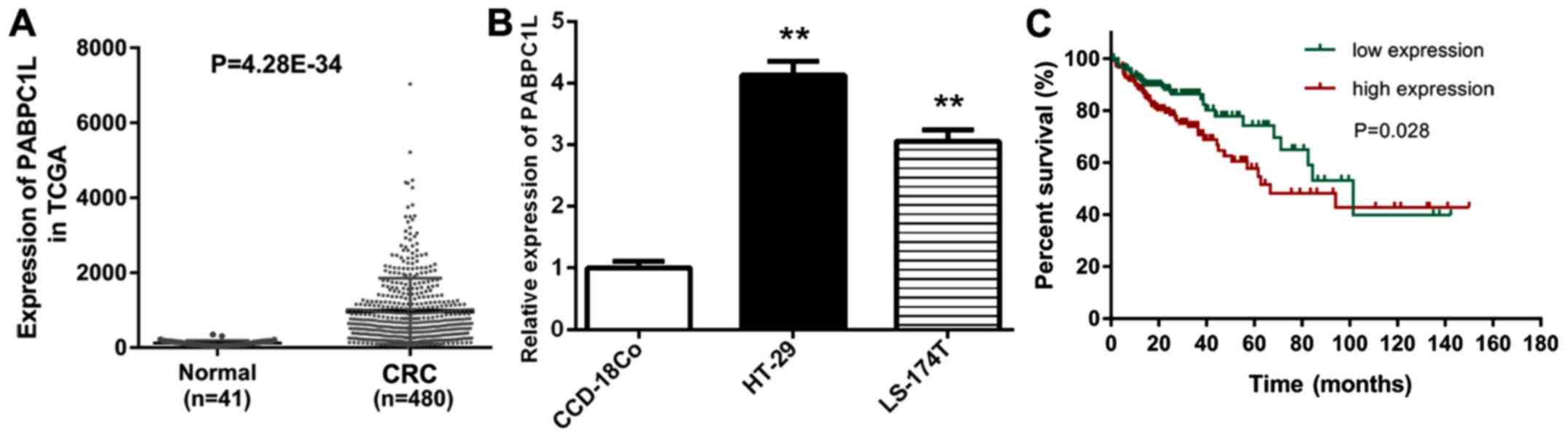
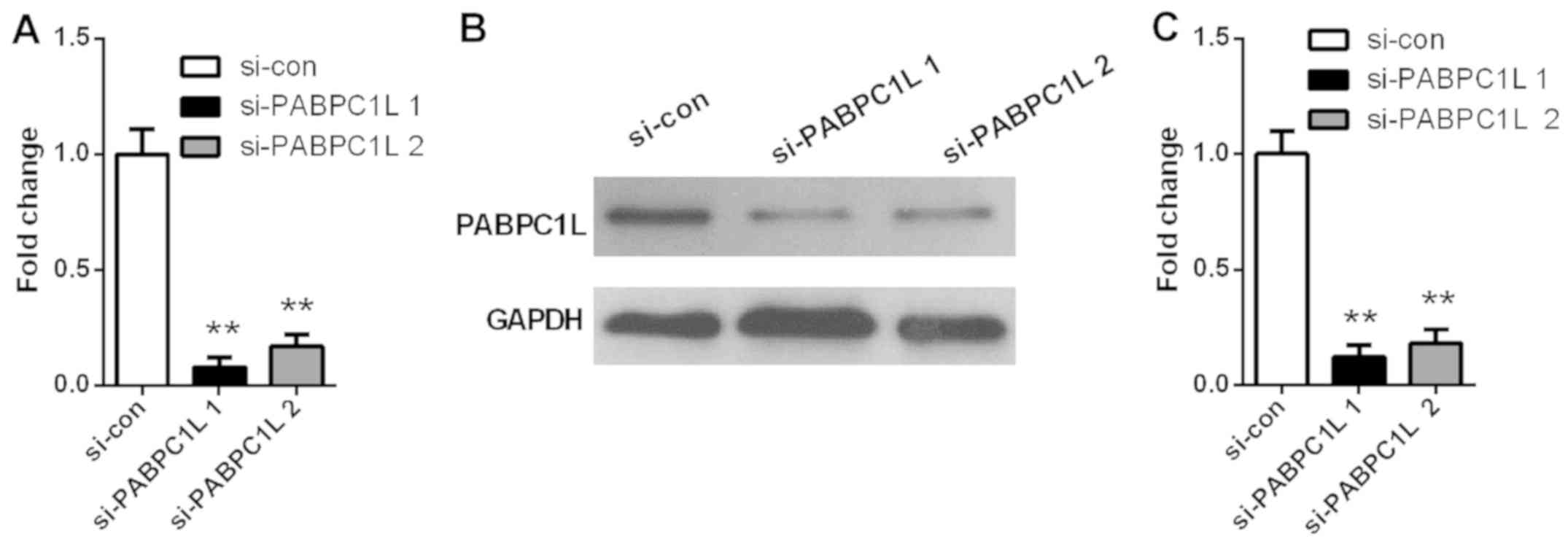
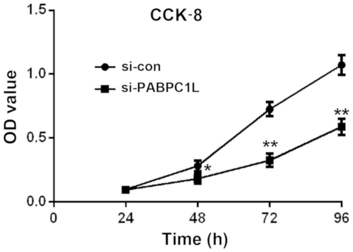
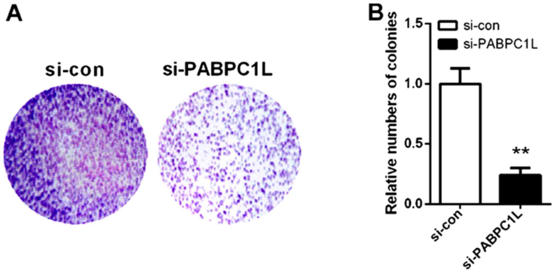
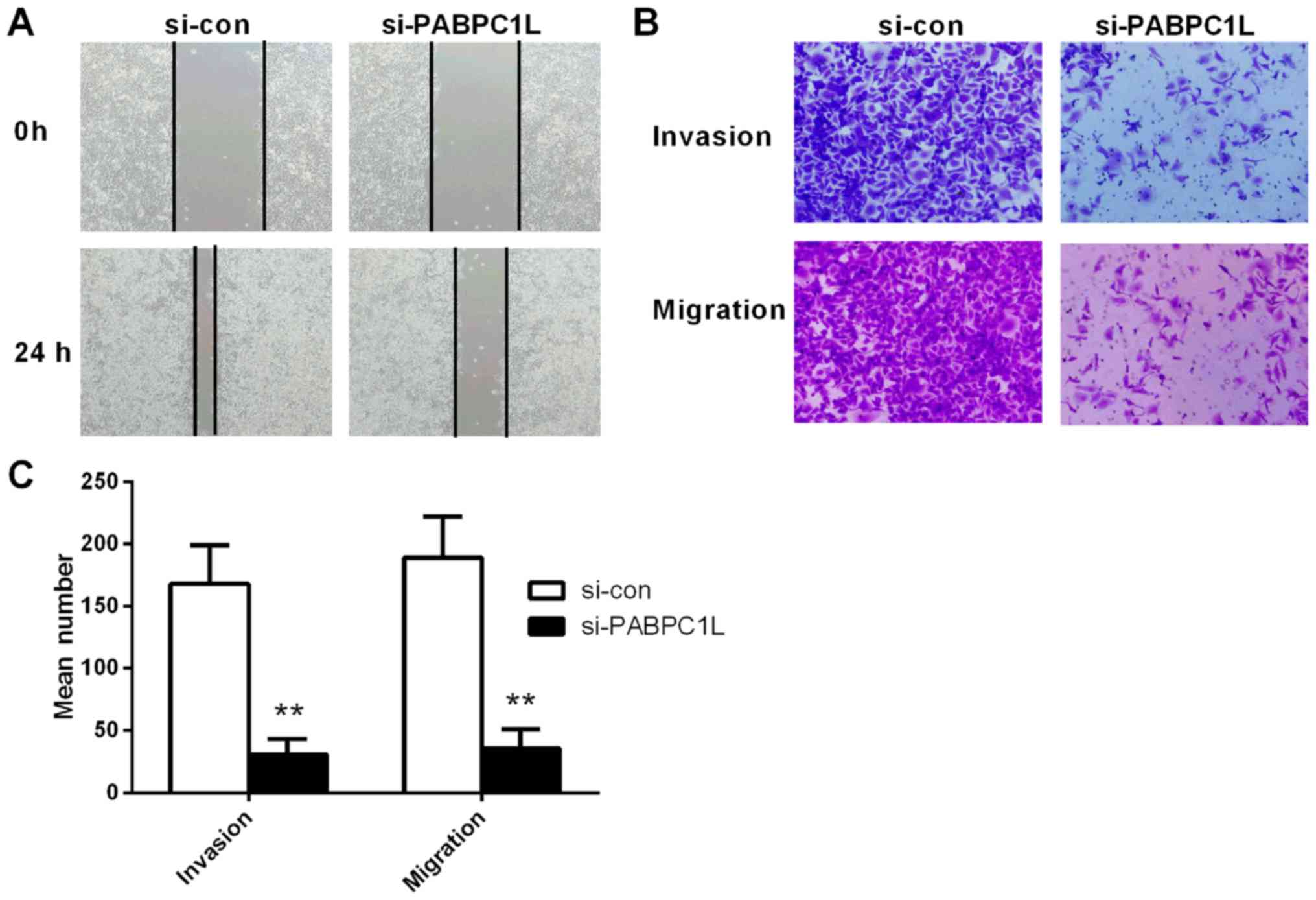
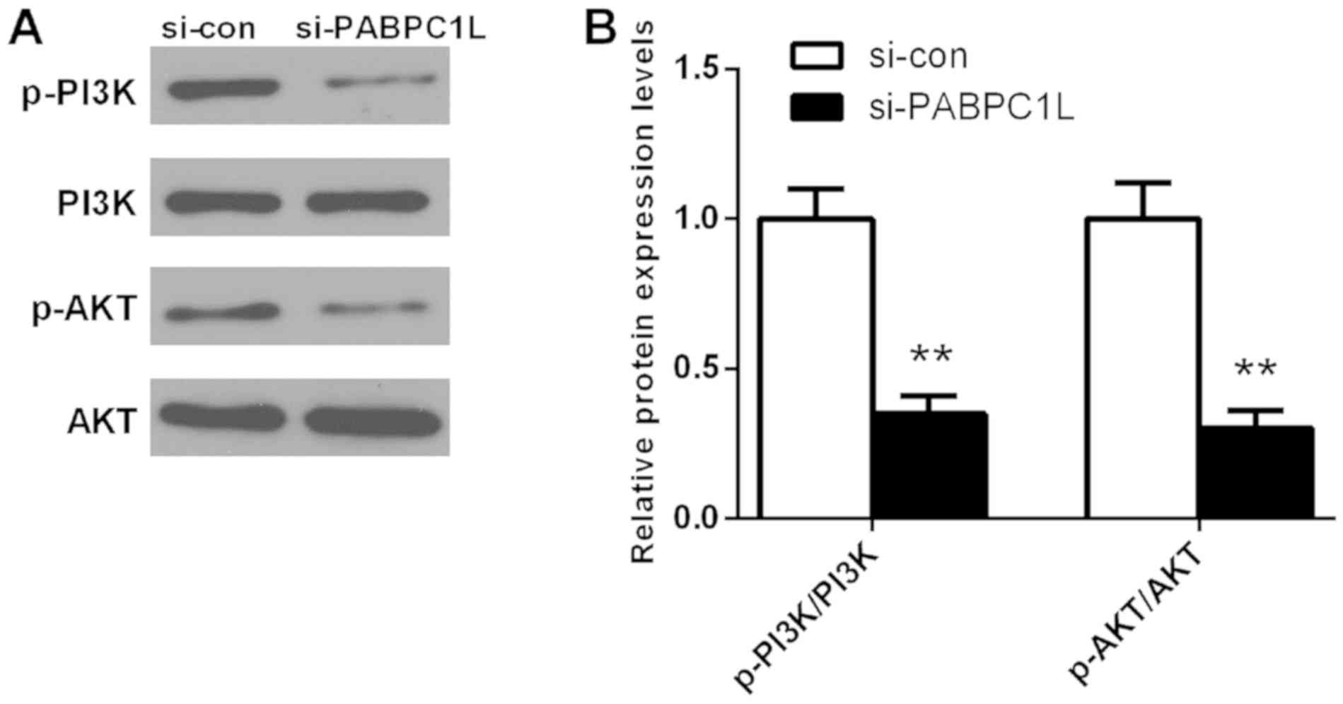
|
1
|
Siegel RL, Ma J, Zou Z and Jemal A: Cancer
Statistics, 2014. CA Cancer J Clin. 64:102014. View Article : Google Scholar
|
|
2
|
Siegel RL, Miller KD and Jemal A: Cancer
statistics, 2015. CA Cancer J Clin. 65:11–30. 2015. View Article : Google Scholar
|
|
3
|
Weitz J, Koch M, Debus J, Höhler T, Galle
PR and Büchler MW: Colorectal cancer. Lancet. 365:153–165. 2005.
View Article : Google Scholar : PubMed/NCBI
|
|
4
|
Villalba A, Coll O and Gebauer F:
Cytoplasmic polyadenylation and translational control. Curr Opin
Genet Dev. 21:452–457. 2011. View Article : Google Scholar : PubMed/NCBI
|
|
5
|
Grange T, de Sa CM, Oddos J and Pictet R:
Human mRNA polyadenylate binding protein: Evolutionary conservation
of a nucleic acid binding motif. Nucleic Acids Res. 15:4771–4787.
1987. View Article : Google Scholar : PubMed/NCBI
|
|
6
|
Silvera D, Arju R, Darvishian F, Levine
PH, Zolfaghari L, Goldberg J, Hochman T, Formenti SC and Schneider
RJ: Essential role for eIF4GI overexpression in the pathogenesis of
inflammatory breast cancer. Nat Cell Biol. 11:903–908. 2009.
View Article : Google Scholar : PubMed/NCBI
|
|
7
|
Zhu J, Ding H, Wang X and Lu Q: PABPC1
exerts carcinogenesis in gastric carcinoma by targeting miR-34c.
Int J Clin Exp Pathol. 8:3794–3802. 2015.PubMed/NCBI
|
|
8
|
Eisermann K, Dar JA, Dong J, Wang D,
Masoodi KZ and Wang Z: Poly (A) binding protein cytoplasmic 1 is a
novel co-regulator of the androgen receptor. PLoS One.
10:e01284952015. View Article : Google Scholar : PubMed/NCBI
|
|
9
|
Takashima N, Ishiguro H, Kuwabara Y,
Kimura M, Haruki N, Ando T, Kurehara H, Sugito N, Mori R and Fujii
Y: Expression and prognostic roles of PABPC1 in esophageal cancer:
Correlation with tumor progression and postoperative survival.
Oncol Rep. 15:667–671. 2006.PubMed/NCBI
|
|
10
|
Ritchie ME, Phipson B, Wu D, Hu Y, Law CW,
Shi W and Smyth GK: limma powers differential expression analyses
for RNA-sequencing and microarray studies. Nucleic Acids Res.
43:e472015. View Article : Google Scholar : PubMed/NCBI
|
|
11
|
Livak KJ and Schmittgen TD: Analysis of
relative gene expression data using real-time quantitative PCR and
the 2(-Delta Delta C(T)) method. Methods. 25:402–408. 2001.
View Article : Google Scholar : PubMed/NCBI
|
|
12
|
Li L, Wang L, Song P, Geng X, Liang X,
Zhou M, Wang Y, Chen C, Jia J and Zeng J: Critical role of histone
demethylase RBP2 in human gastric cancer angiogenesis. Mol Cancer.
13:812014. View Article : Google Scholar : PubMed/NCBI
|
|
13
|
Jiang CG, Lv L, Liu FR, Wang ZN, Liu FN,
Li YS, Wang CY, Zhang HY, Sun Z and Xu HM: Downregulation of
connective tissue growth factor inhibits the growth and invasion of
gastric cancer cells and attenuates peritoneal dissemination. Mol
Cancer. 10:1222011. View Article : Google Scholar : PubMed/NCBI
|
|
14
|
Tian Y, Pan Q, Shang Y, Zhu R, Ye J, Liu
Y, Zhong X, Li S, He Y, Chen L, et al: MicroRNA-200 (miR-200)
cluster regulation by achaete scute-like 2 (Ascl2): Impact on the
epithelial-mesenchymal transition in colon cancer cells. J Biol
Chem. 289:36101–36115. 2014. View Article : Google Scholar : PubMed/NCBI
|
|
15
|
Assinder SJ, Dong Q, Kovacevic Z and
Richardson DR: The TGF-beta, PI3K/Akt and PTEN pathways:
Established and proposed biochemical integration in prostate
cancer. Biochem J. 417:411–421. 2009. View Article : Google Scholar : PubMed/NCBI
|
|
16
|
Osaki M, Oshimura M and Ito H: PI3K-Akt
pathway: Its functions and alterations in human cancer. Apoptosis.
9:667–676. 2004. View Article : Google Scholar : PubMed/NCBI
|
|
17
|
Kim SM, Park JH, Kim KD, Nam D, Shim BS,
Kim SH and Ahn KS, Choi SH and Ahn KS: Brassinin induces apoptosis
in PC-3 human prostate cancer cells through the suppression of
PI3K/Akt/mTOR/S6K1 signaling cascades. Phytother Res. 28:423–431.
2014. View
Article : Google Scholar : PubMed/NCBI
|
|
18
|
Liu YZ, Wu K, Huang J, Liu Y, Wang X, Meng
ZJ, Yuan SX, Wang DX, Luo JY, Zuo GW, et al: The PTEN/PI3K/Akt and
Wnt/β-catenin signaling pathways are involved in the inhibitory
effect of resveratrol on human colon cancer cell proliferation. Int
J Oncol. 45:104–112. 2014. View Article : Google Scholar : PubMed/NCBI
|
|
19
|
Jiang QG, Li TY, Liu DN and Zhang HT:
PI3K/Akt pathway involving into apoptosis and invasion in human
colon cancer cells LoVo. Mol Biol Rep. 41:3359–3367. 2014.
View Article : Google Scholar : PubMed/NCBI
|
|
20
|
Fruman DA and Rommel C: PI3K and cancer:
Lessons, challenges and opportunities. Nat Rev Drug Discov.
13:140–156. 2014. View
Article : Google Scholar : PubMed/NCBI
|
|
21
|
Josse C, Bouznad N, Geurts P, Irrthum A,
Huynh-Thu VA, Servais L, Hego A, Delvenne P, Bours V and Oury C:
Identification of a microRNA landscape targeting the PI3K/Akt
signaling pathway in inflammation-induced colorectal
carcinogenesis. Am J Physiol Gastrointest Liver Physiol.
306:G229–G243. 2014. View Article : Google Scholar : PubMed/NCBI
|
|
22
|
Xiao ZM, Wang XY and Wang AM: Periostin
induces chemoresistance in colon cancer cells through activation of
the PI3K/Akt/survivin pathway. Biotechnol Appl Biochem. 62:401–406.
2015. View
Article : Google Scholar : PubMed/NCBI
|
|
23
|
Malinowsky K, Nitsche U, Janssen KP, Bader
FG, Späth C, Drecoll E, Keller G, Höfler H, Slotta-Huspenina J and
Becker KF: Activation of the PI3K/AKT pathway correlates with
prognosis in stage II colon cancer. Br J Cancer. 110:2081–2089.
2014. View Article : Google Scholar : PubMed/NCBI
|
|
24
|
Nuvoli B, Santoro R, Catalani S,
Battistelli S, Benedetti S, Canestrari F and Galati R: CELLFOOD™
induces apoptosis in human mesothelioma and colorectal cancer cells
by modulating p53, c-myc and pAkt signaling pathways. J Exp Clin
Cancer Res. 33:242014. View Article : Google Scholar : PubMed/NCBI
|
|
25
|
Yu C, Yu J, Yao X, Wu WK, Lu Y, Tang S, Li
X, Bao L, Li X, Hou Y, et al: Discovery of biclonal origin and a
novel oncogene SLC12A5 in colon cancer by single-cell sequencing.
Cell Res. 24:701–712. 2014. View Article : Google Scholar : PubMed/NCBI
|