Introduction
Gastric cancer (GC) remains one of the leading
causes of cancer-related mortality in Eastern Asia, Europe and
South America (1). In fact, >70%
of GC patients are diagnosed at an advanced disease stage (2), despite the discovery of numerous
chemotherapeutics [including fluorouracil
(FU)/leucovorin/oxaliplatin], which have improved the treatment of
patients with advanced GC, the overall prognosis remains poor
(3). Novel prognostic indicators are
urgently required to facilitate improvements in the treatment and
diagnosis of GC.
Replication protein A (RPA) is a single-stranded
DNA-binding gene family consisting of four members, RPA1-4
(4). RPAs play an essential role in
DNA replication, repair, recombination and cell-cycle regulation
(4–8). Previously, RPAs were thought to serve
as prognostic biomarkers in several tumor types (5–8).
Specifically, high protein expression levels of RPA1 (detected
using immunohistochemistry) were associated with poorer outcomes in
patients with esophageal carcinoma, compared with those with normal
levels of expression (5).
Moreover, RPA2 was identified as an independent
prognostic indicator of astrocytic tumors (6). In addition, decreased expression of
both RPA1 and 2 resulted in an adverse prognosis for patients with
muscle-invasive urothelial carcinoma and colon cancer (7,8).
Nonetheless, the specific prognostic values of RPAs in GC are yet
to be fully determined. The present in silico study
characterized both the prognostic and immunological potential of
RPAs in GC, using bioinformatics strategies and public online
resources.
Materials and methods
Oncomine database analysis
The Student's t-test was used to compare the
differences in the expression levels of RPAs between GC and normal
control tissues, using three datasets (GSE13911, GSE13861 and
PMID:19081245) (9–11) retrieved from the Oncomine online
database (https://www.oncomine.org). P<0.01
and a fold-change >2 were selected as cut-off values and
considered to indicate statistically significant differences
(12).
Kaplan-Meier (KM) analysis
To investigate the prognostic value of RPA family
mRNA expression levels in patients with GC, KM analysis was
performed (www.kmplot.com) (13) to evaluate the differences in overall
survival (OS) time between the high- and low-expression groups. The
hazard ratio (HR) with 95% confidence interval (CI), and log-rank
P-values were calculated, and are presented on each KM survival
plot. P<0.05 was considered to indicate a statistically
significant difference. The following datasets were retrieved from
the Gene Expression Omnibus: GSE14210 (n=146) (14), GSE15459 (n=200) (15), GSE22377 (n=43) (16), GSE29272 (n=268) (17), GSE51105 (n=94) (18) and GSE62254 (n=300) (19). However, according to the
recommendation of the KM plotter administrators, GSE62254 was
excluded from KM analysis due to markedly different survival times
and expression profiles, compared with the other datasets (13). No other inclusion or exclusion
criteria were specified. The mRNA expression profiles of RPA family
members 1–4 were collected from each dataset, and normalization
procedures were performed (13). The
optimal significance cut-off value between the high- and
low-expression groups was calculated based on the imbedded
algorithm of the KM plotter (13).
Determination of the prognostic value
of the RPA family signature using SurvExpress
The prognostic value of the RPA family signature was
evaluated with STAD datasets retrieved from The Cancer Genome Atlas
(TCGA), via bioinformatics analysis using the SurvExpress biomarker
validation tool (http://bioinformatica.mty.itesm.mx:8080/Biomatec/SurvivaX.jsp)
(20). A maximized risk score
algorithm was used to categorize the data into thigh- and low-risk
groups.
Tumor-immunological features of RPAs
in the Tumor Immune Estimation Response (TIMER)
The correlation between RPA expression and tumor
immune infiltrating cell (TIIC; B cells, CD4+ T cells, CD8+ T
cells, neutrophils, macrophages and dendritic cells) activity was
analyzed via the TIMER platform (https://cistrome.shinyapps.io/timer/) (21,22),
which is a comprehensive resource used for the systematic
evaluation of immunological features, based on the datasets
retrieved from TGCA (21,22). The correlation between the expression
of each gene and TIICs was determined using the purity-corrected
partial Spearman's rank correlation coefficient. Negative
association with tumor purity indicated high expression in the
microenvironment (21,22).
Results
mRNA expression levels of RPA1-4 in GC
tissues
The relative mRNA expression levels of RPA1-4 in GC
tissues was elucidated by comparing data on GC and normal tissues,
retrieved from the Oncomine database. The mRNA expression levels of
RPA1 and 2 in gastric intestinal type adenocarcinoma (GITA),
diffuse gastric adenocarcinoma (DGA) and gastric mixed
adenocarcinoma (GMA) were all significantly higher than those in
normal gastric mucosal tissues (Fig.
1). The mRNA expression levels of RPA3 in gastric intestinal
type adenocarcinoma and diffuse gastric adenocarcinoma were higher
compared with those in normal gastric mucosal tissues, whereas no
significance was observed in gastric mixed adenocarcinoma. However,
RPA4 did not exhibit significantly differential expression between
the GC and normal tissue groups.
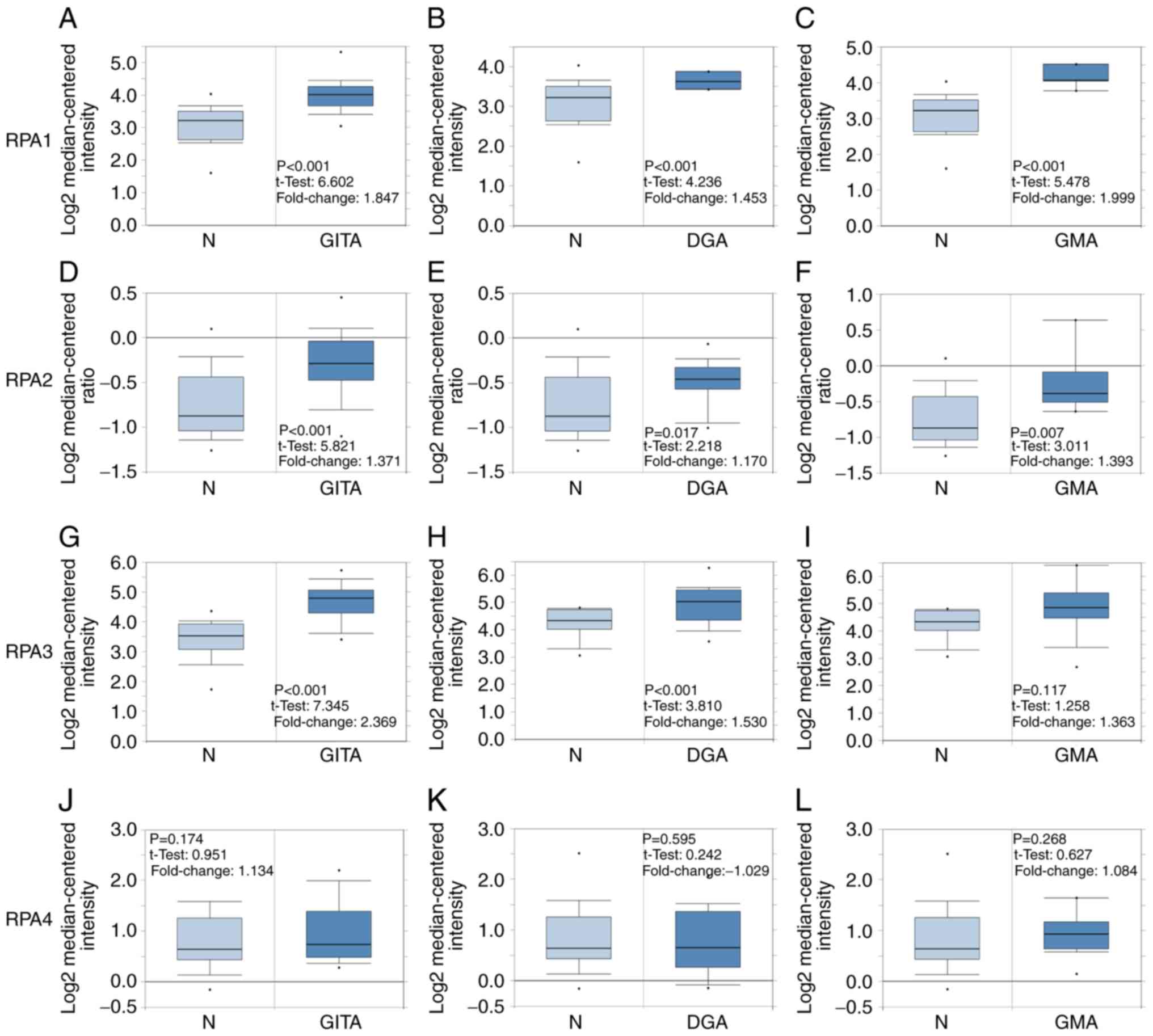 | Figure 1.RPA family analysis in patients with
GC. Box plots comparing the expression of specific RPA family
members in normal and GC tissues, based on datasets retrieved from
the Oncomine database. (A-C) Comparison of RPA1 mRNA expression
levels between normal tissues and those in (A) GITA, (B) DGA and
(C) GMA. Comparison of RPA2 mRNA expression between normal and (D)
GITA, (E) DGA and (F) GMA tissues. (G-I) Comparison of RPA3 mRNA
expression between normal and (G) GITA, (H) DGA and (I) GMA
tissues. Comparison of RPA4 mRNA expression between normal and (J)
GITA, (K) DGA and (L) GMA tissues. GC, gastric cancer; GITA,
gastric intestinal type adenocarcinoma; DGA, diffuse gastric
adenocarcinoma; GMA, gastric mixed adenocarcinoma; RPA, replication
protein A. |
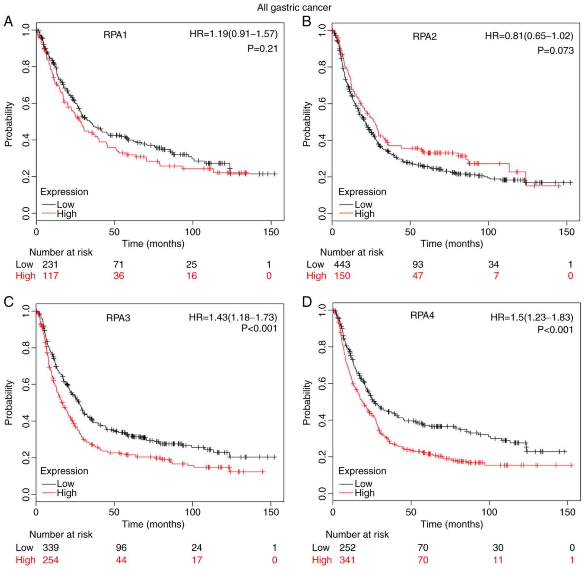
Prognostic values of RPA1-4 in GC
High mRNA expression levels of RPA3 (Fig. 2C; HR, 1.43; 95% CI, 1.18–1.73;
P=2.9×10⁻⁴) and RPA4 (Fig.
2D; HR=1.5; 95% CI, 1.23–1.83; P=7×10⁻⁵) were
significantly correlated with a poor prognosis in patients with GC.
By contrast, RPA1 (Fig. 2A; P=0.21)
and RPA2 (Fig. 2B, P=0.073) did not
exert a significant correlation between high-expression level and
poor prognosis.
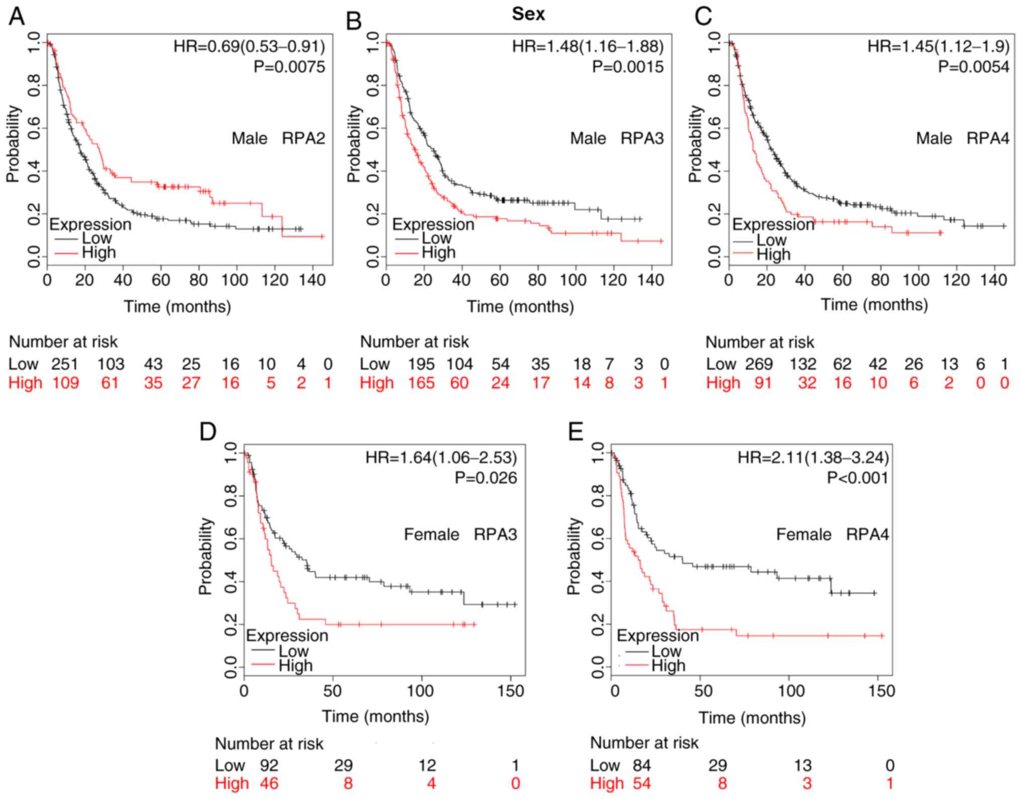
The association between the prognostic values of
RPA1-4 and various clinicopathological features (including sex,
HER2 expression, differentiation, Lauren classification, treatments
and TNM stage) was also evaluated following stratification into
subsets. The difference in prognostic value between the two sexes
is detailed in Fig. 3. High RPA2
mRNA expression was associated with improved OS in male patients
(Fig. 3A; HR, 0.69; 95% CI,
0.53–0.91; P=0.0075). Furthermore, high mRNA expression of RPA3 and
RPA4 were significantly associated with a poorer prognosis in both
male and female patients with GC (Fig.
3B-E).
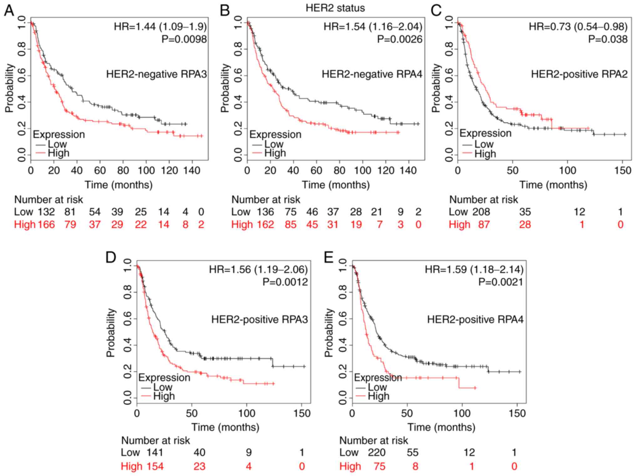
HER2 status analysis determined that a high RPA2
mRNA expression level was associated with improved OS time in
HER2-positive patients (Fig. 4C; HR,
0.73; 95% CI, 0.54–0.98; P=0.038). By contrast, high RPA3 and 4
mRNA expression were significantly associated with a poor prognosis
in both HER2-negative and -positive patients with GC (Fig. 4A, P=0.0098; Fig. 4B, P=0.0026; Fig. 4D, P=0.0012; Fig. 4E, P=0.0021).
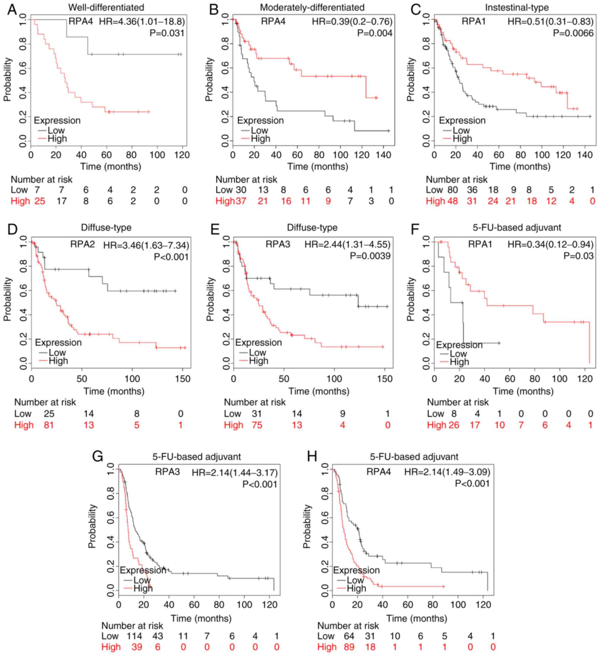
High RPA4 mRNA expression was significantly
correlated with poorer OS in GC patients with well-differentiated
tumors (Fig. 5A; HR, 4.36; 95% CI,
1.01–18.8; P=0.031). However, the high-expression GC group with
moderately-differentiated tumors were also significantly associated
with better prognosis (Fig. 5B; HR,
0.39; 95% CI, 0.2–0.76; P=0.004).
A high-expression level of RPA1 was significantly
correlated with improved prognosis in patients with intestinal-type
GC (Fig. 5C; HR, 0.51; 95% CI,
0.31–0.83; P=0.0066), whilst high-expression levels of RPA2 and 3
were significantly associated with poor survival in patients with
diffuse-type GC (Fig. 5D,
P=6.4×10⁻⁴; Fig. 5E,
P=0.0039).
High mRNA expression of RPA1 was significantly
associated with an improved prognosis in patients with GC receiving
5-FU-based adjuvant chemotherapy (Fig.
5F; HR, 0.34; 95% CI, 0.12–0.94; P=0.03). By contrast, high
RPA3 and 4 mRNA expression was significantly associated with poor
OS time in GC patients that had received 5-FU-based adjuvant
chemotherapy (Fig. 5G,
P=1×10⁻⁴; Fig. 5H,
P=2.9×10⁻⁵).
Subset analysis of tissues from patients at
different TNM stages indicated that patients with high RPA1 mRNA
expression levels with stage I GC, and high RPA4 mRNA expression in
tissues from stage IV patients, were both significantly associated
with a favorable prognosis (Table
I). Conversely, high RPA1 and 4 mRNA expression levels in stage
II patients, and high RPA3 mRNA expression levels in stage III
patients were both significantly associated with a poor prognosis
relative to the low-expression groups. High mRNA expression of RPA2
and 4 correlated with favorable OS times in lymph node
(LN)-negative patients. RPA2 and 3 mRNA expression was found to be
associated with an unfavorable OS in LN-positive patients, while
RPA4 was found to be associated with favorable OS in LN-positive
patients. Regarding metastatic status, high RPA3 mRNA expression
was found to be associated with poorer OS times in M0 patients.
Additionally, high RPA 4 mRNA expression correlated with improved
OS times in GC patients at the M0 metastatic stage (Table I).
 | Table I.Association of RPA family mRNA
expression with various clinical stages in patients with GC. |
Table I.
Association of RPA family mRNA
expression with various clinical stages in patients with GC.
| Clinical stage | RPA | Cases, n | Hazard ratio (95%
CI) | P-value |
|---|
| TNM stage |
|
|
|
|
| I | RPA1 | 34 | 0.29
(0.08–1.05) | 0.045a |
|
| RPA2 | 39 | 0.52
(0.17–1.56) | 0.230 |
|
| RPA3 | 39 | 0.61
(0.2–1.87) | 0.380 |
|
| RPA4 | 39 | 0.41
(0.14–1.21) | 0.094 |
| II |
|
|
|
|
|
| RPA1 | 44 | 313921208.06
(0-inf) | 0.007a |
|
| RPA2 | 49 | 1.64
(0.7–3.87) | 0.250 |
|
| RPA3 | 49 | 0.68
(0.29–1.59) | 0.380 |
|
| RPA4 | 49 | 2.45
(1.02–5.89) | 0.039a |
|
III |
|
|
|
|
|
| RPA1 | 109 | 0.77
(0.48–1.24) | 0.280 |
|
| RPA2 | 217 | 0.77
(0.56–1.07) | 0.120 |
|
| RPA3 | 217 | 1.43
(1.03–1.99) | 0.032a |
|
| RPA4 | 217 | 1.26
(0.87–1.81) | 0.210 |
| IV |
|
|
|
|
|
| RPA1 | 66 | 1.36
(0.74–2.51) | 0.330 |
|
| RPA2 | 74 | 1.24
(0.71–2.19) | 0.450 |
|
| RPA3 | 74 | 0.63
(0.34–1.15) | 0.130 |
|
| RPA4 | 74 | 0.50
(0.27–0.9) | 0.020a |
| LN
(−) |
|
|
|
|
|
| RPA1 | 38 | 1.87
(0.68–5.18) | 0.220 |
|
| RPA2 | 38 | 0.34
(0.13–0.92) | 0.027a |
|
| RPA3 | 38 | 0.43
(0.15–1.27) | 0.120 |
|
| RPA4 | 38 | 0.32
(0.11–0.94) | 0.030a |
| LN
(+) |
|
|
|
|
|
| RPA1 | 175 | 1.40
(0.89–2.2) | 0.140 |
|
| RPA2 | 175 | 1.83
(1.2–2.78) | 0.004a |
|
| RPA3 | 175 | 1.53
(1.05–2.22) | 0.027a |
|
| RPA4 | 175 | 0.50
(0.34–0.72) |
<0.001a |
| M0 |
|
|
|
|
|
| RPA1 | 186 | 0.69
(0.47–1.02) | 0.064 |
|
| RPA2 | 186 | 0.70
(0.44–1.11) | 0.130 |
|
| RPA3 | 186 | 1.72
(1.1–2.71) | 0.017a |
|
| RPA4 | 186 | 0.67
(0.45–0.98) | 0.038a |
| M1 |
|
|
|
|
|
| RPA1 | 31 | 1.60
(0.73–3.49) | 0.240 |
|
| RPA2 | 31 | 1.80
(0.66–4.89) | 0.240 |
|
| RPA3 | 31 | 0.42
(0.16–1.06) | 0.059 |
|
| RPA4 | 31 | 0.54
(0.24–1.2) | 0.130 |
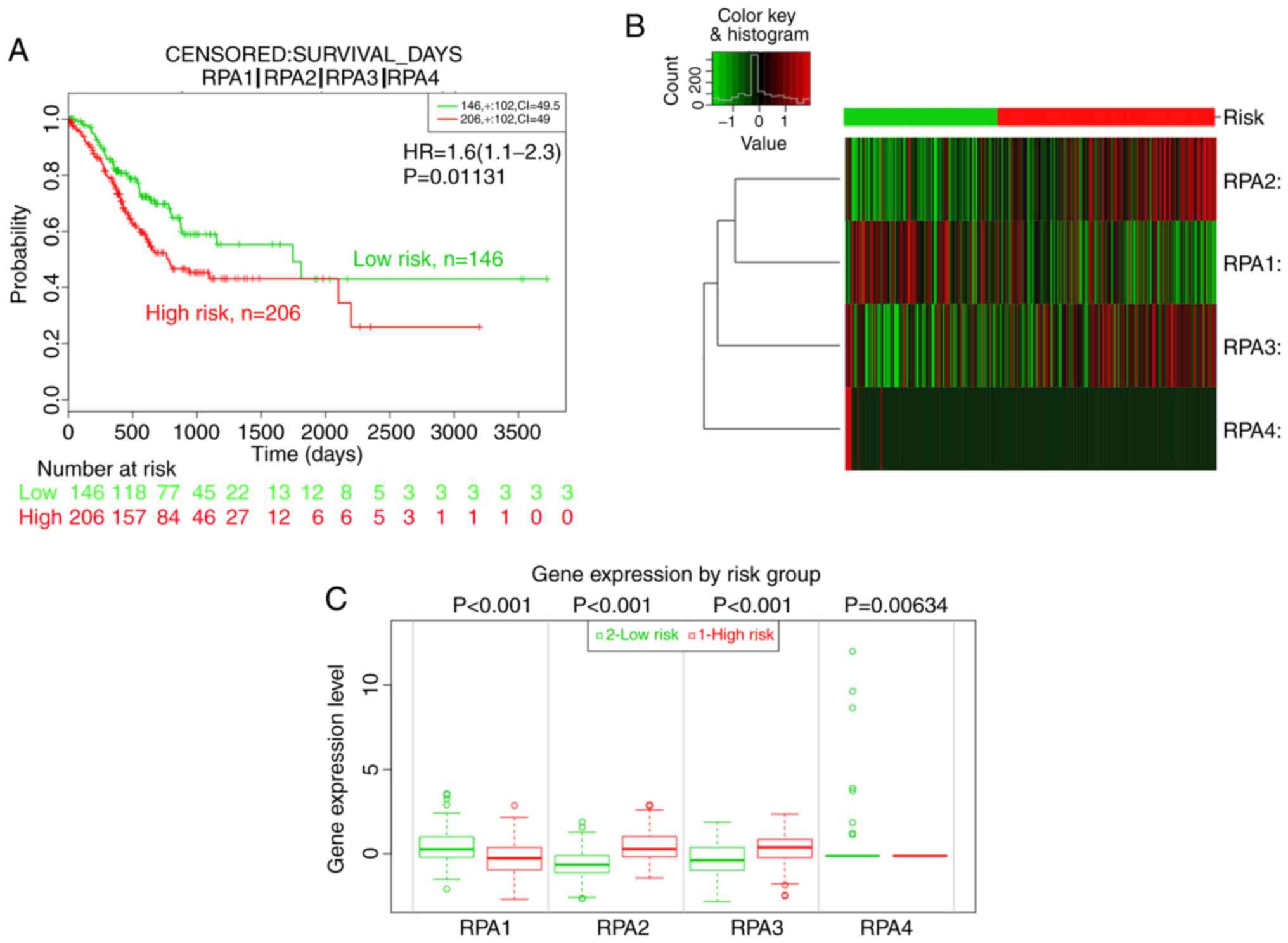
Prognostic value of RPA family
signatures
Given the increasing focus on the application of
gene signatures for prognostic analysis, the prognostic value of
the RPA family signature was further assessed using SurvExpress.
Divided by the maximized risk group algorithm, the high-risk group
(n=206) showed a poorer outcome than the low-risk group (n=146; HR,
1.6; 95% CI, 1.11–2.30; P=0.0113; Fig.
6A-C). In addition, RPA2 and 3 mRNA expression in the high-risk
groups was significantly elevated compared with those of the
low-risk groups, whist RPA1 expression was lower in the high-risk
groups (Fig. 6B and C).
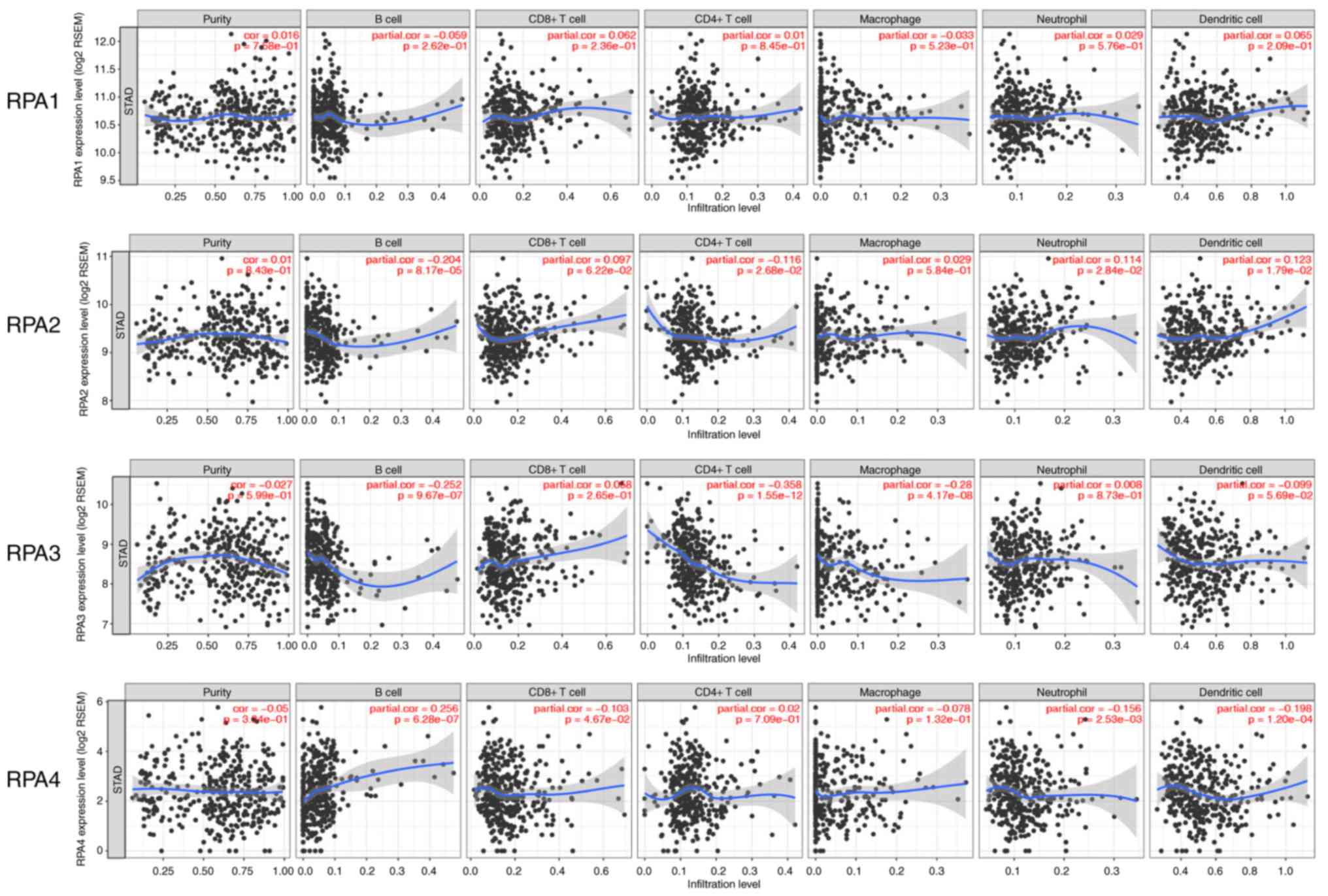
Correlation between RPA expression and
TIICs
To further examine the immunological influence of
RPAs in GC tissues, the TIMER platform was utilized to analyze the
correlation between RPAs and the prevalence of various immune
infiltrating cell types. RPA3 expression exhibited a significant
correlation with CD4+ T cells (partial.cor=−0.358,
P=1.55×10⁻¹²; Fig. 6). As
presented in Fig. 7, no significant
correlation was observed between RPA1 and the tested cell types. In
RPA2, significant correlations were identified with B cells, CD4+ T
cells, neutrophils and dendritic cells (partial.cor=−0.204,
P=8.17×10⁻⁵; partial.cor=−0.116, P=2.68×10⁻²;
partial.cor=0.114, P=2.84×10⁻² and partial.cor=0.123,
P=1.79×10⁻², respectively). In RPA3, significant
correlations were identified with B cells, CD4+ T cells and
macrophages (partial.cor=−0.252, P=9.67×10⁻⁷;
partial.cor=−0.358, P=1.55×10⁻¹² and partial.cor=−0.28,
P=4.17×10⁻⁸, respectively). In RPA4, significant
correlations were identified with B cells, CD8+ T cells,
neutrophils and dendritic cells (partial.cor=0.256,
P=6.28×10⁻⁷; partial.cor=−0.103, P=4.67×10;
partial.cor=−0.156, P=2.53×10⁻³ and partial.cor=−0.198,
P=1.20×10⁻⁴, respectively) (Fig. 7).
Discussion
The majority of studies on RPAs have focused on
their structure, function and mechanism of action. The members of
the RPA family are essential components of multiple pathways that
influence DNA replication, recombination, repair and damage
signaling (4–8,23,24).
Clinical studies have determined that RPAs are upregulated in
various cancer types, including hepatocellular and esophageal
carcinoma, and colon and bladder cancer (5,7–8,25–27), and
that this frequently correlates with poor prognosis (28–30).
Nonetheless, the influence of RPAs on GC progression is yet to be
elucidated. To the best of our knowledge, the present study
represents the first comprehensive analysis of the prognostic
potential and immunological influence of RPAs in GC. The present
results indicate that the expression levels of RPA1, 2 and 3 mRNA
were all significantly higher in GC, compared with normal tissues.
Furthermore, high mRNA expression levels of RPA3 and 4 were
significantly associated with unfavorable OS time in patients with
GC.
Notably, the present study investigated the effects
of different RPAs on various clinicopathological features using
subgroup stratification and OS analysis. HER2 expression status
(positive/negative) in GC patients is a well-characterized risk
factor for GC progression. High expression levels of HER2 in GC
have resulted in the development of trastuzumab, a drug that
specifically targets HER2 upregulation. Moreover, HER2 status is
widely used in subgroup analysis. To the best of our knowledge, the
relationship between HER2 and the OS times of patients with high or
low RPA expression has not yet been clarified in gastric cancer.
The results of the present study may provide insights into the
development of individualized therapeutic strategies.
In the present study, high expression levels of RPA1
mRNA were significantly associated with improved prognosis in
subgroups of patients with i) intestinal-type GC; ii) 5-FU-based
adjuvant chemotherapy treatment; and iii) a stage I tumor. However,
high RPA1 mRNA expression predicted significantly poorer survival
outcome in GC patients with stage II tumors.
A high level of RPA2 mRNA expression was
significantly associated with a favorable OS rate in male patients
with GC, as well as patients with N-stage disease, while indicating
an unfavorable OS rate in patients with diffuse-type, as well as N+
stage disease. Previous reports have highlighted the value of RPA2
as an indicator of poor prognosis in numerous malignancies,
including astrocytic tumor, colon cancer and muscle-invasive
urothelial carcinomas (6–8). Mechanistically, hyperphosphorylation of
RPA2 (occurring in response to DNA damage) was associated with
single-stranded DNA and Rab51, and mutant phosphorylation-deficient
RPA2 could increase chromosomal aberration (31). However, the biological roles of RPA2,
in terms of tumor invasion, metastasis and histological
differentiation, have not been fully characterized. Therefore,
based on its intrinsic correlation, it is possible that RPA2 may be
either a poor or favorable prognostic indicator in different subset
analyses.
Notably, in the present study, high RPA2 expression
predicted HER2-positive status in the GC group. This suggests that
RPA2 may inhibit tumor cell invasion or proliferation by
interacting with the HER2 signaling pathway. However, further
clinical validation of the prognostic value of RPA2 is
required.
High RPA3 mRNA expression levels were significantly
associated with poor OS in patients with GC, indicating the
potential prognostic value of RPA3 in GC. The results of the
present study were consistent with a previous study (32), which reported that RPA3 expression
was upregulated in GC compared with normal tissues, and also
predicted poor patient survival rate. Therefore, RPA3 may be useful
as a potential biomarker to predict the OS of patients with GC.
Moreover, the current study illustrated that a high mRNA expression
level of RPA3 indicated poor OS in male and female, HER2-negative
and positive, M0, N+ and stage III patients. Patients with
diffuse-type GC, and those receiving 5-FU-based adjuvant treatment
also exhibited poorer outcomes when RPA3 was upregulated.
RPA4, a human homolog of RPA2, is involved in
maintaining the genomic integrity of the cell (4). Previous studies have reported that RPA4
possesses 47% structural homology with RPA2, and that it interacts
with both RPA1 and 3 (33). However,
characterization of the role of RPA4 expression in various
malignancies remains limited. In the present study, high RPA4 mRNA
expression was significantly associated with poor OS in patients
with GC, particularly in well-differentiated, male and female,
HER2-negative and positive patients, in addition to those receiving
5-FU based adjuvant treatment, and those with TNM stage II disease.
Conversely, higher RPA4 expression levels were correlated with
improved prognosis in patients at stage IV, N- and + stage, and M0
stage. Increased RPA4 expression correlated with a significantly
high risk of a lower OS time in all patients with GC. However,
during subset analysis; in patients with a well-differentiated
tumor, RPA4 upregulation represented only a mildly significantly
high risk of a lower OS. Nonetheless, patients with a moderately
differentiated tumor and high RPA4 expression represented a
significantly low risk factor for OS. By contrast, in patients that
presented with lymph node-positive and -negative cases, stage IV
and M0, RPA4 was determined to be a favorable indicator of OS.
This apparent contradiction may be explained by the
low population size of each subset. Similar research conducted in
the future would be more reliable if univariate/multivariate cox
regression analysis was incorporated. Mechanistically, RPA4 may be
associated with tumor progression, lymph node metastasis and
histological differentiation. Particularly given the contradictory
results for RPA4 in the lymph node positive/negative and the
general group, which included all patients with GC regardless of
the subtype. It may be worth investigating the association between
RPA4 expression and lymph node metastasis in a larger cohort.
Correlation analysis was subsequently performed to
investigate the associations between TIICs and the expression
levels of RPA family members. Intriguingly, B cells were highly
correlated with RPA2, 3 and 4 expression, indicating the
interaction between B cells and the immune features of the RPA
family. To date, and to the best of our knowledge, this is the
first study that has investigated RPA-associated TIICs as well as
their prognostic values in GC.
The present study had certain limitations; it was
solely based on bioinformatics analysis and would benefit from
experimental or clinical validation. Subsequent investigations
should focus on validation of the prognostic value of the RPA
family members. Future investigations require a larger cohort of
samples to validate the clinical significance of RPAs in GC
prognosis, as well as functional and mechanistic exploration.
Several directions for future research have been made apparent.
Firstly, the prognostic value of the mRNA/protein expression levels
of RPAs using GC tissue samples. Secondly, the prognostic value of
the RPA signature may represent a cost-effective biomarker.
Thirdly, the correlation between RPA family members and TIICs
requires further in-depth characterization.
In conclusion, RPA1, 2 and 3 mRNA expression levels
are significantly higher in GC vs. normal tissues. Furthermore,
high mRNA expression levels of RPA3 and 4 are associated with poor
prognosis in patients with GC. RPAs should therefore be considered
for use as potential prognostic biomarkers of GC progression.
Acknowledgements
The authors would like to thank Dr. Ernest Johann
Helwig (Tongji Hospital, Tongji Medical College, Huazhong
University of Science and Technology) for his contribution to the
manuscript revision and general guidance.
Funding
No funding was received.
Availability of data and materials
All data generated or analyzed during this study are
included in this published article.
Authors' contributions
YZ and CY performed the data analysis, drafted the
manuscript, participated in the study design and data collection,
and read and approved the final manuscript.
Ethics approval and consent to
participate
Not applicable.
Patient consent for publication
Not applicable.
Competing interests
The authors declare that they have no competing
interests.
Glossary
Abbreviations
Abbreviations:
|
RPA
|
replication protein A
|
|
KM
|
Kaplan-Meier
|
|
OS
|
overall survival
|
|
HER2
|
human epidermal growth factor receptor
2
|
|
TIMER
|
tumor immune estimation response
|
References
|
1
|
Siegel RL, Miller KD and Jemal A: Cancer
statistics, 2017. CA Cancer J Clin. 67:7–30. 2017. View Article : Google Scholar : PubMed/NCBI
|
|
2
|
Song Z, Wu Y, Yang J, Yang D and Fang X:
Progress in the treatment of advanced gastric cancer. Tumour Biol.
39:10104283177146262017. View Article : Google Scholar : PubMed/NCBI
|
|
3
|
Mayer RJ, Venook AP and Schilsky RL:
Progress against GI cancer during the American society of clinical
oncology's first 50 years. J Clin Onco. 32:1521–1530. 2014.
View Article : Google Scholar
|
|
4
|
Haring SJ, Humphreys TD and Wold MS: A
naturally occurring human RPA subunit homolog does not support DNA
replication or cell-cycle progression. Nucleic Acids Res.
38:846–858. 2010. View Article : Google Scholar : PubMed/NCBI
|
|
5
|
Dahai Y, Sanyuan S, Hong L, Di Z and Chong
Z: A relationship between replication protein A and occurrence and
prognosis of esophageal carcinoma. Cell Biochem Biophys.
67:175–180. 2013. View Article : Google Scholar : PubMed/NCBI
|
|
6
|
Kanakis D, Levidou G, Gakiopoulou H,
Eftichiadis C, Thymara I, Fragkou P, Trigka EA, Boviatsis E,
Patsouris E and Korkolopoulou P: Replication protein A: A reliable
biologic marker of prognostic and therapeutic value in human
astrocytic tumors. Hum Pathol. 42:1545–1553. 2011. View Article : Google Scholar : PubMed/NCBI
|
|
7
|
Levidou G, Gakiopoulou H, Kavantzas N,
Saetta AA, Karlou M, Pavlopoulos P, Thymara I, Diamantopoulou K,
Patsouris E and Korkolopoulou P: Prognostic significance of
replication protein A (RPA) expression levels in bladder urothelial
carcinoma. BJU Int. 108:E59–E65. 2011. View Article : Google Scholar : PubMed/NCBI
|
|
8
|
Givalos N, Gakiopoulou H, Skliri M,
Bousboukea K, Konstantinidou AE, Korkolopoulou P, Lelouda M,
Kouraklis G, Patsouris E and Karatzas G: Replication protein A is
an independent prognostic indicator with potential therapeutic
implications in colon cancer. Mod Pathol. 20:159–166. 2007.
View Article : Google Scholar : PubMed/NCBI
|
|
9
|
D'Errico M, de Rinaldis E, Blasi MF, Viti
V, Falchetti M, Calcagnile A, Sera F, Saieva C, Ottini L, Palli D,
et al: Genome-wide expression profile of sporadic gastric cancers
with microsatellite instability. Eur J Cancer. 45:461–469. 2009.
View Article : Google Scholar
|
|
10
|
Chen X, Leung SY, Yuen ST, Chu KM, Ji J,
Li R, Chan AS, Law S, Troyanskaya OG, Wong J, et al: Variation in
gene expression patterns in human gastric cancers. Mol Biol Cell.
14:3208–3215. 2003. View Article : Google Scholar : PubMed/NCBI
|
|
11
|
Cho JY, Lim JY, Cheong JH, Park YY, Yoon
SL, Kim SM, Kim SB, Kim H, Hong SW, Park YN, et al: Gene expression
signature-based prognostic risk score in gastric cancer. Clin
Cancer Res. 17:1850–1857. 2011. View Article : Google Scholar : PubMed/NCBI
|
|
12
|
Rhodes DR, Yu J, Shanker K, Deshpande N,
Varambally R, Ghosh D, Barrette T, Pandey A and Chinnaiyan AM:
ONCOMINE: A cancer microarray database and integrated data-mining
platform. Neoplasia. 6:1–6. 2004. View Article : Google Scholar : PubMed/NCBI
|
|
13
|
Lánczky A, Nagy Á, Bottai G, Munkácsy G,
Szabó A, Santarpia L and Győrffy B: miRpower: A web-tool to
validate survival-associated miRNAs utilizing expression data from
2178 breast cancer patients. Breast Cancer Res Treat. 160:439–446.
2016. View Article : Google Scholar : PubMed/NCBI
|
|
14
|
Kim HK, Choi IJ, Kim CG, Kim HS, Oshima A,
Michalowski A and Green JE: A gene expression signature of acquired
chemoresistance to cisplatin and fluorouracil combination
chemotherapy in gastric cancer patients. PLoS One. 6:e166942011.
View Article : Google Scholar : PubMed/NCBI
|
|
15
|
Ooi CH, Ivanova T, Wu J, Lee M, Tan IB,
Tao J, Ward L, Koo JH, Gopalakrishnan V, Zhu Y, et al: Oncogenic
pathway combinations predict clinical prognosis in gastric cancer.
PLoS Genet. 5:e10006762009. View Article : Google Scholar : PubMed/NCBI
|
|
16
|
Förster S, Gretschel S, Jöns T, Yashiro M
and Kemmner W: THBS4, a novel stromal molecule of diffuse-type
gastric adenocarcinomas, identified by transcriptome-wide
expression profiling. Mod Pathol. 24:1390–1403. 2011. View Article : Google Scholar : PubMed/NCBI
|
|
17
|
Wang G, Hu N, Yang HH, Wang L, Su H, Wang
C, Clifford R, Dawsey EM, Li JM, Ding T, et al: Comparison of
global gene expression of gastric cardia and noncardia cancers from
a high-risk population in China. PLoS One. 8:e638262013. View Article : Google Scholar : PubMed/NCBI
|
|
18
|
Busuttil RA, George J, Tothill RW,
Ioculano K, Kowalczyk A, Mitchell C, Lade S, Tan P, Haviv I and
Boussioutas A: A signature predicting poor prognosis in gastric and
ovarian cancer represents a coordinated macrophage and stromal
response. Clin Cancer Res. 20:2761–2772. 2014. View Article : Google Scholar : PubMed/NCBI
|
|
19
|
Cristescu R, Lee J, Nebozhyn M, Kim KM,
Ting JC, Wong SS, Liu J, Yue YG, Wang J, Yu K, et al: Molecular
analysis of gastric cancer identifies subtypes associated with
distinct clinical outcomes. Nat Med. 21:449–456. 2015. View Article : Google Scholar : PubMed/NCBI
|
|
20
|
Aguirre-Gamboa R, Gomez-Rueda H,
Martínez-Ledesma E, Martínez-Torteya A, Chacolla-Huaringa R,
Rodriguez-Barrientos A, Tamez-Peña JG and Treviño V: SurvExpress:
An online biomarker validation tool and database for cancer gene
expression data using survival analysis. PLoS One. 8:e742502013.
View Article : Google Scholar : PubMed/NCBI
|
|
21
|
Li T, Fan J, Wang B, Traugh N, Chen Q, Liu
JS, Li B and Liu XS: TIMER: A web server for comprehensive analysis
of tumor-infiltrating immune cells. Cancer Res. 77:e108–e110. 2017.
View Article : Google Scholar : PubMed/NCBI
|
|
22
|
Li B, Severson E, Pignon JC, Zhao H, Li T,
Novak J, Jiang P, Shen H, Aster JC, Rodig S, et al: Comprehensive
analyses of tumor immunity: Implications for cancer immunotherapy.
Genome Biol. 17:1742016. View Article : Google Scholar : PubMed/NCBI
|
|
23
|
Bochkarev A and Bochkareva E: From RPA to
BRCA2: Lessons from single-stranded DNA binding by the OB-fold.
Curr Opin in Struct Biol. 14:36–42. 2004. View Article : Google Scholar
|
|
24
|
Zou Y, Liu Y, Wu X and Shell SM: Functions
of human replication protein A (RPA): From DNA replication to DNA
damage and stress responses. J Cell Physiol. 208:267–273. 2006.
View Article : Google Scholar : PubMed/NCBI
|
|
25
|
Wang J, Yang T, Chen H, Li H and Zheng S:
Oncogene RPA1 promotes proliferation of hepatocellular carcinoma
via CDK4/Cyclin-D pathway. Biochem Biophys Res Commun. 498:424–430.
2018. View Article : Google Scholar : PubMed/NCBI
|
|
26
|
Qu C, Zhao Y, Feng G, Chen C, Tao Y, Zhou
S, Liu S, Chang H, Zeng M and Xia Y: RPA3 is a potential marker of
prognosis and radioresistance for nasopharyngeal carcinoma. J Cell
Mol Med. 21:2872–2883. 2017. View Article : Google Scholar : PubMed/NCBI
|
|
27
|
Xiao W, Zheng J, Zhou B and Pan L:
Replication protein A 3 is associated with hepatocellular carcinoma
tumorigenesis and poor patient survival. Dig Dis. 36:26–32. 2018.
View Article : Google Scholar : PubMed/NCBI
|
|
28
|
Jekimovs C, Bolderson E, Suraweera A,
Adams M, O'Byrne KJ and Richard DJ: Chemotherapeutic compounds
targeting the DNA double-strand break repair pathways: The good,
the bad, and the promising. Front Oncol. 4:862014. View Article : Google Scholar : PubMed/NCBI
|
|
29
|
Mishra AK, Dormi SS, Turchi AM, Woods DS
and Turchi JJ: Chemical inhibitor targeting the replication protein
A-DNA interaction increases the efficacy of Pt-based chemotherapy
in lung and ovarian cancer. Biochem Pharmacol. 93:25–33. 2015.
View Article : Google Scholar : PubMed/NCBI
|
|
30
|
Patrone JD, Kennedy JP, Frank AO, Feldkamp
MD, Vangamudi B, Pelz NF, Rossanese OW, Waterson AG, Chazin WJ and
Fesik SW: Discovery of protein-protein interaction inhibitors of
replication protein A. ACS Med Chem Lett. 4:601–605. 2013.
View Article : Google Scholar : PubMed/NCBI
|
|
31
|
Shi W, Feng Z and Zhang J, Gonzalez-Suarez
I, Vanderwaal RP, Wu X, Powell SN, Roti Roti JL, Gonzalo S and
Zhang J: The role of RPA2 phosphorylation in homologous
recombination in response to replication arrest. Carcinogenesis.
31:994–1002. 2010. View Article : Google Scholar : PubMed/NCBI
|
|
32
|
Dai Z, Wang S, Zhang W and Yang Y:
Elevated expression of RPA3 is involved in gastric cancer
tumorigenesis and associated with poor patient survival. Dig Dis
Sci. 62:2369–2375. 2017. View Article : Google Scholar : PubMed/NCBI
|
|
33
|
Keshav KF, Chen C and Dutta A: Rpa4, a
homolog of the 34-kilodalton subunit of the replication protein A
complex. Mol Cell Biol. 15:3119–3128. 1995. View Article : Google Scholar : PubMed/NCBI
|