Introduction
Squamous cell carcinoma of the oral cavity
represents the most common type of oral cancer (1). The 5-year survival for oral squamous
cell carcinoma (OSCC) was ~50% in 2016, worldwide despite advances
in surgical treatment (robotic surgery), postoperative
chemoradiotherapy or targeted therapy in locally advanced and
recurrent/metastatic disease (2,3).
Loco-regional invasion and metastasis to cervical lymph nodes are
considered to be the reasons for the poor prognosis of patients
with OSCC. It is therefore crucial to understand the underlying
mechanisms of metastasis in OSCC and to identify potential novel
biomarkers that would allow the earlier detection of cancer and
could be used as agents for targeted therapy in OSCC.
Studies from the past 40 years have reported that
matrix metalloproteinases (MMPs), a family of zinc-dependent
endopeptidases, and the tissue inhibitor of metalloproteinases
(TIMPs), are the principal mediators of the alterations observed in
the tumor microenvironment during cancer progression, indicating
that they are directly involved in the majority of biological
processes leading to metastasis (4).
MMP expression is controlled by various hormones (estrogen and
progesterone), cytokines (interleukins and interferons) and growth
factors (epidermal growth factor, fibroblast growth factor,
vascular endothelial growth factor and tumor growth factors)
(4,5). Loco-regional metastases are hallmarks
of OSCC. MMPs degrade the components of the extracellular matrix
(ECM) and the basement membranes, allowing tumor cells to migrate
and metastasize into other organs via the vascular and lymphatic
systems (4). TIMPs are known to
inhibit the catalytic activity of MMPs in a 1:1 stoichiometric
relationship (6). It has been
previously demonstrated that expression levels of MMP-9, also known
as gelatinase-B, MMP-2, also known as gelatinase-A, TIMP-1 and
TIMP-2 are associated with invasion and metastasis in OSCC
(6–9).
Melatonin (N-acetyl-5-methoxytryptamine) is an
indolic compound primarily secreted by the pineal gland that
regulates the sleep-wake cycle (10). Small amounts of melatonin are also
synthesized in the retina (11),
gastrointestinal tract (12), skin
(13), bone marrow (14), lymphocytes (14) and gut (12). Melatonin was also recently discovered
in serous cells of human salivary glands (15). Previous studies have demonstrated
that melatonin is not only a hormone, but is also involved in tumor
suppression in various types of cancer, including breast (16), prostate (17), ovarian (18), cervical (18), endometrial (18) and colon (19) cancer, through its anti-proliferative,
pro-apoptotic and anti-angiogenic functions. The anti-metastatic
role of melatonin in OSCC was mainly investigated using preclinical
in vitro and in vivo models (20–23).
Numerous studies have reported that melatonin decreases oral cancer
cell proliferation in vivo and in vitro by inhibiting
MMP-9 activation (21–23). However, to the best of our knowledge,
the association between circulating melatonin levels and the
aggressive behavior of OSCC in humans has not yet been
investigated. In addition, whether melatonin may be a hormone
capable of regulating MMP expression remains unknown.
The present study hypothesized that the serum
melatonin level may be associated with MMP and TIMP expression in
patients with OSCC. Therefore, this study aimed to determine
whether serum melatonin level may be associated with MMP-9, MMP-2,
TIMP-1, TIMP-2 expression levels and the clinicopathological
characteristics of patients with OSCC.
Materials and methods
Patients
A total of 40 men with OSCC (mean age, 57±7 years;
age range, 46–70 years), scheduled to undergo resection surgery at
the Coltea Clinical Hospital (Bucharest, Romania) between November
2014 and March 2015 were included in the present study. Samples
analyses were performed at the Institute of Oncology Bucharest. The
diagnosis of OSCC was based on patient history, physical
examination, routine laboratory tests, endoscopy, tissue sampling
and cross-sectional imaging (CT and MRI) or functional imaging with
18F-fluorodeoxyglucose positron emission tomography. The inclusion
criteria were as follows: Histological diagnosis of OSCC and
surgical treatment with curative intent. The exclusion criteria
were as follows: i) Patients with acute or chronic infection; ii)
patients with immune deficiencies; iii) patients ongoing treatments
with beta-adrenergic blocking drugs (sympathetic innervation via
noradrenaline has a significant role in the regulation of melatonin
secretion), corticosteroids and heparin (known as MMP inhibitors)
(24); iv) patients with endocrine
disorders; v) patients with schizophrenia; vi) patients with burn
injuries and vii) patients with previous history of
chemoradiotherapy. All patients underwent primary tumor excision
with adequate margins (≥5 mm). Radical neck dissection (functional
removal of lymph nodes) was performed based on the clinical and
surgical findings, which did not apply to all patients. The
treatment strategies for patients were carried out according to the
Coltea Clinical Hospital guidelines. Anesthesia was induced by
midazolam (0.2 mg/kg), propofol (2–2.5 mg/kg), sufentanil
(0.01–0.025 mg) and sevoflurane (1–2%). Atracurium (0.6–1 mg/kg)
facilitated the tracheal intubation. Anesthesia was maintained with
sufentanil infusion (0.0005 mg/kg/h) and sevoflurane (1–2%),
whereas neuromuscular blockade was maintained with the
administration of atracurium 50 mg every 40 min.
A total of 30 healthy men (mean age, 56±5 years; age
range, 43–69 years) with no clinical evidence of ear, nose, and
throat disorders were recruited during the same period. The
exclusion criteria that were applied to the patients with OSCC were
also used to select the volunteers.
OSCC is more common in men compared with women, with
a ratio ranging between 2:1 and 4:1 (25). Only men were included in the present
study (patients and control groups) to avoid intersex variations.
This study followed the principles of the Declaration of Helsinki
and was approved by the Coltea Clinical Hospital Ethics Committee.
All patients and volunteers signed informed consent prior to the
study.
Histopathology
Clinical and histopathological data were collected
from patient medical records. In the 8th edition of the American
Joint Cancer Committee Cancer Staging Manual (25), an additional feature for primary
tumor characterization has been introduced (26); this updated classification system has
developed a modified staging system that integrates the depth of
invasion (DOI) into the tumor size (T) category. Clinically,
measurement of DOI requires the recognition of the basement
membrane level of the closest adjacent normal mucosa and dropping
of a ‘plumb line’ to the deepest point of tumor invasion (25,26). New
cut-offs have therefore been introduced according to the size and
extension of the tumor (4 cm for size and 10 mm for depth). These
boundary lines were used to classify patients into two subgroups as
follows: T-DOI I (n=15) [small tumors with less invasive lesions
(T≤4 cm; DOI<10 mm)] and T-DOI II (n=25) [large tumors with
invasive depth (T>4 cm; DOI>10 mm)]. The number of patients
with positive lymph node metastases was 23 [N1 (n=8), N2 (n=14), N3
(n=1)], whereas 17 patients were diagnosed with lymph node-negative
disease (N0). N1, N2 and N3 patients were analyzed as ‘N1 + N2 +
N3’ or ‘Yes’ subgroup (lymph-node positive subgroup), whereas N0
remained as a single ‘N0’ or ‘No’ (lymph-node negative) subgroup.
Tumors were graded as well (G1), moderately (G2) and poorly (G3)
differentiated accordingly to the World Health Organization
Classification of Head and Neck Tumors (27). According to the tumor grade, patients
were classified as follows: G1 (n=22), G2 (n=8) and G3 (n=10).
Blood sample collection and
processing
Blood specimens were collected by venipuncture into
Vacutainer serum separation tubes (6 ml; Becton, Dickinson and
Company) before surgery and 2 days following surgery. Serum samples
were obtained by blood clotting for 30 min at room temperature and
centrifugation at 1,000 × g for 15 min at 4°C. Lipemic, icteric or
hemolytic specimens were excluded from this study. Following
exclusion, 40 serum samples from patients with OSCC and 30 samples
from healthy controls remained in the study. Serum samples were
immediately aliquoted into labeled cryo-vials and stored at −70°C
for further analyses.
Melatonin pre-purification by
solid-phase extraction
Due to the low concentration of melatonin and the
coexistence of numerous other endogenous compounds in blood, such
as N-acetyl serotonin and 5-methoxi tryptamine, the determination
of melatonin was an analytical challenge. Melatonin was therefore
extracted from serum samples using C18 columns (R-Biopharm AG) with
a recovery ranging between 87.5 and 94.8% for 10–200 pg
melatonin/ml. The solid-phase extraction procedure consisted of the
following steps: i) Column conditioning with 1 ml of bidistilled
water, followed by 1 ml of pure methanol; ii) sample application
where a 0.5 ml serum sample was passed through a C18 column that
was washed with 0.5 ml of bidistilled water and 2 ml of
water-methanol (90:10, v/v); iii) elution of the extract where
melatonin was eluted from the column with pure methanol and iv)
evaporation and reconstitution of the extract where the eluate was
evaporated to dryness and resuspended in 0.15 ml of bidistilled
water for further analysis. Melatonin pre-purification from serum
samples was essential for high sensitivity of the analysis method
(28). The procedure described above
allowed melatonin detection with high sensitivity and without
interference from other components in the serum.
Detection of serum melatonin, MMP-9,
MMP-2, TIMP-1 and TIMP-2 by ELISA
Serum concentrations of melatonin, MMP-9, MMP-2,
TIMP-1 and TIMP-2 were measured using commercially available
quantitative ELISA kits from R&D Systems, Inc. for human MMP-9
(cat. no. DMP900), MMP-2 (cat. no. DMP2F0), TIMP-1 (cat. no.
DTM100) and TIMP-2 (cat. no. DTM200) and from DRG International
Inc. for melatonin (cat. no. EIA-1431), according to the
manufacturer's instructions.
The precision (intra-assay variation) of the assay
was tested by 20 measurements of three different samples of known
concentrations. The reproducibility (inter-assay variation) for the
same three samples was also tested. The values of the inter-assay
imprecision study were higher compared with those from the
intra-assay study with coefficients of variations ranging from
3.8–16.4 and 1.9–10.3%, respectively. The lower limits of detection
were 1.6 pg/ml for melatonin, 0.156 ng/ml for MMP-9, 0.047 ng/ml
for MMP-2, 0.08 ng/ml for TIMP-1 and 0.011 ng/ml for TIMP-2.
Contamination from saliva can cause falsely elevated
concentrations. Since melatonin, MMP-9, TIMP-1 and TIMP-2 are
present in saliva (6,15), protective measures in the form of
masks were taken whilst assessing the samples to prevent
contamination of kit reagents while running the assay.
The serum levels of MMP-9, MMP-2, TIMP-1 and TIMP-2
were measured, and the MMP-9/TIMP-1 and MMP-2/TIMP-2 ratios were
calculated. Each sample was assessed in duplicate as recommended by
manufacturers in order to minimize the effects of repeated
freeze-thaw cycles.
Statistical analysis
Patient data processing was performed using
Microsoft Office Excel 2007 SP2, including Data Analysis Tools
(Microsoft Corporation). Statistical analysis was performed using
Statistica software (version 8.0; StatSoft, Inc.). Continuous
variables were expressed as the means ± standard deviation or
median value with interquartile range when appropriate. The
distribution of all variables was verified with the
Kolmogorov-Smirnov test. The non-parametric Kruskal-Wallis test was
used to compare distribution of continuous variables between
different categories for independent samples (groups: OSCC vs.
healthy controls; subgroups: T-DOI I vs. T-DOI II, N0 vs. N1 + N2 +
N3 or G1 vs. G2 vs. G3), whereas the Wilcoxon test was used for
paired samples (OSCC group before and 2 days after surgery).
Pairwise comparisons with Bonferroni post hoc corrections were used
with the Kruskal-Wallis test. The correlation between circulating
biomarkers was assessed using Pearson's correlation coefficient,
and one-way ANOVA was used for association analysis between
biomarkers and histopathological characteristics. P<0.05 was
considered to indicate a statistically significant difference.
Results
Melatonin concentration is lower in
patients with OSCC compared with healthy controls
The patient distribution according to the primary
tumor site was as follows: i) Tongue and base of tongue, 57.5% (23
out of 40; 15 with mucosal SCC and 8 with salivary SCC); ii)
gingiva, 15% (6 out of 40; all with salivary SCC); iii) buccal
mucosa (including buccal sulcus/mucobuccal fold), 10% (4 out of 40;
3 with mucosal SCC and 1 with salivary SCC); iv) soft palate and
tonsil, 12.5% (5 out of 40; 3 with mucosal SCC and 2 with salivary
SCC) and v) lip, 5% (2 out of 40, all with mucosal SCC) (Table I). Data from the analysis of the
serum obtained from patients with OSCC (before and 2 days after
surgery) and healthy controls are summarized in Table II. No significant difference was
observed in patient age between OSCC and healthy controls. The
circulating level of melatonin was significantly lower in patients
with OSCC, both before and 2 days after surgery, compared with
healthy controls (P<0.001). In addition, serum melatonin
concentration decreased by 34.6% (from 18.2–11.9 pg/ml) 2 days
after surgery, although this decrease was not statistically
significant. Altered levels of the circulating ECM degradation
biomarkers MMP-9, TIMP-1 and TIMP-2 (P<0.001) were detected in
patients with OSCC compared with the controls (Table II). The serum levels of MMP-9,
TIMP-1, and MMP-9/TIMP-1 and MMP-2/TIMP-2 ratios were higher, while
those of TIMP-2 were lower both before and 2 days after surgery,
compared with controls. No differences were observed in MMP-2 serum
level between patients with OSCC and healthy controls. Furthermore,
serum concentrations of MMP-9, MMP-2, and MMP-9/TIMP-1 and
MMP-2/TIMP-2 ratios decreased 2 days post-surgery compared to the
pre-surgical concentrations, while the levels of TIMP-1 and TIMP-2
increased (Table II).
 | Table I.Patient characteristics. |
Table I.
Patient characteristics.
|
| Patient, n |
|
|---|
|
|
|
|
|---|
| Primary tumor
site | (Total=40) | % |
|---|
| Tongue and base of
tongue |
| Mucosal
SCC | 15 | 37.5 |
|
Salivary SCC | 8 | 20.0 |
| Gingiva |
|
Salivary SCC | 6 | 15.0 |
| Buccal mucosa |
| Mucosal
SCC | 3 | 7.5 |
|
Salivary SCC | 1 | 2.5 |
| Soft palate and
tonsil |
| Mucosal
SCC | 3 | 7.5 |
|
Salivary SCC | 2 | 5.0 |
| Lip |
| Mucosal
SCC | 2 | 5.0 |
| Table II.Biochemical data in patients with
OSCC and healthy controls. |
Table II.
Biochemical data in patients with
OSCC and healthy controls.
|
| OSCC |
|
|
|
|
|---|
|
|
|
|
|
|
|
|---|
| Variables | Pre-surgery | Post-surgery | Control |
P-valuec |
P-valued |
P-valuee |
|---|
| Number, n | 40 | 40 | 30 | >0.999 | 0.452 | 0.452 |
| Age,
yearsa | 57±7 | 57±7 | 56±5 | >0.999 | 0.670 | 0.670 |
| Melatonin,
pg/mlb | 18.2
(11.0–39.2) | 11.9
(8.7–31.4) | 47.6
(37.7–66.4) | 0.230 |
<0.001f |
<0.001f |
| MMP-9,
ng/mlb | 1,619
(914–1,964) | 1,402
(877–2,001) | 291 (211–330) | 0.939 |
<0.001f |
<0.001f |
| TIMP-1,
ng/mlb | 262 (213–430) | 351 (255–452) | 164 (121–201) | 0.132 |
<0.001f |
<0.001f |
|
MMP-9/TIMP-1b | 4.8 (3.1–7.4) | 3.9 (2.6–6.2) | 1.7 (1.5–1.8) | 0.129 |
<0.001f |
<0.001f |
| MMP-2,
ng/mlb | 160 (130–197) | 149 (127–196) | 150 (122–180) | 0.632 | 0.658 | 0.991 |
| TIMP-2,
ng/mlb | 57.5
(51.6–70.5) | 62.5 (49.9–76) | 89.5
(79.7–101) | 0.289 |
<0.001f |
<0.001f |
|
MMP-2/TIMP-2b | 2.8 (2.4–3.2) | 2.6 (2.2–2.9) | 1.6 (1.3–1.9) | 0.392 |
<0.001f |
<0.001f |
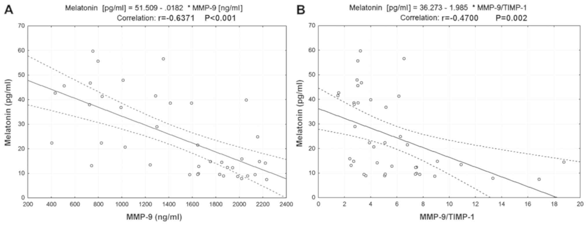
Serum melatonin level is negatively
correlated with MMP-9 and MMP-9/TIMP-1 ratio in patients with
OSCC
Scatter plots presented in Fig. 1 indicated a negative correlation
between serum melatonin and MMP-9 levels (r=−0.6371, P<0.001;
Fig. 1A) and between serum melatonin
level and the MMP-9/TIMP-1 ratio (r=−0.47, P=0.002; Fig. 1B). No correlations between serum
melatonin and TIMP-1, MMP-2 and TIMP-2 levels or the MMP-2/TIMP-2
ratio were identified (Table
III).
| Table III.Association between circulating
biomarkers and clinicopathological characteristics of patients with
OSCC. |
Table III.
Association between circulating
biomarkers and clinicopathological characteristics of patients with
OSCC.
| Variables | Melatonin | P-value | MMP-9 | P-value | MMP-2 | P-value | TIMP-1 | P-value | TIMP-2 | P-value |
|---|
| Melatonin | – | – | −0.6371 | <0.001 | −0.1766 | 0.276 | 0.0238 | 0.884 | −0.2947 | 0.065 |
| MMP-9 | −0.6371 | <0.001 | – | – | 0.0006 | 0.997 | 0.2780 | 0.082 | 0.2093 | 0.195 |
| TIMP-1 | 0.0238 | 0.884 | 0.2780 | 0.082 | 0.1853 | 0.252 | – | – | 0.2455 | 0.127 |
| MMP-9/TIMP-1 | −0.4700 | 0.002 | 0.4813 | 0.002 | −0.1735 | 0.284 | −0.5345 | <0.001 | −0.0035 | 0.983 |
| MMP-2 | −0.1766 | 0.276 | 0.0009 | 0.997 | – | – | 0.1853 | 0.252 | 0.6796 | <0.001 |
| TIMP-2 | −0.2947 | 0.065 | 0.2093 | 0.195 | 0.6796 | <0.001 | 0.2455 | 0.127 | – | – |
| MMP-2/TIMP-2 | 0.1760 | 0.277 | −0.2307 | 0.152 | 0.3380 | 0.033 | −.0.0822 | 0.614 | −0.4350 | 0.005 |
| T-DOI | −0.3494 | 0.027 | 0.3321 | 0.032 | 0.2494 | 0.121 | 0.1669 | 0.303 | 0.0852 | 0.601 |
| Nodal
involvement | −0.5583 | <0.001 | 0.3422 | 0.031 | 0.1760 | 0.277 | −.0160 | 0.922 | 0.1180 | 0.468 |
|
Differentiation | −0.1309 | 0.421 | 0.1646 | 0.310 | 0.4072 | 0.009 | 0.2257 | 0.161 | 0.1448 | 0.373 |
Low serum levels of melatonin and high
serum levels of MMP-9 are associated with large tumors with
invasive depth
Serum melatonin and MMP-9 levels were associated
with T-DOI (r=−0.35, P=0.027 and r=0.33, P=0.032, respectively) in
patients with OSCC (Table III).
Furthermore, as presented in Table
IV, serum levels of melatonin and MMP-9 were significantly
lower and higher, respectively, in the T-DOI II subgroup compared
with the T-DOI I subgroup (P=0.012 and P=0.033, respectively).
| Table IV.Association between biomarkers serum
level and clinicopathological characteristics of patients with
OSCC. |
Table IV.
Association between biomarkers serum
level and clinicopathological characteristics of patients with
OSCC.
|
| T-DOI |
| Nodal
Involvement |
|
Differentiationb |
|---|
|
|
|
|
|
|
|
|---|
| Variables | T-DOI I, n=15 | T-DOI II, n=25 | P-value | No, n=17 | Yes, n=23 | P-value | G1, n=22 | G2, n=8 | G3, n=10 |
|---|
| Melatonin,
pg/mla | 36.8
(14.5–46.7) | 13.4
(9.4–28.9) | 0.012c | 37.9
(21.5–46.7) | 12.4
(9.4–22.3) |
<0.001c | 21.4
(13.1–42.7) | 14.3
(9.0–30.0) | 18.5
(9.4–38.6) |
| MMP-9,
ng/mla | 995 (742–1851) | 1,647
(1,299–1,987) | 0.033c | 1,029
(798–1,593) | 1,833
(1,413–2,021) | 0.029c | 1,271
(827–1,851) | 1,651
(1,496–2,003) | 1,756
(724–2,070) |
| TIMP-1,
ng/mla | 247 (192–342) | 264 (220–463) | 0.493 | 264 (222–344) | 261 (167–452) | 0.902 | 245 (192–296) | 373 (233–651) | 298 (239–515) |
|
MMP-9/TIMP-1a | 3.3 (2.9–6.3) | 5.1 (3.5–7.5) | 0.307 | 3.3 (2.8–6.2) | 5.1 (3.5–7.7) | 0.082 | 5.1 (3.0–7.4) | 4.5 (2.7–7.8) | 3.9 (3.0–5.1) |
| MMP-2,
ng/mla | 135 (128–174) | 182 (136–211) | 0.096 | 142 (129–192) | 180 (133–211) | 0.279 | 135 (128–180) | 178 (139–208) | 197 (138–231) |
| TIMP-2,
ng/mla | 53.8
(51.6–65.8) | 58.7
(51.7–72.1) | 0.442 | 53.8
(51.6–65.8) | 58.7
(51.7–72.1) | 0.476 | 54.4
(50.8–65.8) | 67.8
(52.7–71.7) | 60 (53.5–74) |
|
MMP-2/TIMP-2a | 2.5 (2.1–3.1) | 2.9 (2.5–3.3) | 0.175 | 2.5 (2.2–3.1) | 2.9 (2.5–3.2) | 0.451 | 2.5 (2.3–3.1) | 2.6 (2.4–2.9) | 3.2 (3.1–3.5) |
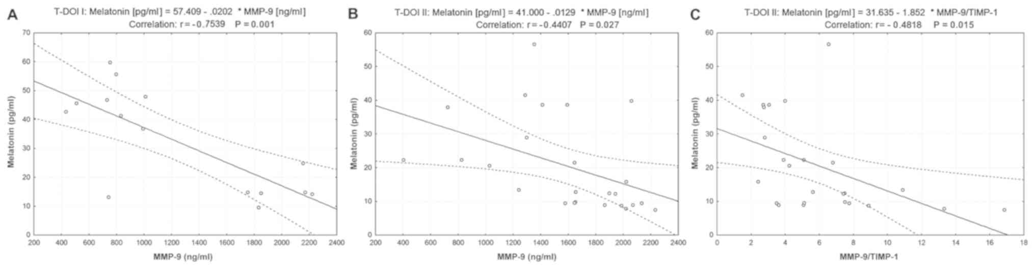
The correlation between serum melatonin and ECM
biomarker levels was also investigated in the two subgroups of
patients with OSCC. The results presented in Fig. 2 demonstrated a negative correlation
between serum melatonin and MMP-9 levels (r=−0.7539, P=0.001 in
T-DOI I and r=−0.4407, P=0.027 in T-DOI II). Furthermore, melatonin
was negatively correlated with MMP-9/TIMP-1 ratio in the T-DOI II
subgroup (r=−0.4818, P=0.015; Fig.
2C).
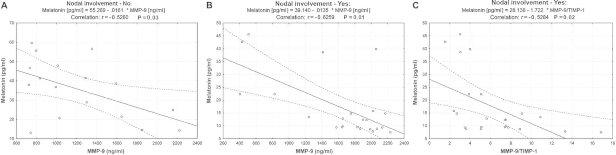
Low serum levels of melatonin and high
serum levels of MMP-9 are associated with lymph node
metastasis
Melatonin and MMP-9 serum levels were associated
with lymph node involvement (r=−0.56, P<0.001 and r=0.34,
P=0.031, respectively) in patients with OSCC (Table III). Serum levels of melatonin and
MMP-9 were significantly lower and higher, respectively, in the
lymph node-positive subgroup compared with the lymph node-negative
subgroup (P<0.001 and P=0.029, respectively; Table IV). Serum TIMP-1, MMP-2 and TIMP-2
levels and the MMP-2/TIMP-2 ratio were not significantly different
in patients with negative lymph node metastases compared with
patients with positive lymph node metastases. Regarding the
analysis of patients with OSCC according to tumor size or the
extent of the tumor, melatonin was negatively correlated with serum
MMP-9 levels in both subgroups (r=−0.5260, P=0.03 in lymph
node-negative subgroup; Fig. 3A;
r=−0.6259, P=0.01 in lymph node-positive subgroup; Fig. 3B) and with the MMP-9/TIMP-1 ratio
only in the subgroup of patients with positive lymph node
metastases (r=−0.5284, P=0.02 in lymph node-positive subgroup;
Fig. 3C).
Association between melatonin, ECM
biomarkers and tumor grading
No significant differences were observed between the
serum levels of all biomarkers measured in G1, G2 or G3 subgroups
of patients with OSCC, with the exception of MMP-2 (P=0.028;
Table IV). Regarding correlations
between melatonin and ECM degradation biomarkers, strong negative
correlations were identified between serum melatonin and MMP-9
levels in the G1 and G2 subgroups (r=−0.65, P=0.001 and r=−0.79,
P=0.018, respectively) and between serum melatonin levels and the
MMP-9/TIMP-1 ratio only in the G1 subgroup (r=−0.47, P=0.025)
(Table V).
| Table V.Correlation between circulating
biomarkers in G1, G2 and G3 subgroups of patients with OSCC. |
Table V.
Correlation between circulating
biomarkers in G1, G2 and G3 subgroups of patients with OSCC.
| Subgroups | G1 | G2 | G3 |
|---|
|
|
|
|
|---|
| Variables | Melatonin | MMP-9 | MMP-9/TIMP-1 | Melatonin | MMP-9 | MMP-9/TIMP-1 | Melatonin | MMP-9 | MMP-9/TIMP-1 |
|---|
| Melatonin | – | −0.6576 | −0.4755 | – | −0.7948 | −0.6149 | – | −0.5379 | −0.6185 |
|
|
| P=0.001 | P=0.025 |
| P=0.018 | P=0.105 |
| P=0.109 | P=0.057 |
| MMP-9 | −0.6576 | – | 0.5641 | −0.7948 | – | 0.6386 | −0.5379 | – | 0.06051 |
|
| P=0.001 |
| P=0.006 | P=0.018 |
| P=0.038 | P=0.109 |
| P=0.024 |
| MMP-9/ | −0.4755 | 0.5641 | – | −0.6149 | 0.6386 | – | −0.6185 | 0.06051 | – |
| TIMP-1 | P=0.025 | P=0.006 |
| P=0.105 | P=0.038 |
| P=0.057 | P=0.024 |
|
No correlations were identified between serum
melatonin and TIMP-1, MMP-2 and TIMP-2 levels or the MMP-2/TIMP-2
ratio in all subgroups characterized according to tumor size and
DOI, lymph node involvement or tumor grading (data not shown).
Discussion
The present study demonstrated that serum melatonin
levels were significantly lower in patients with OSCC before and 2
days after surgery compared with healthy controls. In addition,
serum melatonin level was negatively correlated with serum MMP-9
level and the MMP-9/TIMP-1 ratio in patients with OSCC. Low serum
levels of melatonin and high serum levels of MMP-9 were associated
with invasion depth and lymph node metastasis.
Previous studies have reported that the disruption
of blood melatonin level is associated with an increased risk of
various types of cancer occurrence, including breast, prostate,
ovarian and colon cancer (16–19). The
results of the present study demonstrated that serum melatonin
level was significantly lower in patients with OSCC compared with
healthy controls, which was consistent with results from previous
studies reporting that low serum melatonin level was associated
with an increased risk of breast (16), prostate (17), ovarian (18) or colon (19) cancer. In addition, median circulating
melatonin concentration in the T DOI I (≤36.8 pg/ml) or negative
lymph node metastasis (≤37.9 pg/ml) subgroups of patients with OSCC
were similar to those obtained by Yang et al (16) in patients with breast cancer (≤39.5
pg/ml).
Previous studies have reported a modest reduction in
melatonin secretion on the first day following different types of
surgery, such as orthopedic and abdominal, compared with
pre-surgery levels, as well as a high increase in melatonin
secretion on the second day following surgery (29,30). The
decrease in melatonin and MMP-9 serum level one day post-surgery
may be due to the effects of general anesthesia, particularly the
administration of propofol (31,32). To
eliminate the influence of propofol on the circadian rhythm of
melatonin and MMP-9 in patients with OSCC in the present study,
blood was collected on the second day post-surgery. In contrast to
previous studies (29,30), the present study demonstrated that
serum melatonin level decreased by 34.6% 2 days following surgery,
although this decrease was not statistically significant. These
differences may be due to the various sites of surgery [oral cavity
in the present study vs. knee (29)
and abdomen (30) in previous
studies]. OSCC affects the preferential sites of melatonin
reactivity in the oral cavity that are the salivary glands,
including parotid, submandibular and sublingual glands. In the
present study, 42.5% of patients with OSCC (17 out of 40) were
diagnosed with salivary SCC.
Regarding MMP-9, the results of the present study
were consistent with previous studies (7,9) that
reported a reduction in serum MMP-9 levels in patients with OSCC
following surgery. The present study demonstrated a decrease in
serum MMP-9 levels 2 days post-surgery, suggesting that MMP-9 may
be considered as a marker of tumor presence, which has been
previously demonstrated (7,9,33);
however, the difference between serum MMP-9 level before and after
surgery was not significant. In addition, previous studies have
demonstrated that MMP-9 serum levels may reflect its expression in
resected tumor tissue (7,9,33). The
lack of statistical significance in the present study may be due to
the fact that the 2-day post-surgical interval may be too short to
observe a total recovery of a normal serum MMP-9 concentration. A
significant decrease in the serum MMP-9 level becomes evident only
1 month following surgery (33).
The results from the present study suggested that
radical surgery with excision of the primary tumor, including
salivary glands, reduced not only serum MMP-9 levels, but also
serum melatonin level. High MMP-9 and melatonin expression levels
have been observed in excised tumor tissues (7,9,15). These findings suggest that melatonin
suppression may be prevented by treatment with melatonin
supplements, and that a prospective clinical trial assessing the
therapeutic potential of melatonin in patients with OSCC may be
useful.
The present study hypothesized that melatonin may be
able to regulate MMP expression and activity. To the best of our
knowledge, the present study was the first to demonstrate the
association between melatonin, MMPs, TIMPs and the
clinicopathological characteristics of patients with OSCC.
Our group previously reported that the imbalance
between MMP-9 and TIMP-1 serum levels may serve a crucial role in
the metastatic spread of head and neck cancer cells via lymphatic
pathways (6,8). Furthermore, MMP-2 and TIMP-2 may be
used as markers of tumor differentiation in head and neck squamous
cell carcinoma (2,6). In the present study, serum MMP-9 level
and the MMP-9/TIMP-1 ratio were significantly higher, whereas serum
melatonin level was significantly lower in patients with OSCC
compared with healthy controls. These results were consistent with
those from Lin et al (34),
who demonstrated that melatonin receptor type 1A (MTNR1A) was
reduced in 618 patients with oral cancer compared with 560
non-cancer controls, as well as those from other studies reporting
that an increase in serum MMP-9 level or the MMP-9/TIMP-1 ratio may
enhance invasion in head and neck cancers (7,9).
Inhibition of MMP-9 activity may therefore reduce cancer metastasis
in patients with OSCC (34,35).
In the present study, serum melatonin level was
negatively correlated with serum MMP-9 level and the MMP-9/TIMP-1
ratio. Similarly, previous studies reported a negative correlation
between melatonin, MTNR1A and MMP-9 expression in healthy kidney
and renal cell carcinoma tissues (36), and a 50% inhibitory effect of
melatonin on the induction and catalytic activity of MMP-9 in the
human gastric adenocarcinoma cell line AGS (29). Lin et al (36) have demonstrated that melatonin can
inhibit NF-κB-mediated MMP-9 transcription and cancer cell invasion
by targeting the Akt-Erk/JNK pathways in renal cell carcinoma.
Furthermore, in a human gastric adenocarcinoma cell line, melatonin
inhibits MMP-9 activity by binding to its active site and by
interacting with key residues within its catalytic site, including
three histidines that form the coordination complex with zinc,
proline 421 and alanine 191 (37).
Numerous in vitro studies have reported that
MMP-9 is a critical target of melatonin in the regulation of oral
cancer metastasis (20–23). A number of mechanisms by which
melatonin could repress MMP-9 expression have been revealed. For
example, melatonin can inhibit the motility of oral cancer cells
(HSC-3 and OECM-1), attenuating MMP-9 expression and activity by
reducing histone acetylation on the promoter of MMP-9 gene
(21). Furthermore, melatonin can
suppress nasopharyngeal carcinoma cell migration by regulating
MMP-9 gene expression and inhibiting the DNA-binding ability of the
transcription factor specificity protein-1 (22). Lu et al (20) have demonstrated that melatonin
treatment of the OSCC cell lines SCC-9 and SCC-25 can reduce the
survival and migration of OSCC-associated neutrophils and suppress
the tumor-associated neutrophil release of C-X-C motif chemokine
ligand 8, C-C motif chemokine ligand 2, C-C motif chemokine ligand
4 and MMP-9 by blocking p38 MAPK and Akt signaling.
To the best of our knowledge, no clinical studies
exploring the effect of melatonin and MMPs mediated ECM remodeling
on the susceptibility of developing OSCC and histological features
of OSCC are presently available. Lu et al (20) have reported that intensive
infiltration of tumor-associated neutrophils in OSCC tissue
specimens is associated with advanced stage, lymphatic metastasis
and poor prognosis of patients with OSCC. Furthermore, the in
vitro study by Lu et al (20) reported that the pro-motility and
pro-angiogenesis effects of OSCC-associated neutrophils decreased
by melatonin were dependent of MMP-9 suppression in OSCC. In the
present study, the results of the assessment of melatonin and MMP-9
serum levels in patients with OSCC demonstrated that low levels of
melatonin and high levels of MMP-9 were associated with advanced
tumor stage and lymphatic metastasis.
Tumor size and the extent of the primary tumor are a
well-known prognostic factor for numerous types of cancer,
including OSCC, and larger tumors predict a worse prognosis
(25,38). The results from a recent study
suggest that patient prognosis worsens when DOI increases, and that
DOI is a better predictive parameter compared with the T category
(26). The results of the present
study demonstrated that low levels of melatonin and high levels of
MMP-9 were associated with large tumors with high invasive depth (T
>4 cm; DOI >10 mm). In addition, serum melatonin and MMP-9
levels were significantly lower and higher in the T-DOI II subgroup
compared with the T-DOI I, respectively. In both T-DOI subgroups,
serum melatonin level was negatively correlated with serum MMP-9
level. Furthermore, melatonin was inversely associated with T-DOI
in patients with OSCC. In line with the findings of the present
study, Nakamura et al (39)
analyzed 50 primary OSCC tumors by immunohistochemistry and
reported that the absence of immunoreactive MTNR1A is negatively
associated with the T category of the Tumor-Node-Metastasis (TNM)
classification.
Regarding metastatic dissemination to regional lymph
nodes, the present study demonstrated that melatonin was inversely
associated with the presence of lymph node metastases in patients
with OSCC. By contrast, Nakamura et al (39) reported that the expression of MTNR1A
was not associated with the N category of the TNM classification,
suggesting that the reduced MTNR1A expression and MTNR1A
inactivation may occur in a specific subgroup of OSCC, even at an
earlier stage, and contribute to the higher proliferative and/or
survival activity of tumor cells in this subgroup. The findings of
the study by Nakamura et al (39) may also be valid in the present study,
although this study demonstrated that melatonin was associated with
the N category, since the loss of melatonin occurred in an earlier
stage in the lymph-node negative subgroup, and the serum
concentration was lower compared with that in controls. In
addition, serum melatonin level was negatively correlated with
serum MMP-9 level even in the lymph node-negative subgroup. The
present study also demonstrated that serum melatonin level in the
lymph node-positive subset of patients was significantly lower
compared with patients in the lymph node-negative subgroup
(P<0.001). These results suggested that a reduced serum level of
melatonin may be associated with the metastatic rate in cervical
lymph nodes in patients with OSCC.
An essential feature of the invasive phenotype of a
tumor is its ability to produce MMPs (4,6,7). In the present study, MMP-2 and MMP-9
serum levels were higher in poorly differentiated tumors,
corresponding to an advanced stage of neoplasia, compared with
well-differentiated tumors, which are normally associated with
better prognosis. Furthermore, serum melatonin level was not
associated with the degree of differentiation of the tumor cells.
However, serum melatonin and MMP-9 levels were negatively
correlated in the low- and intermediate-grade tumor subgroup, but
not in the high-grade tumor subgroup.
The present study presented certain limitations.
Firstly, this study was a single-center, retrospective cohort
analysis, with a small number of patients included. However,
despite the small sample size, the population was homogeneous and
included only men in both groups. The samples were collected during
the same season (winter) and at the same time in the morning in
order to avoid any artefacts. Secondly, immunohistochemistry to
detect melatonin and ECM biomarkers in OSCC tissue specimens could
not be performed, and their association was therefore not
established. Further studies investigating the immunohistochemical
expression of melatonin and ECM biomarkers in OSCC tissue specimens
are required. Thirdly, cell experiments to validate the association
between melatonin and MMP-9 were not conducted. This association
should be investigated in future studies.
In summary, the results of the present study
demonstrated that serum melatonin level was negatively correlated
with serum MMP-9 level, tumor size, depth of invasion and
metastasis in patients with OSCC. The results of this retrospective
study indicated that melatonin may be considered as a predictive
biomarker of cancer cell proliferation and metastasis and a
potential therapeutic agent for patients with OSCC. Further
investigation is required to confirm these findings.
Acknowledgements
Not applicable.
Funding
No funding was received.
Availability of data and materials
The datasets used and/or analyzed during the current
study are available from the corresponding author on reasonable
request.
Authors' contributions
AES and AZCA conceived the study and planned the
experiments. Experiments were performed by AES. AES and MMS
performed statistical analysis. AES, AZCA, MMS, APS, CN and DCG
contributed to the interpretation of the results. AS wrote the
manuscript. All authors provided critical feedback and contributed
to the design of this study, data analysis and manuscript
writing.
Ethics approval and consent to
participate
The present study conformed to the principles of The
Declaration of Helsinki and was approved by the Coltea Clinical
Hospital Ethics Committee (Bucharest, Romania). Written informed
consent was obtained from all patients and volunteers prior to the
study start.
Patient consent for publication
Not applicable.
Competing interests
The authors declare that they have no competing
interests.
References
|
1
|
Yeh CM, Su SC, Lin CW, Yang WE, Chien MH,
Reiter RJ and Yang SF: Melatonin as a potential inhibitory agent in
head and neck cancer. Oncotarget. 8:90545–90556. 2017. View Article : Google Scholar : PubMed/NCBI
|
|
2
|
Stanciu AE, Zamfir-Chiru-Anton A, Stanciu
MM, Popescu CR, Gheorghe DC and Nitipir C: Serum matrix
metalloproteinase-2 in head and neck squamous cell carcinoma is
associated with tumor differentiation. Rom Biotech Lett.
22:12419–12426. 2017.
|
|
3
|
Koziorowski J, Stanciu AE, Gomez-Vallejo V
and Llop J: Radiolabeled nanoparticles for cancer diagnosis and
therapy. Anticancer Agents Med Chem. 17:333–354. 2017. View Article : Google Scholar : PubMed/NCBI
|
|
4
|
Gialeli C, Theocharis AD and Karamanos NK:
Roles of matrix metalloproteinases in cancer progression and their
pharmacological targeting. FEBS J. 278:16–27. 2011. View Article : Google Scholar : PubMed/NCBI
|
|
5
|
Stanciu AE, Vatasescu RG, Stanciu MM,
Serdarevic N and Dorobantu M: The role of pro-fibrotic biomarkers
in paroxysmal and persistent atrial fibrillation. Cytokine.
103:63–68. 2018. View Article : Google Scholar : PubMed/NCBI
|
|
6
|
Stanciu AE, Zamfir-Chiru-Anton A, Stanciu
MM, Popescu CR and Gheorghe DC: Imbalance between matrix
metalloproteinases and tissue inhibitors of metalloproteinases
promotes invasion and metastasis of head and neck squamous cell
carcinoma. Clin Lab. 63:1613–1620. 2017. View Article : Google Scholar : PubMed/NCBI
|
|
7
|
Patel BP, Shah SV, Shukla SN, Shah PM and
Patel PS: Clinical significance of MMP-2 and MMP-9 in patients with
oral cancer. Head Neck. 29:564–572. 2007. View Article : Google Scholar : PubMed/NCBI
|
|
8
|
Stanciu AE, Zamfir-Chiru-Anton A, Stanciu
MM, Popescu CR and Gheorghe DC: Serum level of matrix
metalloproteinase-9 in patients with head and neck squamous cell
carcinoma. Clin Lab. 62:1569–1574. 2016. View Article : Google Scholar : PubMed/NCBI
|
|
9
|
Burduk PK, Sawicki P, Szylberg L, Bodnar M
and Marszalek A: Expression of matrix metalloproteinase-2/9 and
tissue inhibitor of metalloproteinase-1/2 as predictive factors in
oropharyngeal squamous cell carcinoma. Iran J Otorhinolaryngol.
31:153–161. 2019.PubMed/NCBI
|
|
10
|
Hanedan Uslu G, Canyilmaz E, Serdar L and
Ersöz S: Protective effects of genistein and melatonin on mouse
liver injury induced by whole-body ionising radiation. Mol Clin
Oncol. 10:261–266. 2019.PubMed/NCBI
|
|
11
|
Laurent V, Sengupta A, Sánchez-Bretaño A,
Hicks D and Tosini G: Melatonin signaling affects the timing in the
daily rhythm of phagocytic activity by the retinal pigment
epithelium. Exp Eye Res. 165:90–95. 2017. View Article : Google Scholar : PubMed/NCBI
|
|
12
|
Siah KT, Wong RK and Ho KY: Melatonin for
the treatment of irritable bowel syndrome. World J Gastroenterol.
20:2492–2498. 2014. View Article : Google Scholar : PubMed/NCBI
|
|
13
|
Slominski A, Fischer TW, Zmijewski MA,
Wortsman J, Semak I, Zbytek B, Slominski RM and Tobin DJ: On the
role of melatonin in skin physiology and pathology. Endocrine.
27:137–148. 2005. View Article : Google Scholar : PubMed/NCBI
|
|
14
|
Conti A, Conconi S, Hertens E,
Skwarlo-Sonta K, Markowska M and Maestroni JM: Evidence for
melatonin synthesis in mouse and human bone marrow cells. J Pineal
Res. 28:193–202. 2000. View Article : Google Scholar : PubMed/NCBI
|
|
15
|
Isola M and Lilliu MA: Melatonin
localization in human salivary glands. J Oral Pathol Med.
45:510–515. 2016. View Article : Google Scholar : PubMed/NCBI
|
|
16
|
Yang WS, Deng Q, Fan WY, Wang WY and Wang
X: Light exposure at night, sleep duration, melatonin, and breast
cancer: A dose-response analysis of observational studies. Eur J
Cancer Prev. 23:269–276. 2014. View Article : Google Scholar : PubMed/NCBI
|
|
17
|
Tai SY, Huang SP, Bao BY and Wu MT:
Urinary melatonin-sulfate/cortisol ratio and the presence of
prostate cancer: A case control study. Sci Rep. 6:296062016.
View Article : Google Scholar : PubMed/NCBI
|
|
18
|
Zhao M, Wan JY, Zeng K, Tong M, Lee AC,
Ding JX and Chen Q: The reduction in circulating melatonin level
may contribute to the pathogenesis of ovarian cancer: A
retrospective study. J Cancer. 7:831–836. 2016. View Article : Google Scholar : PubMed/NCBI
|
|
19
|
Chok KC, Ng CH, Koh RY, Ng KY and Chye SM:
The potential therapeutic actions of melatonin in colorectal
cancer. Horm Mol Biol Clin Investig. May 29–2019.(Epub ahead of
print). doi: 10.1515/hmbci-2019-0001. View Article : Google Scholar : PubMed/NCBI
|
|
20
|
Lu H, Wu B, Ma G, Zheng D, Song R, Huang
E, Mao M and Lu B: Melatonin represses oral squamous cell carcinoma
metastasis by inhibiting tumor-associated neutrophils. Am J Transl
Res. 9:5361–5374. 2017.PubMed/NCBI
|
|
21
|
Yeh CM, Lin CW, Yang JS, Yang WE, Su SC
and Yang SF: Melatonin inhibits TPA-induced oral cancer cell
migration by suppressing matrix metalloproteinase-9 activation
through the histone acetylation. Oncotarget. 7:21952–21967. 2016.
View Article : Google Scholar : PubMed/NCBI
|
|
22
|
Ho HY, Lin CW, Chien MH, Reiter RJ, Su SC,
Hsieh YH and Yang SF: Melatonin suppresses TPA-induced metastasis
by downregulating matrix metalloproteinase-9 expression through
JNK/SP-1 signaling in nasopharyngeal carcinoma. J Pineal Res.
61:479–492. 2016. View Article : Google Scholar : PubMed/NCBI
|
|
23
|
Yang CY, Lin CK, Tsao CH, Hsieh CC, Lin
GJ, Ma KH, Shieh YS, Sytwu HK and Chen YW: Melatonin exerts
anti-oral cancer effect via suppressing LSD1 in patient-derived
tumor xenograft models. Oncotarget. 8:33756–33769. 2017.PubMed/NCBI
|
|
24
|
Mannello F, Jung K, Tonti GA and
Canestrari F: Heparin affects matrix metalloproteinases and tissue
inhibitors of metalloproteinases circulating in peripheral blood.
Clin Biochem. 41:1466–1473. 2008. View Article : Google Scholar : PubMed/NCBI
|
|
25
|
Amin MB, Edge S, Greene F, Byrd DR,
Brookland RK, Washington MK, Gershenwald JE, Compton CC, Hess KR,
Sullivan DC, et al: AJCC Cancer Staging Manual, eighth edition
(2017) published by Springer Science and Business Media LLC
(springer.com). AJCC Cancer Staging Manual. 8th. Springer; New
York, NY: 2017, View Article : Google Scholar
|
|
26
|
Lydiatt WM, Patel SG, O'Sullivan B,
Brandwein MS, Ridge JA, Migliacci JC, Loomis AM and Shah JP: Head
and Neck cancers-major changes in the American Joint Committee on
cancer eighth edition cancer staging manual. CA Cancer J Clin.
67:122–137. 2017. View Article : Google Scholar : PubMed/NCBI
|
|
27
|
El-Naggar AK, Chan JKC, Grandis JR, Takata
T and Slootweg PJ: WHO Classification of Head and Neck Tumours. WHO
Classification of Tumours. 4th. 9. WHO Press; Geneva: 2017
|
|
28
|
Kennaway DJ: A critical review of
melatonin assays: Past and present. J Pineal Res. 67:e125722019.
View Article : Google Scholar : PubMed/NCBI
|
|
29
|
Karkela J, Vakkuri O, Kaukinen S, Huang WQ
and Pasanen M: The influence of anaesthesia and surgery on the
circadian rhythm of melatonin. Acta Anaesthesiol Scand. 46:30–36.
2002. View Article : Google Scholar : PubMed/NCBI
|
|
30
|
Vacas S, Kurien P and Maze M: Sleep and
Anesthesia Common mechanisms of action. Sleep Med Clin. 8:1–9.
2013. View Article : Google Scholar : PubMed/NCBI
|
|
31
|
Norouzi A, Fateh S, Modir H, Kamali A and
Akrami L: Premedication effect of melatonin on propofol induction
dose for anesthesia, anxiety, orientation and sedation after
abdominal surgery: A double-blinded randomized trial. Med Gas Res.
9:62–67. 2019.PubMed/NCBI
|
|
32
|
Wang G, Liu J, Gao J and Zheng X:
Comparison of the effects of sevoflurane and propofol anesthesia on
pulmonary function, MMP-9 and postoperative cognition in patients
receiving lung cancer resection. Oncol Lett. 17:3399–3405.
2019.PubMed/NCBI
|
|
33
|
Vasaturo F, Solai F, Malacrino C, Nardo T,
Vincenzi B, Modesti M and Scarpa S: Plasma levels of matrix
metalloproteinases 2 and 9 correlate with histological grade in
breast cancer patients. Oncol Lett. 5:316–320. 2013. View Article : Google Scholar : PubMed/NCBI
|
|
34
|
Lin FY, Lin CW, Yang SF, Lee WJ, Lin YW,
Lee LM, Chang JL, Weng WC, Lin CH and Chien MH: Interactions
between environmental factors and melatonin receptor type 1A
polymorphism in relation to oral cancer susceptibility and
clinicopathologic development. PLoS One. 10:e01216772015.
View Article : Google Scholar : PubMed/NCBI
|
|
35
|
Lin FY, Hsieh YH, Yang SF, Chen CT, Tang
CH, Chou MY, Chuang YT, Lin CW and Chen MK: Resveratrol suppresses
TPA-induced matrix metalloproteinase-9 expression through the
inhibition of MAPK pathways in oral cancer cells. J Oral Pathol
Med. 44:699–706. 2015. View Article : Google Scholar : PubMed/NCBI
|
|
36
|
Lin YW, Lee LM, Lee WJ, Chu CY, Tan P,
Yang YC, Chen WY, Yang SF, Hsiao M and Chien MH: Melatonin inhibits
MMP-9 transactivation and renal cell carcinoma metastasis by
suppressing Akt-MAPKs pathway and NF-κB DNA-binding activity. J
Pineal Res. 60:277–290. 2016. View Article : Google Scholar : PubMed/NCBI
|
|
37
|
Rudra DS, Pal U, Maiti NC, Reiter RJ and
Swarnakar S: Melatonin inhibits matrix metalloproteinase-9 activity
by binding to its active site. J Pineal Res. 54:398–405. 2013.
View Article : Google Scholar : PubMed/NCBI
|
|
38
|
Stanciu AE, Zamfir-Chiru-Anton A, Stanciu
MM, Pantea Stoian A, Jinga V, Nitipir C, Bucur A, Pituru TS, Arsene
AL, Dragoi CM, et al: Clinical significance of serum melatonin in
predicting the severity of oral squamous cell carcinoma. Oncol
Lett. 19:1537–1543. 2019.PubMed/NCBI
|
|
39
|
Nakamura E, Kozaki K, Tsuda H, Suzuki E,
Pimkhaokham A, Yamamoto G, Irie T, Tachikawa T, Amagasa T, Inazawa
J and Imoto I: Frequent silencing of a putative tumor suppressor
gene melatonin receptor 1 A (MTNR1A) in oral squamous-cell
carcinoma. Cancer Sci. 99:1390–400. 2008. View Article : Google Scholar : PubMed/NCBI
|