Introduction
Renal cell carcinoma (RCC), is among the most common
urological tumor, and accounts for 3.8% of adult malignancies in
the United States (1). Clear cell
renal cell carcinoma (ccRCC), which is the most common RCC subtype,
associates with early distant metastasis (2). Accumulating knowledge and scientific
research have demonstrated that localized ccRCC (even partly
advanced stages) may be curable, however, up to 17% of patients
with ccRCCs exhibit distant metastases at time of diagnosis
(3,4). For patients with distant metastasis,
chemotherapy and radiotherapy are common treatments. However, not
all patients respond to these treatments, and the long-term
survival rate for patients with ccRCC and distant metastasis
remains poor (5). The poor long-term
survival rate has been associated with late detection of advanced
stage (6–8). Therefore, the determination of novel
diagnostic and prognostic biomarkers for this disease is
required.
LncRNA AGAP2-AS1 (AGAP2 Antisense 1), which is also
known as PUNISHER ENSG00000255737, is located at 12q14.1 and has
been identified to be associated with a variety of cancer types,
including non-small cell lung cancer (9,10) and
malignant glioma (11). Recent
research has demonstrated that the overexpression of AGAP2-AS1
occurs in breast cancer compared with paired adjacent noncancerous
tissues, and promotes cell growth and trastuzumab resistance
(12). In pancreatic cancer,
AGAP2-AS1 was indicated to be associated with highly metastatic
tumor characters by recruiting zeste homolog 2 (13). Additionally, further studies have
revealed that the upregulated expression of AGAP2-AS1 markedly
correlates with clinical features in hepatocellular carcinoma (HCC)
and promotes the effects of hypoxia on metastasis and EMT (14). AGAP2-AS1 has also been detected in
tissues samples of patients with gastric cancer and gastric cancer
cell lines, which suggests that AGAP2-AS1 may be a potential
prognostic biomarker (15). However,
few studies have reported the expression of AGAP2-AS1 and the
prognosis of ccRCC until recently.
The aim of the current study was to assess the
correlation between the expression of AGAP2-AS1 and ccRCC based on
data obtained from The Cancer Genome Atlas (TCGA). Transcriptomes
and clinical documents of 539 patients with ccRCC were downloaded
from TCGA and the expression of AGAP2-AS1 was analyzed with
clinical characters of ccRCC. Furthermore, the biological pathways
associated with ccRCC were determined using gene set enrichment
analysis (GSEA).
The results of the current study demonstrated that
the upregulated expression of AGAP-AS1 was associated with poor
prognosis of ccRCC, and that epithelial mesenchymal transition
(EMT), angiogenesis, notch pathway, ECM receptor interaction,
stromal stimulation, basal cell carcinoma and the high recurrence
of bladder cancer were associated with AGAP2-AS1 expression.
Materials and methods
RNA-sequencing patient data and
bioinformatics analysis
The gene expression data (total of 611 samples; 539
ccRCC samples and 72 samples of normal adjacent noncancerous
tissues; workflow type: HTSeq-FPKM) and corresponding clinical
information were downloaded from TCGA. The expression of AGAP2-AS1
in ccRCC was analyzed and compared with adjacent healthy tissues.
The characteristics of patients, including age, gender, grade,
clinical stage and TNM stage, were recorded. Some data were not
available, so these were considered to be missing values.
GSEA
In the present study, according to the expression of
AGAP2-AS1, all cases were divided into the high-AGAP2-AS1
expression group and low-AGAP2-AS1 expression group, then GSEA was
subsequently performed to assess the significant survival
difference that was observed between high- and low-AGAP2-AS1
groups. Gene set permutations were performed 1,000 times for each
analysis. The expression of AGAP2-AS1 was used as a phenotype
label. The nominal P-value and normalized enrichment score (NES)
were used to analyze the enriched pathways.
Statistical analysis
All data were conducted using R (v.3.5.3; http://cran.r-project.org/). The comparison of the
expression of AGAP2-AS1 between ccRCC and normal groups was
performed using Wilcoxon rank sum tests, ccRCC and adjacent groups
were analyzed using Wilcoxon signed-rank tests. Subjects were
divided into two groups: Gene expression above the median value vs.
subjects with gene expression below the median value. The
relationship between AGAP2-AS1 and age, gender, M classification, N
classification were analyzed using the Wilcoxon rank sum test,
AGAP2-AS1 and T classification, stage, grade was used
Kruskal-Wallis test. Clinicopathological characteristics associated
with OS in patients with AGAP2-AS1 were assessed using Cox
regression and the Kaplan-Meier method. Multivariate Cox analysis
was used to compare the influence of AGAP2-AS1 expression on
survival and other clinical characteristics.
Results
Patient characteristics
The clinical characteristics of patients, including
age, gender, grade, TNM stage and clinical stage, were collected
and are presented in Table I. A
total of 191 female patients and 346 male patients were included in
the current study. A total of 173 patients were aged >55 year
old (32.52%) and 359 patients were aged >=55 years old (67.48%).
Clinical stage included stage I (269; 50.37%), stage II (57;
10.67%), stage III (125; 23.41%) and stage IV (83; 15.55%). The
topography distribution included T1 (275; 51.21%), T2 (69; 12.85%),
T3 (182; 33.89%) and T4 (11; 2.05%). A total of 240 patients
(93.39%) exhibited no lymph node metastases. A total of 79 (15.64%)
patients had distant metastases. A number of case files of lymph
node metastases and distant metastases were not available, and
these were treated as missing cases.
 | Table I.Characteristics of patients with clear
cell renal carcinoma based on The Cancer Genome Atlas database. |
Table I.
Characteristics of patients with clear
cell renal carcinoma based on The Cancer Genome Atlas database.
| Clinical
characteristics | Nο. of cases | Percentage |
|---|
| Topography |
|
|
| T1 | 275 | 51.21 |
| T2 | 69 | 12.85 |
| T3 | 182 | 33.89 |
| T4 | 11 | 2.05 |
| Lymph node |
|
|
| N0 | 240 | 93.39 |
| N1 | 17 | 6.61 |
| Metastasis |
|
|
| M0 | 426 | 84.36 |
| M1 | 79 | 15.64 |
| Stage |
|
|
| I | 269 | 50.37 |
| II | 57 | 10.67 |
| III | 125 | 23.41 |
| IV | 83 | 15.55 |
| Age, years |
|
|
|
<55 | 173 | 32.52 |
|
≥55 | 359 | 67.48 |
| Sex |
|
|
|
Female | 191 | 35.57 |
|
Male | 346 | 64.43 |
AGAP2-AS1 were highly expressed in
renal tissues
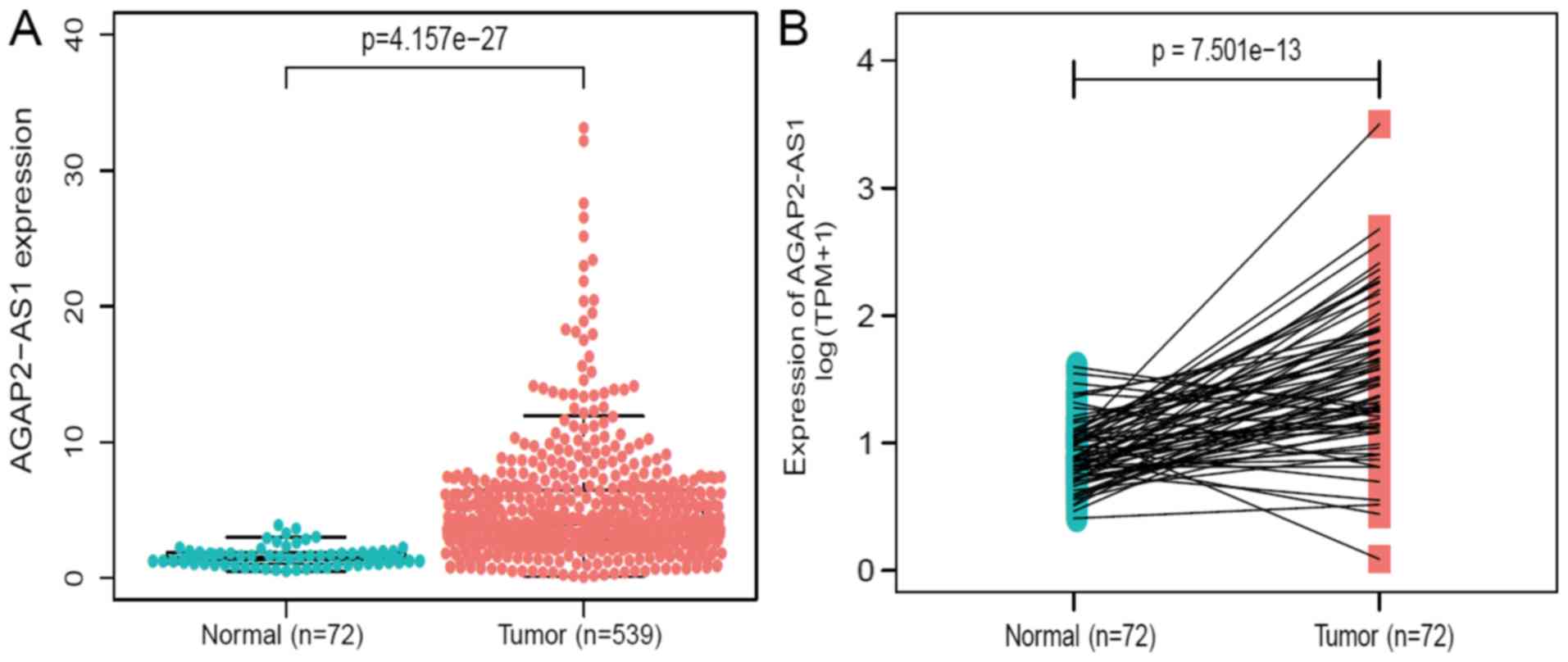
The expression of AGAP2-AS1 was detected in 539
ccRCC tissues and 72 adjacent healthy tissues using Wilcoxon rank
sum test. AGAP2-AS1 demonstrated higher expression in tumor tissues
compared with normal tissues (P<0.001; Fig. 1A). Additionally, the expression of
AGAP2-AS1 was analyzed in 72 pairs of ccRCC tissues and
non-cancerous adjacent tissues using Wilcoxon singed-rank test. The
results indicated that AGAP2-AS1 was significantly overexpressed in
ccRCC tumors (P<0.001; Fig. 1B),
indicating AGAP2-AS1 may be associated with ccRCC
carcinogenesis.
Correlation between the expression of
AGAP2-AS1 and clinical characteristics in patients with ccRCC
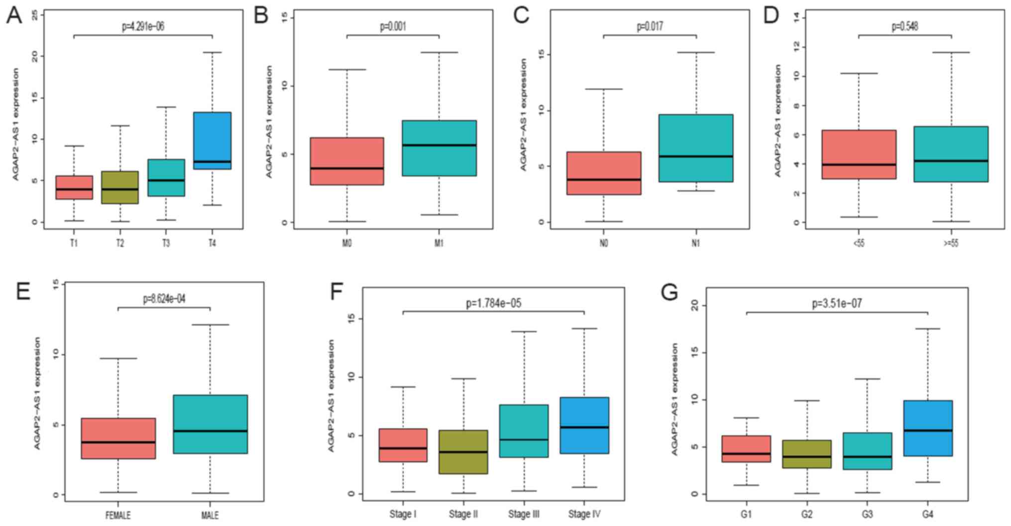
A total of 539 ccRCC samples with AGAP2-AS1
expression data were analyzed from TCGA. Upregulated expression of
AGAP2-AS1 was significantly correlated with clinical stage
(P<0.001; Fig. 2A), the grade of
topography distribution (P<0.001; Fig. 2B), topography distribution
(P<0.001; Fig. 2C), gender
(P<0.001; Fig. 2D), distance
metastasis (P<0.001; Fig. 2E) and
lymph node metastasis (P<0.001; Fig.
2F). Univariate analysis revealed that increased AGAP2-AS1
expression (based on median value) was associated with poor
prognostic clinicopathologic characteristics using logistic
regression (Table II). Upregulated
AGAP2-AS1 expression in ccRCC was significantly associated with TNM
stage (OR, 1.75 for T3/T4 vs. T1/T2; P=0.002; OR, 3.23 for N1 vs.
N0; P=0.047; OR, 1.98 for M1 vs. M0; P=0.007) and stage (OR, 1.77
for stage III/IV vs. stage I/II; P=0.001).
| Table II.lncRNA AGAP2-AS1 expression is
associated with clinicopathological characteristics (logistic
regression). |
Table II.
lncRNA AGAP2-AS1 expression is
associated with clinicopathological characteristics (logistic
regression).
| Clinical
characteristics | Total, n | Odds ratio (95%
CI) | P-value |
|---|
| Age (≥55 vs.
<55) | 530 | 1.16
(0.81–1.68) | 0.405 |
| Sex (male vs.
female) | 530 | 1.83
(1.27–2.63) | 0.001 |
| T (T3/T4 vs.
T1/T2) | 530 | 1.75
(1.23–2.52) | 0.002 |
| N (N1 vs. N0) | 255 | 3.23
(1.09–11.8) | 0.047 |
| M (M1 vs. M0) | 498 | 1.98
(1.21–3.31) | 0.007 |
| Grade (G3/G4 vs.
G2/G1) | 521 | 1.33
(0.94–1.88) | 0.104 |
| Stage (III/IV vs.
I/II) | 527 | 1.77
(1.24–2.52) | 0.001 |
Diagnostic value of AGAP2-AS1
expression in clear cell renal carcinoma
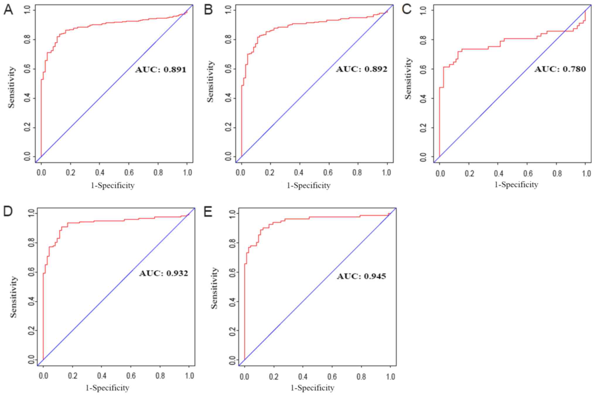
To assess the diagnostic value of AGAP2-AS1 in
ccRCC, a ROC curve analysis was performed by testing the expression
stage between patients with ccRCC and healthy, adjacent cases
(Fig. 3A). The area under the ROC
curve (AUC) was 0.891, which indicated an excellent diagnostic
value. Subgroup analysis demonstrated the diagnostic value of
AGAP2-AS1 expression in different stages of ccRCC, with AUC values
of 0.892 for clinical stage I, 0.780 for clinical stage II, 0.932
for clinical stage III and 0.945 for clinical stage IV (Fig. 3B-3E).
Survival curve, univariate and
multivariate analysis of AGAP2-AS1 in ccRCC
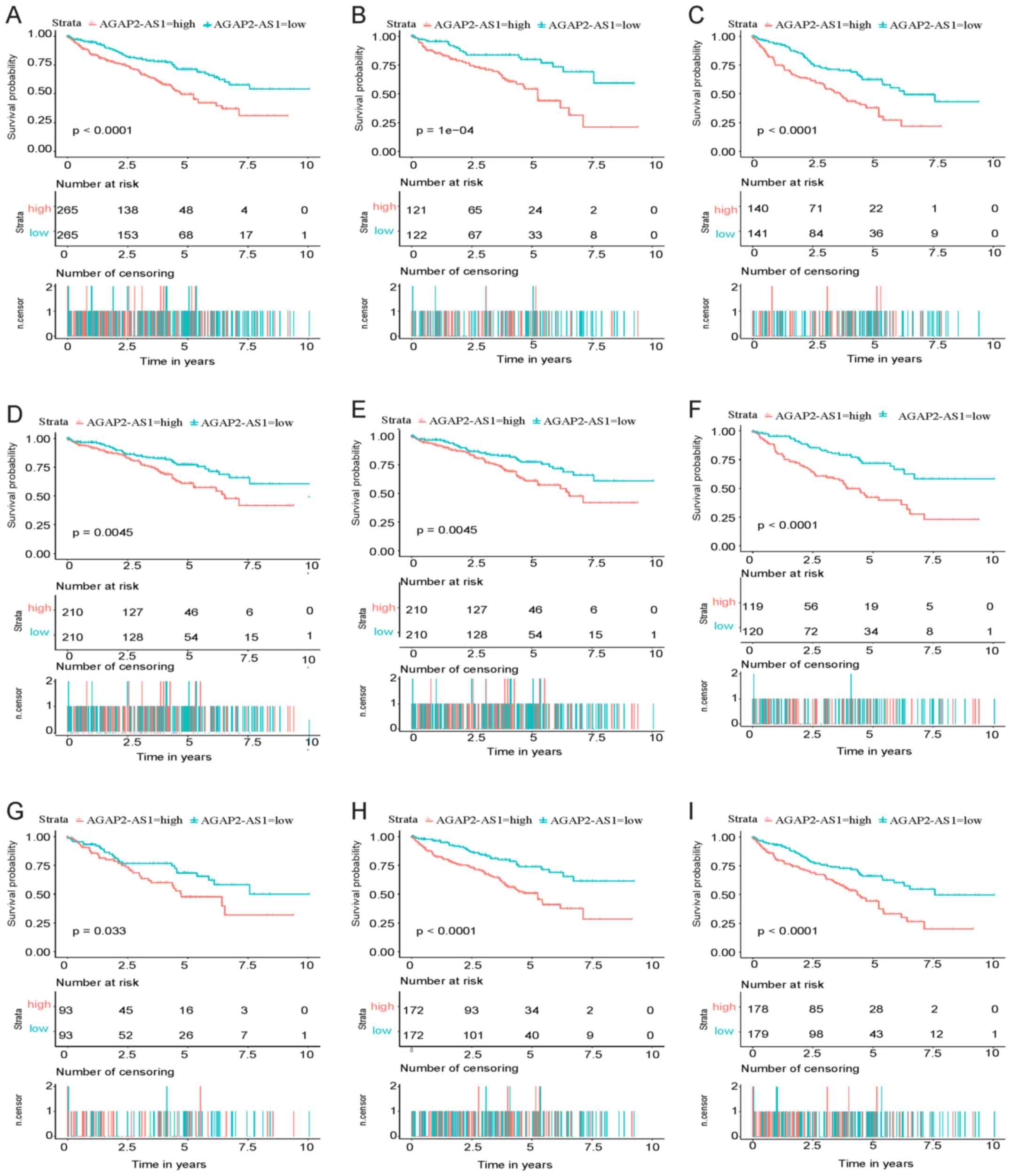
As presented in Fig.
4A, Kaplan-Meier survival analysis indicated that
high-AGAP2-AS1 in ccRCC exhibited a worse prognosis compared with
low-AGAP2-AS1 (P<0.001). The univariate analysis, which was
performed using a Cox regression, revealed that high AGAP2-AS1
correlated significantly with poor OS [hazard ratio (HR), 1.85; 95%
confidence interval (CI), 1.48–2.33; P<0.001]. Other
clinicopathological variables were also indicated to be associated
with poor survival included age, advanced stage and TNM stage
(Table III).
| Table III.Univariate regression and
multivariate survival model of prognostic covariates in patients
with clear cell renal carcinoma (Cox regression). |
Table III.
Univariate regression and
multivariate survival model of prognostic covariates in patients
with clear cell renal carcinoma (Cox regression).
| Clinicopathologic
variables | Hazard ratio (95%
CI) | P-value |
|---|
| Univariate
analysis |
|
|
| Age
(≥55 vs. <55) | 1.58
(0.98–2.56) | 0.060 |
| Sex
(male vs. female) | 1.01
(0.66–1.54) | 0.951 |
| T
(T3/T4 vs. T1/T2) | 1.82
(1.47–2.25) | <0.001 |
| N (N1
vs. N0) | 2.93
(1.52–5.67) | 0.001 |
| M (M1
vs. M0) | 4.07
(2.63–6.30) | <0.001 |
| Grade
(G3/G4 vs. G1/G2) | 1.62
(1.29–2.04) | <0.001 |
| Stage
(III/IV vs. I/II) | 1.92
(1.54–2.34) | <0.001 |
|
AGAP2-AS1 | 1.85
(1.48–2.33) | <0.001 |
| Multivariate
analysis |
|
|
| T
(T3/T4 vs. T1/T2) | 1.41
(0.62–3.25) | 0.412 |
| N (N1
vs. N0) | 1.24
(0.62–2.49) | 0.548 |
| M (M1
vs. M0) | 2.22
(1.32–3.73) | 0.003 |
| Grade
(G3/G4 vs. G1/G2) | 1.67
(1.01–2.75) | 0.045 |
| Stage
(III/IV vs. I/II) | 1.48
(0.58–3.75) | 0.409 |
|
AGAP2-AS1 | 1.57
(1.21–2.03) | 0.001 |
These results indicated ccRCC with increased
AGAP2-AS1 expression correlated with the development into a more
advanced stage (grade 3/4), lymph node metastasis and distance.
Multivariate analysis was subsequently performed (Table III). The results demonstrated that
high expression of AGAP2-AS1 was associated with poor OS in
patients with ccRCC and a high HR.
AGAP2-AS1-associated biological
pathways identified by GSEA
GSEA was performed to identify biological pathways
in ccRCC by comparing the aberrant AGAP2-AS1 expression data sets.
The significant differences (FDR <0.25; NOM P<0.05) in
enrichment of MSigDB Collection (including h.all.v6.2.symbols.gmt
and c2.cp.kegg.v6.2.symbols.gmt). A total of 7 critical biological
pathways, including stromal stimulation, angiogenesis, epithelial
to mesenchymal transition, basal cell carcinoma, ECM receptor
interaction and the Notch signaling pathway indicated significantly
differential enrichment in AGAP2-AS1 high expression phenotype
based on NES, NOM P-value and FDR value (Figs. 5A–5I;
Table IV), indicating the potential
role of AGAP2-AS1 in the development of ccRCC.
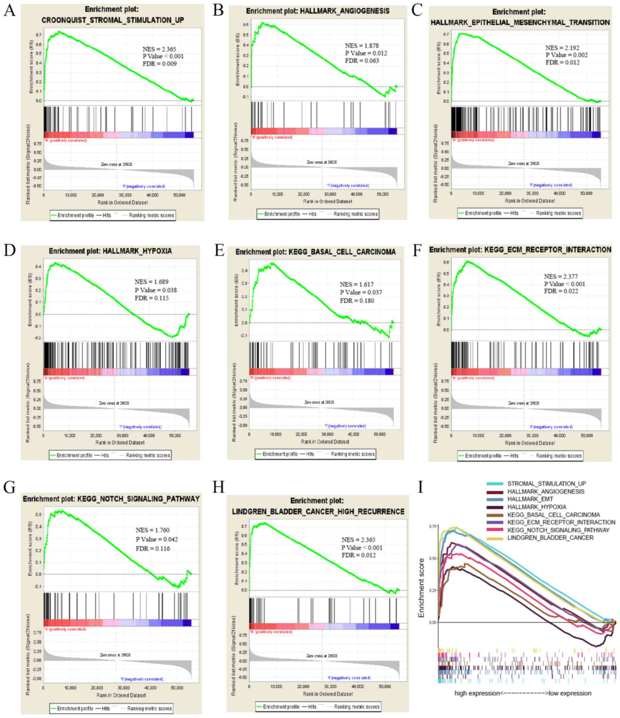 | Figure 5.Enrichment plots from GSEA. GSEA
results showing (A) stromal stimulation, (B) angiogenesis, (C)
epithelial to mesenchymal transition, (D) hypoxia, (E) basal cell
carcinoma, (F) ECM receptor interaction, (G) Notch signaling
pathway, (H) bladder cancer high recurrence and (I) summary of
GSEA. KEGG, Kyoto Encyclopedia of Genes and Genomes; NES,
normalized enrichment score; FDR, false discovery rate; GSEA, gene
set enrichment analysis. |
| Table IV.Gene sets enriched in phenotype high
vs. low of AGAP2-AS1. |
Table IV.
Gene sets enriched in phenotype high
vs. low of AGAP2-AS1.
| MSigDB
collection | Gene xet name | NES | NOM P-value | FDR q-value |
|---|
|
h.all.v6.2.symbols.gmt |
HALLMARK_EPITHELIAL_MESENCHYMAL_TRANSITION_6 | 2.192 | 0.002 | 0.011 |
|
|
HALLMARK_HYPOXIA | 1.688 | 0.038 | 0.115 |
|
|
HALLMARK_ANGIOGENESIS | 1.877 | 0.012 | 0.063 |
|
c2.cp.kegg.v6.2.symbols.gmt |
KEGG_ECM_RECEPTOR_INTERACTION | 2.077 | <0.001 | 0.021 |
|
|
KEGG_NOTCH_SIGNALING_PATHWAY | 1.760 | 0.043 | 0.116 |
|
|
KEGG_BASAL_CELL_CARCINOMA | 1.617 | 0.037 | 0.180 |
|
c2.cgp.v6.2.symbols.gmt |
LINDGREN_BLADDER_CANCER_HIGH_RECURRENCE | 2.367 | <0.001 | 0.012 |
|
|
JECHLINGER_EPITHELIAL_TO_MESENCHYMAL_TRANSITION_UP | 2.215 | <0.001 | 0.020 |
Discussion
ccRCC is a common subtype of kidney cancer, however,
up to 17% of patients with ccRCCs present with primary metastatic
disease (3). Therefore, the
prognosis for patients with ccRCC and distance metastasis remains
difficult to predict (16). lncRNAs
form several groups of molecules that influence the gene expression
of protein-coding genes in different ways. Currently, a large
number of lncRNAs are increasingly considered to be associated with
the recurrence and prognosis of cancer (17,18).
lncRNAs exhibit the potential to be used as therapeutic targets and
prognostic biomarkers. For example, lncRNA SNHG6-003 may function
as a sponge for miR-26a/b to promote the progression of
hepatocellular carcinoma (19).
lncRNA LUCAT1 has been indicated to promote proliferation and
invasion in ccRCC cells via the AKT/GSK-3β signaling pathway
(20). lncRNA AGAP2-AS1 ectopic
expression has been indicated in numerous types of carcinoma,
including in hepatocellular carcinoma (14), non-small cell lung cancer (9,10),
gastric cancer (15) and glioma
(11). Furthermore, AGAP2-AS1 has
been indicated to be co-expressed with HDGF (21) and ANGPTL4 (13), which are associated with tumor
angiogenesis. However, few studies have reported the expression of
AGAP2-AS1 and the effect of its expression on ccRCC prognosis.
Therefore, in the current study, to determine the
clinicopathological and prognostic value of AGAP2-AS1 in ccRCC, the
mRNA expression of AGAP2-AS1 in ccRCC was assessed through
bioinformatics analysis of data from TCGA database. Receiver
operating characteristic (ROC) curve analysis, GSEA and other
methods were also used to assess the diagnostic and prognostic
value of AGAP2-AS1.
In the present study, high throughput RNA sequencing
data of ccRCC were downloaded from the TCGA database, and the
outcomes demonstrated that high lncRNA AGAP2-AS1 was significantly
associated with worse survival status. Kaplan-Meier curves for OS
also indicated that higher expression of lncRNA AGAP2-AS1 was
associated with different genders, levels of ages, high clinical
stage, and advanced TNM stage in patients with ccRCC. Logistic
analyses, univariate and multivariate Cox analyses indicated that
AGAP2-AS1 expression may be a potential indicator for ccRCC
prognosis, and ROC analysis affirmed the diagnostic value of
AGAP2-AS1 expression in ccRCC.
AGAP2-AS1, as a non-coding RNA, has been
demonstrated to promote anaplastic glioma cells proliferation,
migration and invasion, and the knockdown of AGAP2-AS1 has been
indicated to increase apoptosis cell rates (11). Similar outcomes have been revealed in
the human metastatic pancreatic cancer cell line AsPC-1 (13), and in non-small cell lung cancer cell
lines A549 and SK-MES-1 (10).
Consistent with previous studies, the results of the current study
demonstrated that increased AGAP2-AS1 expression was associated
with poor overall survival and the potential mechanisms governing
this may be the connection with stromal simulation (22,23),
angiogenesis (24,25), epithelial-mesenchymal transition
(26), hypoxia (27) or the notch signaling pathway
(28,29). Angiogenesis, epithelial-mesenchymal
transition and hypoxia are well-known hallmarks of cancers, and
angiogenesis serves an important role in the progression of ccRCC
via VEGF (30), FGF-2, PDGF,
angiopoietins, ephrins, apelin (APLN) and chemokines (31,32). In
the present study, the aberrant expression of AGAP2-AS1 was
enriched in the process of angiogenesis, which may be activated by
the PI3K/Akt pathway (33). These
previous studies indicated that lncRNA AGAP2-AS1 may function via
the VEGF and Akt pathway, which could be used as a drug target for
ccRCC. Furthermore, the prognostic value of AGAP2-AS1 expression
was examined in different subgroups of ccRCC and it was indicated
that high lncRNA AGAP2-AS1 expression was significantly associated
with G1/G2, stage I/II and M0 cases, highlighting the potential
value of AGAP2-AS1 in the development of ccRCC.
Currently, surgery is the most common treatment for
ccRCC. However, the possibility of recurrence, which adversely
impacts patient outcomes, is an important factor in the choice of
treatment (34). The current study
also assessed the correlation between AGAP2-AS1 expression and a
number of clinicopathological characters, and the results revealed
that this potential biomarker may help to guide treatment selection
in patients with ccRCC. High expression of AGAP2-AS1 also
negatively affected OS among patients with histological grade
G1/G2, grade G3/G4 and clinical stage III/IV; but not patients with
histological clinical stage I/II, which further demonstrated the
specific prognostic role of AGAP2-AS1 expression in subgroup
analysis and its potential contribution to precision therapy for
ccRCC.
Although the results in the present study provided
information regarding the relationship between AGAP2-AS1 and ccRCC,
there were some limitations to the present study. All clinical
factors should have been considered, including BMI, the details on
treatments received by patients involved, smoking status and other
biomarkers' levels. However, this information is often missing, or
inconsistent treatments are stated in public databases. Only a
total of 72 healthy samples and 539 ccRCC samples were evaluated in
the current study which may limit the present work. Therefore, the
sample size should be increased in future study.
Overall, the current study demonstrated the
diagnostic and prognostic value of AGAP2-AS1 expression in patients
with ccRCC. However, the current study was performed using RNA-seq
of TCGA database, which lacks protein level files and direct
mechanisms information. Additionally, the number of healthy
subjects and information on later stages are limited. Therefore,
further identification of effective biomarkers in ccRCC cases of
advanced stage is required in the future.
In conclusion, we observed that lncRNA AGAP2-AS1 is
up-regulated in ccRCC, which also correlates with clinical
progression and serves as an independent risk factor for OS in
ccRCC. Our findings partily demonstrated that lncRNA AGAP2-AS1 may
be a potential biomarker in the diagnosis and prognosis of
ccRCC.
Acknowledgements
Not applicable.
Funding
The present study was supported by the Program of
Graduate students innovation fund of Hebei Province (grant no.
CXZZBS2019117).
Availability of data and materials
The datasets used and/or analyzed during the present
study are available from the corresponding author on reasonable
request.
Authors' contributions
LG conceived the study and was the major contributor
in writing the manuscript. AZ participated in its design, analyzed
and interpreted the data. XW participated in design, visualization
and critically revised the manuscript for important intellectual
content. All authors read and approved the final manuscript.
Ethics approval and consent to
participate
Not applicable.
Patient consent for publication
Not applicable.
Competing interests
The authors declare that they have no competing
interests.
References
|
1
|
Siegel RL, Miller KD and Jemal A: Cancer
statistics, 2019. CA Cancer J Clin. 69:7–34. 2019. View Article : Google Scholar : PubMed/NCBI
|
|
2
|
Hsieh JJ, Purdue MP, Signoretti S, Swanton
C, Albiges L, Schmidinger M, Heng DY, Larkin J and Ficarra V: Renal
cell carcinoma. Nat Rev Dis Primers. 3:170092017. View Article : Google Scholar : PubMed/NCBI
|
|
3
|
Capitanio U and Montorsi F: Renal cancer.
Lancet. 387:894–906. 2016. View Article : Google Scholar : PubMed/NCBI
|
|
4
|
Gawlik-Jakubczak T, Matuszewski M and
Biernat W: The metastasis of renal cell carcinoma to the urethra
and local tumor recurrence. Urol Int. doi: 10.1159/000501699.
PubMed/NCBI
|
|
5
|
Yu L, Xiang L, Feng J, Li B, Zhou Z, Li J,
Lin Y, Lv Y, Zou D, Lei Z, et al: miRNA-21 and miRNA-223 expression
signature as a predictor for lymph node metastasis, distant
metastasis and survival in kidney renal clear cell carcinoma. J
Cancer. 9:3651–3659. 2018. View Article : Google Scholar : PubMed/NCBI
|
|
6
|
Blandin Knight S, Crosbie PA, Balata H,
Chudziak J, Hussell T and Dive C: Progress and prospects of early
detection in lung cancer. Open Biol. 7:1700702017. View Article : Google Scholar : PubMed/NCBI
|
|
7
|
Rivera-Franco MM and Leon-Rodriguez E:
Delays in breast cancer detection and treatment in developing
countries. Breast Cancer (Auckl).
12:11782234177526772018.PubMed/NCBI
|
|
8
|
Li P, Wong YN, Armstrong K, Haas N, Subedi
P, Davis-Cerone M and Doshi JA: Survival among patients with
advanced renal cell carcinoma in the pretargeted versus targeted
therapy eras. Cancer Med. 5:169–181. 2016. View Article : Google Scholar : PubMed/NCBI
|
|
9
|
Fan KJ, Liu Y, Yang B, Tian XD, Li CR and
Wang B: Prognostic and diagnostic significance of long non-coding
RNA AGAP2-AS1 levels in patients with non-small cell lung cancer.
Eur Rev Med Pharmacol Sci. 21:2392–2396. 2017.PubMed/NCBI
|
|
10
|
Li W, Sun M, Zang C, Ma P, He J, Zhang M,
Huang Z, Ding Y and Shu Y: Upregulated long non-coding RNA
AGAP2-AS1 represses LATS2 and KLF2 expression through interacting
with EZH2 and LSD1 in non-small-cell lung cancer cells. Cell Death
Dis. 7:e22252016. View Article : Google Scholar : PubMed/NCBI
|
|
11
|
Wang W, Yang F, Zhang L, Chen J, Zhao Z,
Wang H, Wu F, Liang T, Yan X, Li J, et al: LncRNA profile study
reveals four-lncRNA signature associated with the prognosis of
patients with anaplastic gliomas. Oncotarget. 7:77225–77236.
2016.PubMed/NCBI
|
|
12
|
Dong H, Wang W, Mo S, Chen R, Zou K, Han
J, Zhang F and Hu J: SP1-induced lncRNA AGAP2-AS1 expression
promotes chemoresistance of breast cancer by epigenetic regulation
of MyD88. J Exp Clin Cancer Res. 37:2022018. View Article : Google Scholar : PubMed/NCBI
|
|
13
|
Hui B, Ji H, Xu Y, Wang J, Ma Z, Zhang C,
Wang K and Zhou Y: RREB1-induced upregulation of the lncRNA
AGAP2-AS1 regulates the proliferation and migration of pancreatic
cancer partly through suppressing ANKRD1 and ANGPTL4. Cell Death
Dis. 10:2072019. View Article : Google Scholar : PubMed/NCBI
|
|
14
|
Liu Z, Wang Y, Wang L, Yao B, Sun L, Liu
R, Chen T, Niu Y, Tu K and Liu Q: Long non-coding RNA AGAP2-AS1,
functioning as a competitive endogenous RNA, upregulates ANXA11
expression by sponging miR-16-5p and promotes proliferation and
metastasis in hepatocellular carcinoma. J Exp Clin Cancer Res.
38:1942019. View Article : Google Scholar : PubMed/NCBI
|
|
15
|
Qi F, Liu X, Wu H, Yu X, Wei C, Huang X,
Ji G, Nie F and Wang K: Long noncoding AGAP2-AS1 is activated by
SP1 and promotes cell proliferation and invasion in gastric cancer.
J Hematol Oncol. 10:482017. View Article : Google Scholar : PubMed/NCBI
|
|
16
|
Motzer RJ and Molina AM: Targeting renal
cell carcinoma. J Clin Oncol. 27:3274–3276. 2009. View Article : Google Scholar : PubMed/NCBI
|
|
17
|
Li X, Wu Z, Fu X and Han W: Long noncoding
RNAs: Insights from biological features and functions to diseases.
Med Res Rev. 33:517–553. 2013. View Article : Google Scholar : PubMed/NCBI
|
|
18
|
Seles M, Hutterer GC, Kiesslich T, Pummer
K, Berindan-Neagoe I, Perakis S, Schwarzenbacher D, Stotz M, Gerger
A and Pichler M: Current Insights into Long Non-Coding RNAs in
Renal Cell Carcinoma. Int J Mol Sci. 17:5732016. View Article : Google Scholar : PubMed/NCBI
|
|
19
|
Cao C, Zhang T, Zhang D, Xie L, Zou X, Lei
L, Wu D and Liu L: The long non-coding RNA, SNHG6-003, functions as
a competing endogenous RNA to promote the progression of
hepatocellular carcinoma. Oncogene. 36:1112–1122. 2017. View Article : Google Scholar : PubMed/NCBI
|
|
20
|
Zheng Z, Zhao F, Zhu D, Han J, Chen H, Cai
Y, Chen Z and Xie W: Long non-coding RNA LUCAT1 promotes
proliferation and invasion in clear cell renal cell carcinoma
through AKT/GSK-3β signaling pathway. Cell Physiol Biochem.
48:891–904. 2018. View Article : Google Scholar : PubMed/NCBI
|
|
21
|
Zheng Y, Lu S, Xu Y and Zheng J: Long
non-coding RNA AGAP2-AS1 promotes the proliferation of glioma cells
by sponging miR-15a/b-5p to upregulate the expression of HDGF and
activating Wnt/β-catenin signaling pathway. Int J Biol Macromol.
128:521–530. 2019. View Article : Google Scholar : PubMed/NCBI
|
|
22
|
Chang WK, Carmona-Fontaine C and Xavier
JB: Tumour-stromal interactions generate emergent persistence in
collective cancer cell migration. Interface Focus. 3:201300172013.
View Article : Google Scholar : PubMed/NCBI
|
|
23
|
Hong M, Cheng H, Song L, Wang W, Wang Q,
Xu D and Xing W: Wogonin suppresses the activity of matrix
metalloproteinase-9 and inhibits migration and invasion in human
hepatocellular carcinoma. Molecules. 23:3842018. View Article : Google Scholar
|
|
24
|
Ramjiawan RR, Griffioen AW and Duda DG:
Anti-angiogenesis for cancer revisited: Is there a role for
combinations with immunotherapy? Angiogenesis. 20:185–204. 2017.
View Article : Google Scholar : PubMed/NCBI
|
|
25
|
Viallard C and Larrivée B: Tumor
angiogenesis and vascular normalization: Alternative therapeutic
targets. Angiogenesis. 20:409–426. 2017. View Article : Google Scholar : PubMed/NCBI
|
|
26
|
Chen T, You Y, Jiang H and Wang ZZ:
Epithelial-mesenchymal transition (EMT): A biological process in
the development, stem cell differentiation, and tumorigenesis. J
Cell Physiol. 232:3261–3272. 2017. View Article : Google Scholar : PubMed/NCBI
|
|
27
|
Manoochehri Khoshinani H, Afshar S and
Najafi R: Hypoxia: A double-edged sword in cancer therapy. Cancer
Invest. 34:536–545. 2016. View Article : Google Scholar : PubMed/NCBI
|
|
28
|
Braune EB and Lendahl U: Notch -- a
goldilocks signaling pathway in disease and cancer therapy. Discov
Med. 21:189–196. 2016.PubMed/NCBI
|
|
29
|
Li L, Tang P, Li S, Qin X, Yang H, Wu C
and Liu Y: Notch signaling pathway networks in cancer metastasis: A
new target for cancer therapy. Med Oncol. 34:1802017. View Article : Google Scholar : PubMed/NCBI
|
|
30
|
Jiang Y, Zhou J, Zou D, Hou D, Zhang H,
Zhao J, Li L, Hu J, Zhang Y and Jing Z: Overexpression of Limb-Bud
and Heart (LBH) promotes angiogenesis in human glioma via
VEGFA-mediated ERK signalling under hypoxia. EBioMedicine.
48:36–48. 2019. View Article : Google Scholar : PubMed/NCBI
|
|
31
|
Lugano R, Ramachandran M and Dimberg A:
Tumor angiogenesis: Causes, consequences, challenges and
opportunities. Cell Mol Life Sci. 2019.https://doi.org/10.1007/s00018-019-03351-7
View Article : Google Scholar : PubMed/NCBI
|
|
32
|
Chappell JC, Payne LB and Rathmell WK:
Hypoxia, angiogenesis, and metabolism in the hereditary kidney
cancers. J Clin Invest. 129:442–451. 2019. View Article : Google Scholar : PubMed/NCBI
|
|
33
|
Duan MX, Zhou H, Wu QQ, Liu C, Xiao Y,
Deng W and Tang QZ: Andrographolide protects against HG-induced
inflammation, apoptosis, migration, and impairment of angiogenesis
via PI3K/AKT-eNOS signalling in HUVECs. Mediators Inflamm.
2019:61683402019. View Article : Google Scholar : PubMed/NCBI
|
|
34
|
Gallardo E, Méndez-Vidal MJ, Pérez-Gracia
JL, Sepúlveda-Sánchez JM, Campayo M, Chirivella-González I,
García-Del-Muro X, González-Del-Alba A, Grande E and Suárez C: SEOM
clinical guideline for treatment of kidney cancer (2017). Clin
Transl Oncol. 20:47–56. 2018. View Article : Google Scholar : PubMed/NCBI
|