Introduction
Ovarian cancer (OC) is one of the most frequent
cancer types among female patients. There were a total of ~295,414
new OC cases in 2018 that resulted in 184,799 deaths worldwide
(1). OC is a highly heterogeneous
disease, and epithelial OC has been divided into four histological
subtypes, including endometrioid, mucinous, serous and clear cell
adenocarcinoma (2). OC is considered
a mixture of these representative histological types with different
molecular etiologies, tumor progression features and disease
prognoses. This hinders the efficacy of current treatments for OC,
such as platinum-based combination chemotherapy, surgery and
neoadjuvant chemotherapy (3,4). Unfortunately, the majority of patients
with OC (>70%) are diagnosed at an advanced or metastatic stage
(III or IV) and their 5-year overall survival rate is ~40%, while
the 5-year overall survival rate of patients with stage I OC is
~90% (5,6). At present, no effective screening
approaches have been reported that can be used effectively to
decrease the mortality rate of OC, due to low sensitivity and
specificity (7). Prognostic
biomarkers with high sensitivity and specificity can be used to
estimate disease metastasis or recurrence, classify patients at
different risk levels for corresponding outcome assessment and
guide clinical treatments, in this way reducing the mortality rate
of patients with OC (8). Therefore,
it is crucial to identify novel prognostic biomarkers to monitor
cancer progression, overall therapeutic efficiency and risk
stratification in patients with OC.
During the past decades, high-throughput sequencing
technology has been used for the investigation of several types of
cancer. This method can aid the identification of specific patterns
for cancer diagnosis, classification and therapeutic response
(9,10). A large amount of core data obtained
by high-throughput sequencing technology has been stored in public
databases. These data can be integrated and re-analyzed in order to
uncover important targets that can be used for cancer therapy. In
the present study, the GSE66957, GSE6008 and GSE26712 datasets were
selected from the Gene Expression Omnibus (GEO) database to
identify differentially expressed genes (DEGs). In the present
study, a total of 269 DEGs were discovered by bioinformatic
analysis (differentially expressed in at least 2 datasets), and the
most promising prognostic marker for OC was selected for further
study. The expression levels of this gene in OC tissues compared
with those in normal tissues were analyzed using the GEO datasets
and confirmed using quantitative PCR. A predictive nomogram with
high accuracy was established for prognosis prediction. Gene set
enrichment analysis (GSEA) and analysis of the tumor-associated
competing endogenous RNA (ceRNA) network were used to clarify the
potential biological processes associated with this gene.
Materials and methods
Patients and tissue samples
A total of 82 OC tissues (from OC patients who
underwent debulking surgery) and 82 normal ovarian tissues (from
hysteromyoma patients who underwent hysterectomy and oophorectomy)
were obtained from the Department of Obstetrics and Gynecology at
Dongguan Affiliated Hospital, Southern Medical University between
July 2014 and December 2017. OC tissues included 47 serous
adenocarcinomas, 9 mucinous adenocarcinomas, 6 endometrioid
adenocarcinomas, 6 clear-cell tumors, 6 germ-cell tumors and 8
other types. The mean age of the patients was 52.96 years, ranging
from 17 to 86 years. None of the recruited patients in the present
study had received preoperative radiotherapy or preoperative
chemotherapy. Written informed consent was obtained from patients.
This study was approved by the Regional Institutional Review Board
of Dongguan Affiliated Hospital, Southern Medical University and
carried out according to the Declaration of Helsinki. All fresh
tumor tissue specimens were cryopreserved immediately in liquid
nitrogen and stored at −80°C until further use.
GEO datasets and the cancer genome
atlas (TCGA) database
GEO (https://www.ncbi.nlm.nih.gov/geo/query/acc.cgi) is a
public database containing experiments and gene expression profiles
that can be downloaded and used for subsequent analysis. The
expression profiles of the transcriptomes from the following
datasets were systematically analyzed to ensure the credibility of
the present study: GSE66957 (n=69; Moffitt Cancer Center,
Biostatistics and Bioinformatics), GSE6008 (n=103; University of
Michigan) and GSE26712 (n=195; University of Michigan) (11–14). The
sequencing data were collected on Affymetrix (Thermo Fisher
Scientific, Inc.) microarray and Illumina, Inc., platforms by
different researchers. The clinicopathological characteristics of
266 patients with OC were collected from TCGA database via the
cBioPortal (http://www.cbioportal.org), including
the 7th American Joint Committee on Cancer (AJCC) staging indices
(https://www.nationwidechildrens.org/research/resources-infrastructure/core
facilities/biospecimen-core- resource/the- cancer-
genome-atlas/tcga-forms-and-documents), the disease-free survival
time, the overall survival time, the quality of life (https://www.nationwidechildrens.org/
research/resources-infrastructure/core facilities/biospecimen-
core-resource/the-cancer-g enome- atlas/tcga-forms- and- documents)
and the recurrence time. The clinical stages of OC were divided
into 4 stages (I, II, III and IV) according to the origin,
pathogenesis and prognosis by FIGO criteria (15,16).
Identification of DEGs
DEGs were analyzed via the R (v3.34) limma package
(http://www.bioconductor.org/packages/release/bioc/html/limma.html)
between OC and normal tissues. The different levels of gene
expression between OC and normal tissues were evaluated by the log2
fold-change. Genes with a log2 fold-change >1 (absolute value)
and P<0.05 were selected as candidate signature genes for
further analysis. A Venn plot was constructed for identification of
reliable OC-associated genes.
Kaplan-Meier plot
The Kaplan-Meier method was used to evaluate the
association between the survival time of patients with OC and the
DEGs selected from the Venn plot collection. The genes with the
most significantly different log-rank P-values were selected as the
candidate genes. The difference in the expression of the candidate
genes was analyzed via the GEO datasets, as well as the prognostic
value of the candidate genes.
Quantitative PCR (qPCR) assay
Total RNA was extracted from the tumor tissue
specimens of the patients with OC using the RNA isolation kit
(BioTeke Corporation) according to the manufacturer's protocol.
CLDN10 mRNA expression levels were assessed by qPCR using a SYBR
Green PCR kit (Takara Biotechnology Co., Ltd.). The reaction
conditions were as follows: Preincubation at 95°C for 10 min,
followed by 40 cycles of 95°C for 10 sec, 60°C for 10 sec, and 72°C
for 20 sec. CLDN10 expression levels were normalized to those of
GAPDH mRNA levels (ΔCq=Cq CLDN10-Cq GAPDH) and was calculated using
2−ΔΔCq between two groups (17). The primers for CLDN10 and GAPDH
detection were as follows: CLDN10 forward,
5′-CTGTGGAAGGCGTGCGTTA-3′ and reverse, 5′-CAAAGAAGCCCAGGCTGACA-3′;
and GAPDH forward, 5′-GTCTCCTCTGACTTCAACAGCG-3′ and reverse,
5′-ACCACCCTGTTGCTGTAGCCAA-3′.
Prognostic nomogram construction
A nomogram was established to predict the 5-year
survival rate. The prognostic significance of the parameters was
evaluated by multivariate Cox proportional hazard regression
analysis. CLDN10 expression was used as a novel variable for the
establishment of the nomogram. For other variables, several
survival-associated indices and basic medical information were
selected, including age, AJCC staging indices and lymphovascular
invasion index. The predictive accuracy of the nomogram for the
5-year survival rate was assessed via the concordance index
(C-index), which was estimated by the area under the receiver
operating characteristic (ROC) curve for predictive probability.
Additionally, the ROC curve of conventional features for predicting
the 5-year survival rate was displayed for comparison. The
calibration was conducted by comparing the nomogram predicted
probabilities with the observed probabilities. The univariate and
multivariate Cox proportional hazards tests were performed for
conventional clinical feature analysis.
ceRNA network construction
A CLDN10-associated ceRNA network was constructed to
reveal the potential biological processes and associations between
CLDN10 and other RNAs, including long non-coding RNAs (lncRNAs),
circular RNAs (circRNA) and miRNAs. miRTarBase (http://mirtarbase.mbc.nctu.edu.tw/php/index.php) was
used to elucidate the association between CLDN10 and miRNA
expression. This database includes a record of experimental
validated miRNA-mRNA associations. miRNAs with at least one
experimental validation were selected. In addition, the
associations among selected miRNAs, circRNAs and lncRNAs were
revealed via Starbase v.2.0 (http://starbase.sysu.edu.cn/starbase2/index.php). The
ceRNA network was created using Cytoscape (v.3.5.1) (https://cytoscape.org).
GSEA
GSEA and Kyoto Encyclopedia of Genes and Genomes
(KEGG) (https://www.kegg.jp) were used to
investigate the biological insight and signaling pathways
associated with CLDN10 expression in OC. Patients with OC in TCGA
database were separated into high and low expression groups
according to the median value of CLDN10 expression (5.23) and were
retained as the phenotypes. The GSEA software was obtained from the
GSEA website (http://software.broadinstitute.org/gsea/index.jsp). A
total of 1,000 permutations were selected as the statistical
significance to produce valid results. Pathways with a false
discovery rate <0.05 and P<0.05 were selected as the enriched
terms. The molecular signature database from GSEA was utilized as
the annotation file.
Statistical analysis
DEGs were identified using the limma package
(Bioconductor/R v.3.34) and Wilcoxon test. Htseq-counts (TCGA) and
RSEM (GEO, log2 scaled) were used as the sequencing data formats.
The nomogram was established using Regression Modeling Strategies
(R v3.34). Unpaired Student's t-test and Fisher's exact test were
performed in R (v.3.34).
Results
Identification of DEGs and candidate
genes
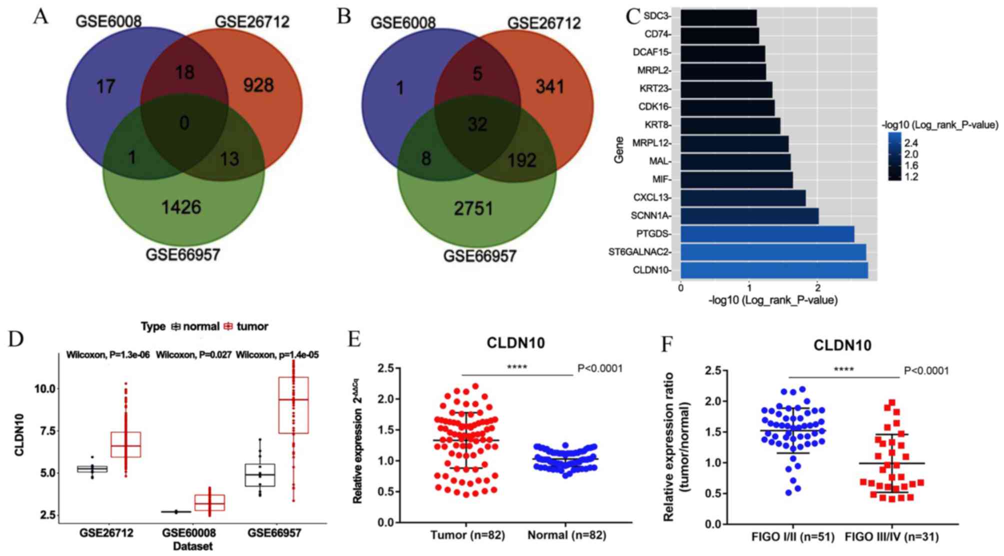
A total of 269 DEGs were discovered by bioinformatic
analysis (differentially expressed in at least 2 datasets),
including 32 upregulated and 237 downregulated genes. Venn plots
were generated to illustrate the collection of DEGs (Fig. 1A and B). To discover the potential
candidate genes to be used for OC prognosis, the Kaplan-Meier
method and the log-rank test were performed on the total number of
DEGs. A total of 15 genes (P<0.05) were selected from the total
number of DEGs as candidate signatures for further analysis. The
median CLDN10 expression level from all OC tissues from the TCGA
database was used as the cut-off value to classify the OC tissues
into high (n=133) and low (n=133) CLDN10 expression groups. CLDN10
expression exhibited the most significant association with survival
time in patients with OC (Fig. 1C).
In addition, the expression levels of CLDN10 were upregulated in OC
tissues compared with those in normal tissues, as determined by the
GEO datasets (Fig. 1D). As shown in
Table I, CLDN10 expression was
significantly associated with age and overall survival time, while
no significant association was identified between CLDN10 expression
and tumor grade, tumor pathological stage, primary site, tumor
status, vascular invasion index, disease-free survival time and
longest diameter. qPCR results demonstrated that CLDN10 expression
was upregulated in OC tissues from the hospital cohort compared
with that in normal tissues (Fig.
1E). Furthermore, the expression levels of CLDN10 were
downregulated in International Federation of Gynecology and
Obstetrics (FIGO) III/IV samples compared with those in FIGO I/II
samples (Fig. 1F).
 | Table I.Association between CLDN10 expression
and clinicopathological characteristics of patients with ovarian
cancer. |
Table I.
Association between CLDN10 expression
and clinicopathological characteristics of patients with ovarian
cancer.
|
| CLDN10
expression |
|
|
|
|---|
|
|
|
|
|
|
|---|
| Clinical
factor | High (n=133) | Low (n=133) | Total (n=266) | 95% CI | P-value |
|---|
| Mean age ± SD,
years | 57.14±10.47 | 60.9±11.04 |
| 1.1513–6.3675 | 0.0049 |
| Tumor pathological
stage, n |
|
|
|
|
|
| Stage
I–II | 9 | 11 | 20 | 0.4495–3.5211 | 0.8167 |
| Stage
III–IV | 123 | 121 | 244 |
|
|
| Grade, n |
|
| 2.8688–3.0024 |
|
|
| G1 | 0 | 1 | 1 | 0.8200 |
|
| G2 | 15 | 17 | 32 |
|
|
| G3 | 113 | 111 | 224 |
|
|
| G4 | 0 | 1 | 1 |
|
|
| GB | 1 | 1 | 2 |
|
|
| GX | 3 | 1 | 4 |
|
|
| Primary site,
n |
|
| 0.3413–0.5238 |
|
|
|
Bilateral | 95 | 85 | 180 | 0.6890 |
|
|
Left | 16 | 19 | 35 |
|
|
|
Right | 18 | 19 | 37 |
|
|
| Tumor status |
|
| 0.7075–1.8518 |
|
|
|
Tumor-free | 34 | 25 | 59 | 0.6426 |
|
| With
tumor | 83 | 91 | 174 |
|
|
| Vascular invasion
indicator |
|
| 0.3081–2.1570 |
|
|
|
Yes | 15 | 18 | 33 | 0.6596 |
|
| No | 21 | 31 | 52 |
|
|
| Mean disease-free
survival time ± SD, months | 20.74±20.23 | 17.139±12.62 |
| −7.4010–0.2000 | 0.0633 |
| Mean overall
survival time ± SD, months | 40.976±30.86 | 31.271±22.46 |
|
−16.5740(−)-2.8350 | 0.0058 |
| Longest dimension,
cm | 1.353077 | 1.485833 |
| 0.0087–0.27426 | 0.0658 |
CLDN10-associated prognostic
nomogram
Patients with OC with low CLDN10 expression had a
poor disease prognosis compared with patients with high expression
(P=0.0018; Fig. 2A). A prognostic
nomogram was established between CLDN10 expression and several
significant clinical factors, including age, vascular invasion
index and AJCC staging rules (Fig.
2C). Furthermore, calibration curves indicated an excellent
performance in prediction (Fig. 2B).
In the current nomogram, all variables were in accordance with the
clinical data. CLDN10 expression was utilized as a new variable
with improved model accuracy. The expression levels of CLDN10 were
positively associated with the risk score. The present nomogram
further indicated that low CLDN10 expression in patients with OC
may result in a low survival rate within 5 years. The C-index of
the model was 0.776.
CLDN10-associated ceRNA network and
biological pathways
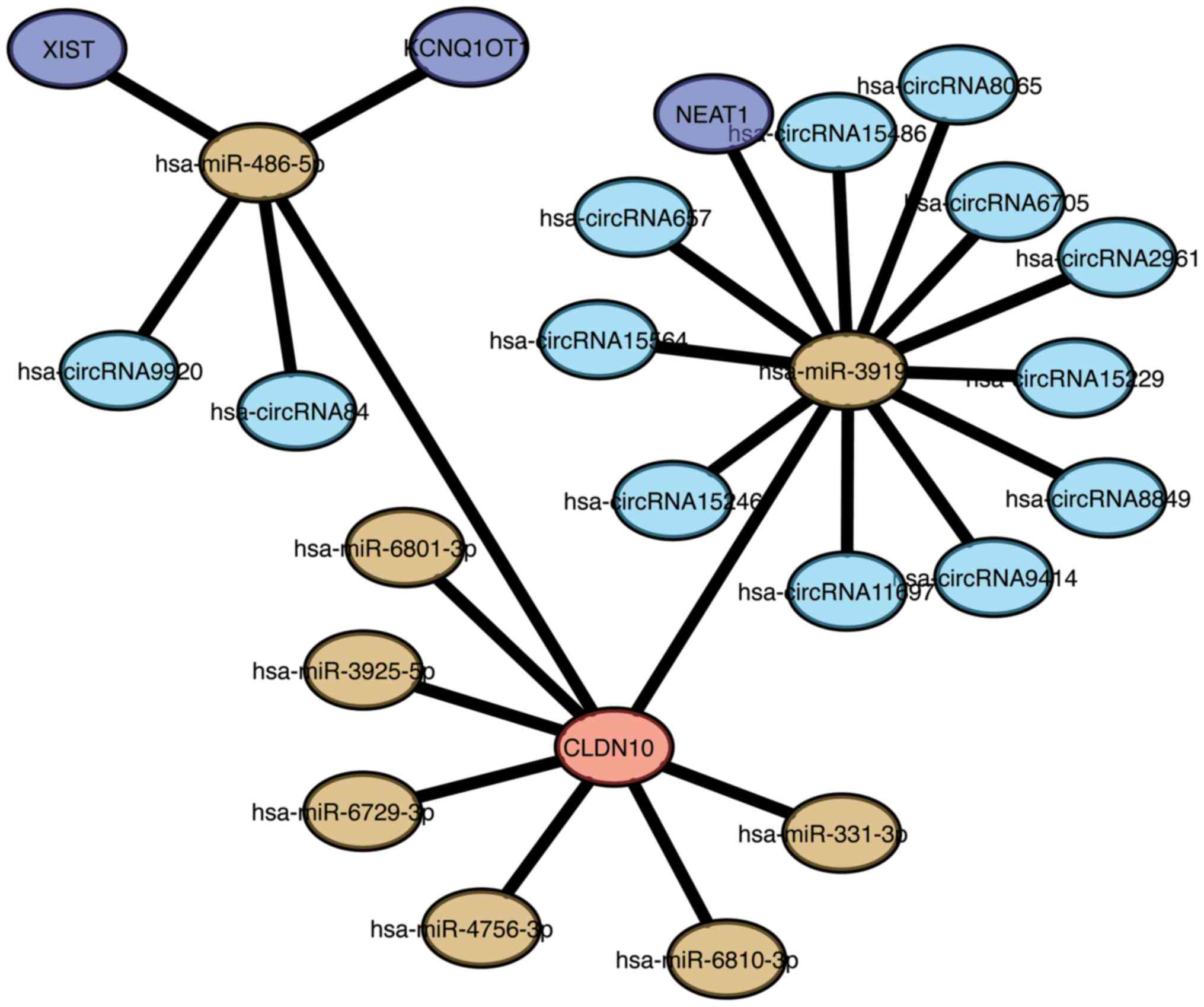
To fully understand the association between CLDN10
expression and lncRNA-miRNA-circRNA pathways, a ceRNA network
associated with CLDN10 expression was established in the present
study. A total of 8 miRNAs were identified to be associated with
CLDN10, including miR-486-5p, miR-3919, miR-6801-3p, miR-3925-5p,
miR-6729-3p, miR-4756-3p, miR-6810-3p and miR-331-3p (Fig. 3). In addition, 13 circRNAs were
associated with CLDN10 expression, namely circRNA9920, circRNA84,
circRNA657, circRNA15564, circRNA15246, circRNA11697, circRNA9414,
circRNA8849, circRNA15229, circRNA2961, circRNA6705, circRNA8065
and circRNA15486. Furthermore, an association between circRNA-miRNA
and lncRNA-miRNA was demonstrated. X-inactive specific transcript,
KCNQ1 overlapping transcript 1 and nuclear paraspeckle assembly
transcript 1 may be considered the key lncRNAs associated with
CLDN10 expression. The ceRNA network revealed that
miR-486-5p-CLDN10 and miR-3919-CLDN10 may be significantly involved
in the signaling pathways of OC.
GSEA
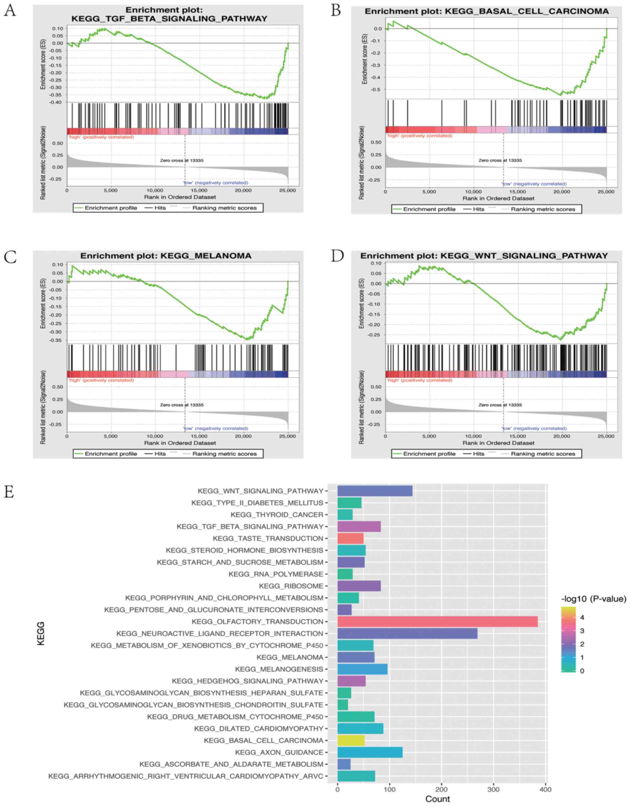
GSEA was applied to elucidate the biological
pathways associated with CLDN10 expression in OC. Four signaling
pathways were associated with CLDN10 expression, including the
‘KEGG TGF beta signaling pathway’ (P=0.00371), the ‘KEGG basal cell
carcinoma’ pathway (P=0.0000513), the ‘KEGG WNT signaling pathway’
(P=0.021978) and the ‘KEGG melanoma’ pathway (P=0.022813). In
addition, 25 signaling pathways were associated with CLDN10
expression as demonstrated by KEGG analysis (Fig. 4E), among which the ‘KEGG TGFβ
signaling pathway’ and the ‘KEGG WNT signaling pathway’ were the
main targets.
Discussion
OC is one of the most fatal gynecological
malignancies in the world, possibly due to the late diagnosis and
low 5-year survival rate of patients with an advanced stage of the
disease (1,18,19). In
addition, poor therapeutic responses of patients with OC have been
reported for specific treatments, including chemotherapeutic agents
based on platinum salts (1,18,19).
Therefore, it is imperative to identify novel prognostic or
diagnostic biomarkers with high sensitivity and specificity for
early OC screening, monitoring of tumor progression and assessment
of the overall therapeutic efficiency or patient risk
stratification.
In the present study, GSE66957, GSE6008 and GSE26712
were selected from the GEO database for DEG analysis. A total of
269 DEGs were identified in OC tissues, of which 32 were
upregulated and 237 were downregulated compared with the
corresponding gene expression levels in normal tissues. The
Kaplan-Meier method was used with the log-rank test and nomogram
analysis, and the results indicated that low CLDN10 expression in
patients with OC may be associated with a low 5-year survival rate.
qPCR analysis demonstrated that CLDN10 expression was upregulated
in OC tissues compared with that in normal tissues. Notably, the
expression levels of CLDN10 were downregulated in FIGO III/IV tumor
samples compared with those in FIGO I/II samples. CLDN10 is a
member of the claudin family, which influences the progression of
several types of cancer, including papillary thyroid cancer, breast
cancer, lung adenocarcinoma, ependymomas, esophageal squamous cell
carcinoma and hepatocellular carcinoma (20–26).
Similarly, lower expression levels of CLDN10 are associated with
higher grade ependymomas and invasive lung adenocarcinoma (23,24).
However, higher CLDN10 expression is associated with a poor
prognosis in papillary thyroid cancer (20). Functional genes serve different roles
in different tumors due to the complexity of the tumor, and
expression levels of claudins seem to change in a tissue-specific
manner (23). Claudins are the major
constituents of tight junctions that function as integral membrane
proteins; their expression levels are absent or decreased in human
neoplastic tissues (27). Tight
junctions could be disrupted or ‘loosened’ in cancer cells in order
to induce dissemination and migration during the cancer metastatic
process. Therefore, claudins, including CLDN1, CLDN3, CLDN4, CLDN6,
CLDN7, CLDN10, CLDN14 and CLDN17, serve a critical role in
modulating carcinogenesis and metastasis (28–37). In
a previous study, CLDN10 was reported to suppress cell metastasis
by phosphorylating the Janus kinase 1/signaling transducer and
activator of transcription 1 signaling pathway in osteosarcoma
cells (38). However, the cellular
mechanisms of CLDN10 in regulating the progression of OC remain
unclear.
In the present study, GSEA was performed to assess
CLDN10 expression, and the results indicated that it was
significantly associated with the TGF-β and WNT signaling pathways.
TGF-β is a multifunctional cytokine that modulates cell
morphogenesis and differentiation, and is the main inducer of EMT
during carcinogenesis and fibrosis (39). In addition, WNT signaling is tightly
associated with cancer progression and is one of the key cascades
responsible for modulating development and stemness of cancer
cells. This pathway is divided into the β-catenin-dependent and
-independent signaling pathways (40). The WNT/β-catenin signaling pathway is
implicated in the carcinogenesis of various types of OC and is
involved in EMT (41). Previous
studies have demonstrated that CLDN1 serves a major role in
regulating EMT and in inducing β-catenin expression in cancer cells
(42,43), indicating that CLDN10 expression in
OC may be associated with EMT or β-catenin. Based on the
aforementioned studies, it was hypothesized that CLDN10 may
regulate the progression of OC via TGF-β- or WNT/β-catenin-induced
EMT, which should be further investigated in future studies.
Furthermore, a CLDN10-associated ceRNA network was constructed in
the present study, and the results revealed that miR-486-5p and
miR-3919 may be the key regulators of CLDN10 expression in OC.
Notably, miR-486 is reported to regulate cancer metastasis in
hepatocellular carcinoma by targeting CLDN10, and miR-486-5p could
inhibit cell proliferation and apoptosis in renal cell carcinoma
via TGF-β-activated kinase 1 (44,45).
Further investigations on the associations among CLDN10,
miR-486-5p, miR-3919, TGF-β, WNT/β-catenin and EMT should be
conducted in future studies, and could provide valuable insight on
the cellular mechanisms underlying OC progression.
In the present study, the results demonstrated that
decreased CLDN10 expression could predict lower overall survival
rate and time in patients with OC. Furthermore, a predictive
nomogram with high accuracy was established for prognosis
prediction. GSEA and ceRNA network analyses were performed to
elucidate the potential biological processes involving CLDN10, and
the results indicated that this gene may regulate the progression
of OC via TGF-β- or WNT/β-catenin-induced EMT. In the present
study, the analyzed datasets displayed limited information about
the histological subtypes of OC, which was insufficient for
statistical analysis. Therefore, the change of expressed genes with
differing histological subtype was not assessed, and the
association between CLDN10 expression and the histological subtype
of OC should be analyzed in future studies. Finally, the
association among CLDN10, miR-486-5p, miR-3919, TGF-β,
WNT/β-catenin and EMT of OC cells should be further explored in
future investigations.
Acknowledgements
Not applicable.
Funding
The present study was financially supported by
grants from the National Natural Science Foundation of China (grant
no. 81702399), the Social Science and Technology Development Key
Project of Dongguan City (grant no. 2018507150011651), the
Guangdong Province Medical Scientific Research Foundation (grant
nos. C2018053 and C2017034) and the National Medical Science and
Technology Foundation (grant no. W2016CWGD05).
Availability of data and materials
The datasets used and/or analyzed during the present
study are available from the corresponding author on reasonable
request. The expression profiles of the transcriptomes from the
following datasets were systematically analyzed to ensure the
credibility of the present study: GSE66957 (n=69; Moffitt Cancer
Center, Biostatistics and Bioinformatics), GSE6008 (n=103;
University of Michigan) and GSE26712 (n=195; University of
Michigan) (https://www.ncbi.nlm.nih.gov/geo/query/acc.cgi). The
clinicopathological characteristics of 266 patients with OC were
collected from TCGA database via the cBioPortal (http://www.cbioportal.org).
Authors' contributions
SH and ZL conceived and designed the experiments.
SH, ZL and WX performed the experiments and data analysis, and
drafted the manuscript. LH, NC, ZH, BL and CW participated in data
collection and analysis. All authors read and approved the final
manuscript.
Ethics approval and consent to
participate
The present study was approved by the Ethics
Committee of Dongguan Affiliated Hospital, Southern Medical
University (Dongguan, China), and informed consent was obtained
from all patients in the hospital cohort. The experiments performed
in the present study involved human specimens and were in
accordance with the Declaration of Helsinki.
Patient consent for publication
Not applicable.
Competing interests
The authors declare that they have no competing
interests.
Glossary
Abbreviations
Abbreviations:
|
OC
|
ovarian cancer
|
|
CLDN10
|
claudin 10
|
|
GSEA
|
gene set enrichment analysis
|
|
ceRNA
|
competing endogenous RNA
|
|
GEO
|
Gene Expression Omnibus
|
|
DEGs
|
differentially expressed genes
|
|
KEGG
|
Kyoto Encyclopedia of Genes and
Genomes
|
References
|
1
|
Siegel RL, Miller KD and Jemal A: Cancer
statistics, 2019. CA Cancer J Clin. 69:7–34. 2019. View Article : Google Scholar : PubMed/NCBI
|
|
2
|
Testa U, Petrucci E, Pasquini L, Castelli
G and Pelosi E: Ovarian cancers: Genetic abnormalities, tumor
heterogeneity and progression, clonal evolution and cancer stem
cells. Medicines (Basel). 5(pii): E162018. View Article : Google Scholar : PubMed/NCBI
|
|
3
|
Meinhold-Heerlein I and Hauptmann S: The
heterogeneity of ovarian cancer. Arch Gynecol Obstet. 289:237–239.
2014. View Article : Google Scholar : PubMed/NCBI
|
|
4
|
Kim S, Han Y, Kim SI, Kim HS, Kim SJ and
Song YS: Tumor evolution and chemoresistance in ovarian cancer. NPJ
Precis Oncol. 2:202018. View Article : Google Scholar : PubMed/NCBI
|
|
5
|
Miller KD, Siegel RL, Lin CC, Mariotto AB,
Kramer JL, Rowland JH, Stein KD, Alteri R and Jemal A: Cancer
treatment and survivorship statistics, 2016. CA Cancer J Clin.
66:271–289. 2016. View Article : Google Scholar : PubMed/NCBI
|
|
6
|
Heintz AP, Odicino F, Maisonneuve P, Quinn
MA, Benedet JL, Creasman WT, Ngan HY, Pecorelli S and Beller U:
Carcinoma of the ovary. FIGO 26th annual report on the results of
treatment in gynecological cancer. Int J Gynaecol Obstet. 95 (Suppl
1):S161–S192. 2006. View Article : Google Scholar
|
|
7
|
Berek JS, Kehoe ST, Kumar L and
Friedlander M: Cancer of the ovary, fallopian tube, and peritoneum.
Int J Gynaecol Obstet. 143 (Suppl 2):S59–S78. 2018. View Article : Google Scholar
|
|
8
|
Altman DG and Riley RD: Primer: An
evidence-based approach to prognostic markers. Nat Clin Pract
Oncol. 2:466–472. 2005. View Article : Google Scholar : PubMed/NCBI
|
|
9
|
Gan Y, Li Y, Li T, Shu G and Yin G: CCNA2
acts as a novel biomarker in regulating the growth and apoptosis of
colorectal cancer. Cancer Manag Res. 10:5113–5124. 2018. View Article : Google Scholar : PubMed/NCBI
|
|
10
|
Kulasingam V and Diamandis EP: Strategies
for discovering novel cancer biomarkers through utilization of
emerging technologies. Nat Clin Pract Oncol. 5:588–599. 2008.
View Article : Google Scholar : PubMed/NCBI
|
|
11
|
Liu J, Meng H, Li S, Shen Y, Wang H, Shan
W, Qiu J, Zhang J and Cheng W: Identification of potential
biomarkers in association with progression and prognosis in
epithelial ovarian cancer by integrated bioinformatics analysis.
Front Genet. 10:10312019. View Article : Google Scholar : PubMed/NCBI
|
|
12
|
Wu R, Zhai Y, Kuick R, Karnezis AN, Garcia
P, Naseem A, Hu TC, Fearon ER and Cho KR: Impact of oviductal
versus ovarian epithelial cell of origin on ovarian endometrioid
carcinoma phenotype in the mouse. J Pathol. 240:341–351. 2016.
View Article : Google Scholar : PubMed/NCBI
|
|
13
|
Yang X, Zhu S, Li L, Zhang L, Xian S, Wang
Y and Cheng Y: Identification of differentially expressed genes and
signaling pathways in ovarian cancer by integrated bioinformatics
analysis. Onco Targets Ther. 11:1457–1474. 2018. View Article : Google Scholar : PubMed/NCBI
|
|
14
|
Liu Y, Kuick R, Hanash S and Richardson B:
DNA methylation inhibition increases T cell KIR expression through
effects on both promoter methylation and transcription factors.
Clin Immunol. 130:213–224. 2009. View Article : Google Scholar : PubMed/NCBI
|
|
15
|
Gomes Ferreira M, Sancho de Salas M,
González Sarmiento R and Doyague Sánchez MJ: Changes in the
management and prognosis of ovarian cancer due to the new FIGO and
WHO classifications: A case series observational descriptive study.
Seven years of follow-up. Int J Gynecol Cancer. 28:1461–1470. 2018.
View Article : Google Scholar : PubMed/NCBI
|
|
16
|
Zeppernick F and Meinhold-Heerlein I: The
new FIGO staging system for ovarian, fallopian tube, and primary
peritoneal cancer. Arch Gynecol Obstet. 290:839–842. 2014.
View Article : Google Scholar : PubMed/NCBI
|
|
17
|
Livak KJ and Schmittgen TD: Analysis of
relative gene expression data using real-time quantitative PCR and
the 2(-Delta Delta C(T)) method. Methods. 25:402–408. 2001.
View Article : Google Scholar : PubMed/NCBI
|
|
18
|
Lengyel E: Ovarian cancer development and
metastasis. Am J Pathol. 177:1053–1064. 2010. View Article : Google Scholar : PubMed/NCBI
|
|
19
|
Torre LA, Trabert B, DeSantis CE, Miller
KD, Samimi G, Runowicz CD, Gaudet MM, Jemal A and Siegel RL:
Ovarian cancer statistics, 2018. CA Cancer J Clin. 68:284–296.
2018. View Article : Google Scholar : PubMed/NCBI
|
|
20
|
Zhou Y, Xiang J, Bhandari A, Guan Y, Xia
E, Zhou X, Wang Y and Wang O: CLDN10 is associated with papillary
thyroid cancer progression. J Cancer. 9:4712–4717. 2018. View Article : Google Scholar : PubMed/NCBI
|
|
21
|
Liao J, Li J, Cheng H, Chen Y and Mo Z:
CLDN10 single nucleotide polymorphism rs1325774 alters the risk of
breast cancer in south chinese women. Medicine (Baltimore).
97:e131872018. View Article : Google Scholar : PubMed/NCBI
|
|
22
|
Huang GW, Ding X, Chen SL and Zeng L:
Expression of claudin 10 protein in hepatocellular carcinoma:
Impact on survival. J Cancer Res Clin Oncol. 137:1213–1218. 2011.
View Article : Google Scholar : PubMed/NCBI
|
|
23
|
Nordfors K, Haapasalo J, Sallinen PK,
Haapasalo H and Soini Y: Expression of claudins relates to tumour
aggressivity, location and recurrence in ependymomas. Histol
Histopathol. 28:1137–1146. 2013.PubMed/NCBI
|
|
24
|
Zhang Z, Wang A, Sun B, Zhan Z, Chen K and
Wang C: Expression of CLDN1 and CLDN10 in lung adenocarcinoma in
situ and invasive lepidic predominant adenocarcinoma. J
Cardiothorac Surg. 8:952013. View Article : Google Scholar : PubMed/NCBI
|
|
25
|
Chattopadhyay I, Singh A, Phukan R,
Purkayastha J, Kataki A, Mahanta J, Saxena S and Kapur S:
Genome-wide analysis of chromosomal alterations in patients with
esophageal squamous cell carcinoma exposed to tobacco and betel
quid from high-risk area in India. Mutat Res. 696:130–138. 2010.
View Article : Google Scholar : PubMed/NCBI
|
|
26
|
Barros-Filho MC, Marchi FA, Pinto CA,
Rogatto SR and Kowalski LP: High diagnostic accuracy based on
CLDN10, HMGA2, and LAMB3 transcripts in papillary thyroid
carcinoma. J Clin Endocrinol Metab. 100:E890–E899. 2015. View Article : Google Scholar : PubMed/NCBI
|
|
27
|
Osanai M, Takasawa A, Murata M and Sawada
N: Claudins in cancer: Bench to bedside. Pflugers Arch. 469:55–67.
2017. View Article : Google Scholar : PubMed/NCBI
|
|
28
|
Nichols LS, Ashfaq R and Iacobuzio-Donahue
CA: Claudin 4 protein expression in primary and metastatic
pancreatic cancer: Support for use as a therapeutic target. Am J
Clin Pathol. 121:226–230. 2004. View Article : Google Scholar : PubMed/NCBI
|
|
29
|
Chao YC, Pan SH, Yang SC, Yu SL, Che TF,
Lin CW, Tsai MS, Chang GC, Wu CH, Wu YY, et al: Claudin-1 is a
metastasis suppressor and correlates with clinical outcome in lung
adenocarcinoma. Am J Respir Crit Care Med. 179:123–133. 2009.
View Article : Google Scholar : PubMed/NCBI
|
|
30
|
Resnick MB, Konkin T, Routhier J, Sabo E
and Pricolo VE: Claudin-1 is a strong prognostic indicator in stage
II colonic cancer: A tissue microarray study. Mod Pathol.
18:511–518. 2005. View Article : Google Scholar : PubMed/NCBI
|
|
31
|
Tokés AM, Kulka J, Paku S, Szik A, Páska
C, Novák PK, Szilák L, Kiss A, Bögi K and Schaff Z: Claudin-1, −3
and −4 proteins and mRNA expression in benign and malignant breast
lesions: A research study. Breast Cancer Res. 7:R296–R305. 2005.
View Article : Google Scholar : PubMed/NCBI
|
|
32
|
Guo Y, Lin D, Zhang M, Zhang X, Li Y, Yang
R, Lu Y, Jin X, Yang M, Wang M, et al: CLDN6-induced apoptosis via
regulating ASK1-p38/JNK signaling in breast cancer MCF-7 cells. Int
J Oncol. 48:2435–2444. 2016. View Article : Google Scholar : PubMed/NCBI
|
|
33
|
Zhou S, Piao X, Wang C, Wang R and Song Z:
Identification of claudin-1, −3, −7 and −8 as prognostic markers in
human laryngeal carcinoma. Mol Med Rep. 20:393–400. 2019.PubMed/NCBI
|
|
34
|
Kominsky SL, Argani P, Korz D, Evron E,
Raman V, Garrett E, Rein A, Sauter G, Kallioniemi OP and Sukumar S:
Loss of the tight junction protein claudin-7 correlates with
histological grade in both ductal carcinoma in situ and invasive
ductal carcinoma of the breast. Oncogene. 22:2021–2033. 2003.
View Article : Google Scholar : PubMed/NCBI
|
|
35
|
Honda H, Pazin MJ, D'Souza T, Ji H and
Morin PJ: Regulation of the CLDN3 gene in ovarian cancer cells.
Cancer Biol Ther. 6:1733–1742. 2007. View Article : Google Scholar : PubMed/NCBI
|
|
36
|
Gao M, Li W, Wang H and Wang G: The
distinct expression patterns of claudin-10, −14, −17 and E-cadherin
between adjacent non-neoplastic tissues and gastric cancer tissues.
Diagn Pathol. 8:2052013. View Article : Google Scholar : PubMed/NCBI
|
|
37
|
Tabariès S and Siegel PM: The role of
claudins in cancer metastasis. Oncogene. 36:1176–1190. 2017.
View Article : Google Scholar : PubMed/NCBI
|
|
38
|
Zhang X, Wang X, Wang A, Li Q, Zhou M and
Li T: CLDN10 promotes a malignant phenotype of osteosarcoma cells
via JAK1/Stat1 signaling. J Cell Commun Signal. 13:395–405. 2019.
View Article : Google Scholar : PubMed/NCBI
|
|
39
|
Willis BC and Borok Z: TGF-beta-induced
EMT: Mechanisms and implications for fibrotic lung disease. Am J
Physiol Lung Cell Mol Physiol. 293:L525–L534. 2007. View Article : Google Scholar : PubMed/NCBI
|
|
40
|
Zhan T, Rindtorff N and Boutros M: Wnt
signaling in cancer. Oncogene. 36:1461–1473. 2017. View Article : Google Scholar : PubMed/NCBI
|
|
41
|
Arend RC, Londoño-Joshi AI, Straughn JJ Jr
and Buchsbaum DJ: The Wnt/β-catenin pathway in ovarian cancer: A
review. Gynecol Oncol. 131:772–779. 2013. View Article : Google Scholar : PubMed/NCBI
|
|
42
|
Suh Y, Yoon CH, Kim RK, Lim EJ, Oh YS,
Hwang SG, An S, Yoon G, Gye MC, Yi JM, et al: Claudin-1 induces
epithelial-mesenchymal transition through activation of the
c-Abl-ERK signaling pathway in human liver cells. Oncogene.
32:4873–4882. 2013. View Article : Google Scholar : PubMed/NCBI
|
|
43
|
Wu X, Xiao J, Zhao C, Zhao C, Han Z, Wang
F, Yang Y, Jiang Y and Fang F: Claudin1 promotes the proliferation,
invasion and migration of nasopharyngeal carcinoma cells by
upregulating the expression and nuclear entry of β-catenin. Exp
Ther Med. 16:3445–3451. 2018.PubMed/NCBI
|
|
44
|
Sun H, Cui C, Xiao F, Wang H, Xu J, Shi X,
Yang Y, Zhang Q, Zheng X, Yang X, et al: miR-486 regulates
metastasis and chemosensitivity in hepatocellular carcinoma by
targeting CLDN10 and CITRON. Hepatol Res. 45:1312–1322. 2015.
View Article : Google Scholar : PubMed/NCBI
|
|
45
|
He Y, Liu J, Wang Y, Zhu X, Fan Z, Li C,
Yin H and Liu Y: Role of miR-486-5p in regulating renal cell
carcinoma cell proliferation and apoptosis via TGF-β-activated
kinase 1. J Cell Biochem. 120:2954–2963. 2019. View Article : Google Scholar : PubMed/NCBI
|