Introduction
Breast cancer is the most common malignant tumor
among women, and its incidence has remained high for many years
(1), accounting for 8–12% of the
malignant tumors in the whole body (2). According to the data, there are more
than one million cases of breast cancer diagnosed globally every
year, and approximately 410,000 deaths (3). Breast cancer is more common in Europe,
North America and other developed cities, among which the United
States has the highest incidence in the world (4). According to Turner et al
(5), in the next 50 years, the
incidence of breast cancer will exceed 50%, becoming the second
most common malignant tumor after gastric cancer. Furthermore, as
early breast cancer has no obvious characteristics, it is often
ignored by patients, making them miss the best treatment period,
leading to high mortality. The fatality of breast cancer in
situ is not high, but once the cancer cells fall off, free
cancer cells can be transferred to any place through blood
circulation and lymph circulation, and the threat of breast cancer
is greatly increased (6). Therefore,
‘early detection and early treatment’ is advocated for the
occurrence of breast cancer in clinical practice, and timely
intervention treatment is conducted to ensure the health of
patients before in situ cancer has spread (7).
Color Doppler ultrasound is a commonly used method
for detection and diagnosis of breast cancer. It can assess the
shape, direction, internal structure and edge of lesions from
multiple planes, and has high resolution in the fat-dominated
breast and the compact gland structure (8). Matrix metalloproteinase (MMP) is a
family of zinc endopeptidases which can lyse almost all components
of the extracellular matrix and many other soluble and cell-related
proteins. In the study of Cheng et al (9), MMP-11 was found as a possible
prognostic marker, and the expression reflects the differentiation
stage and LNM of breast cancer.
Therefore, by studying the diagnostic value of color
Doppler ultrasound parameters combined with MMP-11 in early breast
cancer and benign breast diseases and comparing the diagnostic
accuracy of single diagnosis of breast cancer, this study provides
reference and guidance for clinical practice.
Patients and methods
Basic patient data
A total of 72 patients underwent color Doppler
ultrasound examination in Liaocheng Third People's Hospital
(Liaocheng, China) from March 2015 to August 2018 and were
collected as research subjects, aged 30–65 years, with an average
age of 42.73±12.24 years. Blood samples were collected from 60
healthy subjects, with an average age of 41.9±10.3 years. Both
clinical data collection and this study were approved by the
medical Ethics Committee of the hospital.
Inclusion and exclusion criteria
Inclusion criteria: Patients whose symptoms were
consistent with the clinical manifestations of breast cancer
(10). Patients underwent color
Doppler ultrasound in the hospital. Female patients. Patients aged
30–70 years, with complete case data. Patients who agreed to
cooperate with the arrangement of medical staff in the hospital,
and the patient or immediate family member had signed the informed
consent.
Exclusion criteria: Patients combined with other
tumors. Patients with severe organ failure. Patients with liver and
kidney dysfunction. Patients with mental disease. Patients with a
history of breast plastic surgery. Patients in pregnancy and
lactation. Patients who could not take care of themselves. Patients
who were bedridden. Patients transferred to other hospitals.
Patients with surgical contraindications.
Blood sample processing
On an empty stomach in the morning, venous blood was
extracted and stored at 4°C for 30 min. The serum samples were
centrifuged at 1,500 × g and 25°C for 10 min to extract the
supernatant and stored in a refrigerator at −80°C.
Color ultrasound detection and main
reagents
Color Doppler ultrasound was performed on subjects
using Acuson Sequoia 512 (Siemens AG) with supine position and
upper limb lift, fully exposing chest and both axilla. The couplant
was applied around the nipple, and the quadrants of the breast were
sequentially detected to both axilla. If a lump was found, it was
classified according to the number of lactiferous ducts,
composition of adipose stroma, and proportion of fibrous glandular
tissue in the image under naked eye observation, and the
classification standard was referred to (11). enzyme-linked immunosorbent assay
(ELISA) was used to determine the expression of MMP-11 in serum.
MMP-11 was from Wuhan Fine Biotech Co., Ltd., with the brand of
FineTest and article number of EH0782. Operations were in strict
accordance with the kit instructions.
Outcome measures
Main outcome measures: the expression level of
MMP-11 in serum of breast cancer patients was observed, and the
diagnostic value of color doppler ultrasound combined with MMP-11
in breast cancer was observed.
Secondary outcome measures: the diagnostic results
of color Doppler ultrasound was observed, and the imaging
characteristics of breast cancer patients were evaluated. The
results of biopsy and ultrasound were compared.
Statistical method
In this study, SPSS 20.0 software package was used
for statistical analysis of the collected data, GraphPad 7 software
package was used to draw the required illustrations, and K-S test
was used to analyze the distribution of measurement data, in which
the normal distribution data was expressed as mean ± standard
deviation (mean ± SD). Independent sample t-test was used for
comparison between groups, and paired t-test was used for
inter-group comparison analysis. Enumeration data was expressed by
(%), qualified by Chi-square test and presented by χ2
test. ROC was used to plot the diagnostic value of MMP-11 in breast
cancer, and P<0.05 indicates a statistical difference.
Results
Clinical data of patients
The general data of the 72 cases of breast cancer
patients, included age, height, ethnicity, education, residence,
BMI and fertility circumstance, as shown in Table I.
 | Table I.Basic patient data. |
Table I.
Basic patient data.
| Item | [n(%)] |
|---|
| Age (years) |
|
<42 | 27 (37.50) |
| ≥42 | 45 (62.50) |
| Height (cm) |
|
<155 | 32 (44.44) |
| ≥155 | 40 (55.56) |
| Ethnicity |
| Han | 52 (72.22) |
|
Others | 20 (27.78) |
| Education
background |
|
>Senior high school | 32 (44.44) |
|
<Senior high school | 40 (55.56) |
| Residence |
| City | 55 (76.39) |
|
Countryside | 17 (23.61) |
| BMI |
|
<21 | 31 (43.06) |
| ≥21 | 41 (56.94) |
| Fertility
circumstance |
|
Multipara | 60 (83.33) |
|
Nullipara | 12 (16.67) |
Expression level and diagnostic value
of MMP-11 in serum of breast cancer patients
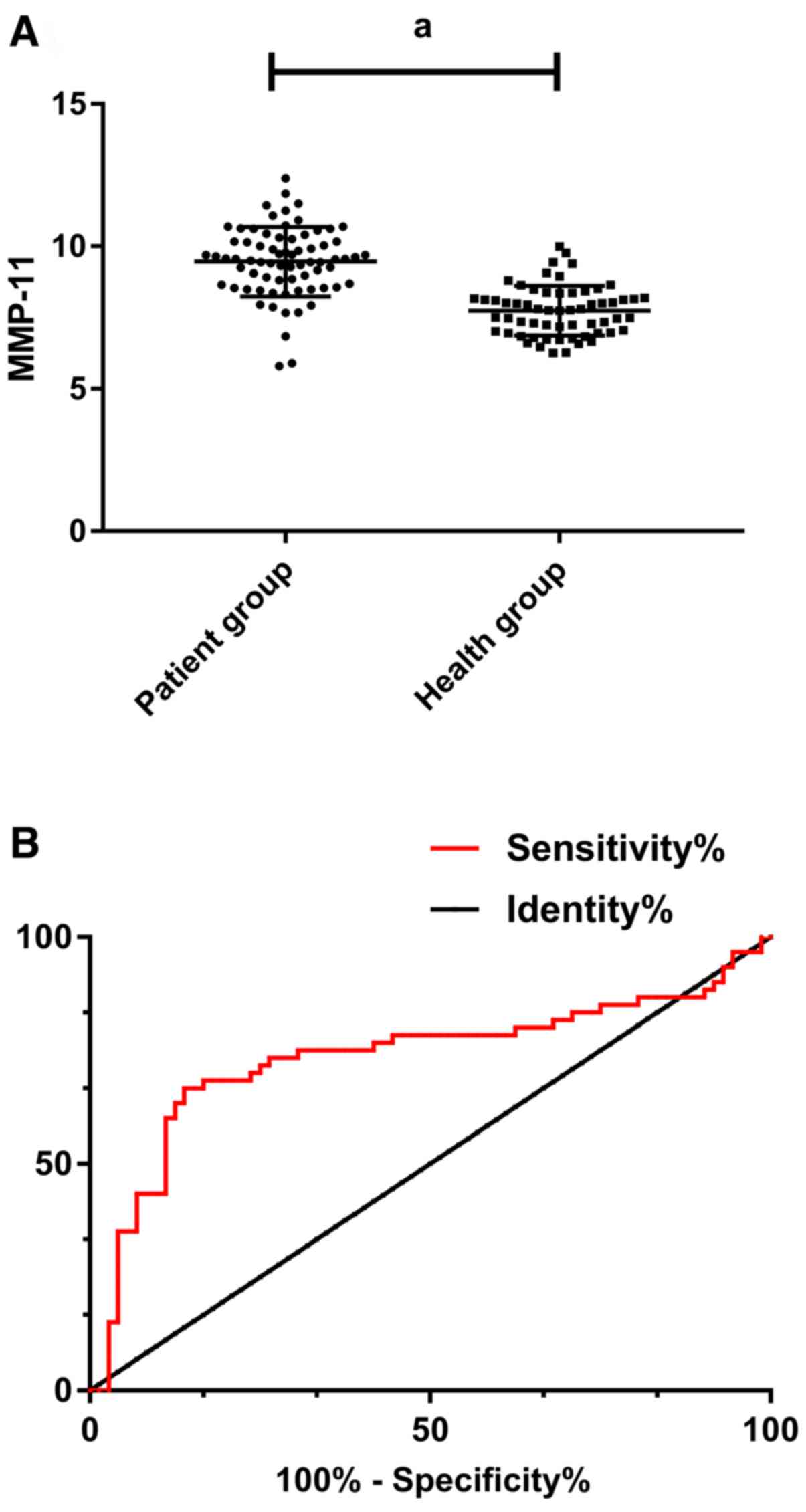
The expression level of MMP-11 was 9.36±1.25 in
breast cancer patients and 7.69±0.82 in serum of healthy subjects.
The expression level of MMP-11 in serum of breast cancer patients
was significantly higher than that of healthy subjects (P<0.05).
Furthermore, by plotting ROC, the AUC of MMP-11 was 0.735, the
sensitivity was 66.67%, and the specificity was 86.11%, P<0.05
(Table II and Fig. 1).
| Table II.ROC curve. |
Table II.
ROC curve.
|
| MMP-11 |
|---|
| AUC | 0.735 |
| Standard error | 0.048 |
| 95% CI | 0.641–0.828 |
| P-value | 0.001 |
| Cut-off | 8.440 |
| Sensitivity
[n(%)] | 66.67% |
| Specificity (%) | 86.11% |
Diagnostic results
Among the 72 patients, there were 41 patients
diagnosed with breast cancer by serum MMP-11 examination, 38
patients diagnosed by ultrasound examination, 33 patients diagnosed
by combined diagnosis, and 30 patients diagnosed by pathology
biopsy. Among the other 42 patients, there were 23 patients
diagnosed with adenosis of breast, 10 patients diagnosed with
galactocele, 4 patients diagnosed with tuberculosis of breast, and
3 patients diagnosed with mammary duct ectasia.
Imaging characteristics
After the ultrasound diagnosis, all the data of the
examination were read by 3 experienced clinicians in the hospital
separately and the diagnosis results were jointly given after
reaching an agreement. The main manifestations of ultrasound
examination in breast cancer patients were: i) Unclear boundary and
irregular shape, mostly showing burr shape and crab-like shape. ii)
Increased anteroposterior diameter of the lesion, and the
thickness/length was >1. iii) Posterior echo attenuation,
multiple medullary cells, loose tissue structure, and no echo
enhancement in the lesion posterior area. iv) Visible microscopic
calcification within the lesion, with a size of 100–500 µm and
acoustic shadowing in the rear. v) Abundant blood flow signals
within the lesion, common high speed and high resistance blood
flow. The main manifestations of mammography examination were: i)
Irregular boundary, high density nodules, uneven density. ii) Burr
shape edge, with visible small clusters of shape and gravel like
calcification.
Diagnostic efficacy assessment
The pathology biopsy was taken as gold standard.
According to the calculation, the sensitivity, specificity and
diagnostic accordance rate of MMP-11 for breast cancer were 58.54,
80.65 and 68.06%, respectively. The sensitivity, specificity and
diagnostic accordance rate of ultrasound for breast cancer were
68.42, 88.24 and 77.78%, respectively. The sensitivity, specificity
and diagnostic accordance rate of combined examination were 87.88,
97.44 and 93.06%, respectively. The diagnostic efficacy of
ultrasound combined with mammography examination was significantly
better than the two single examinations (P<0.050) (Tables III–V).
| Table III.Results of MMP-11 in diagnosis of
breast cancer. |
Table III.
Results of MMP-11 in diagnosis of
breast cancer.
|
| Biopsy (+) | Biopsy (−) |
|
|---|
| MMP-11 (+) | 24 | 17 | 41 |
| MMP-11 (−) | 6 | 25 | 31 |
|
| 30 | 42 |
|
| Table V.Results of combined diagnosis of
breast cancer. |
Table V.
Results of combined diagnosis of
breast cancer.
|
| Biopsy (+) | Biopsy (−) |
|
|---|
| Combined diagnosis
(+) | 29 | 4 | 33 |
| Combined diagnosis
(−) | 1 | 38 | 39 |
|
| 30 | 42 |
|
Discussion
As the most common malignant tumor among women in
the world, the incidence and mortality of breast cancer are on the
rise year by year (12). In order to
improve the diagnosis and treatment rate of breast cancer, clinical
efforts are being made to explore the pathogenic mechanism of
breast cancer from various perspectives for diagnosis and
treatment, but no significant breakthrough has been made so far.
Poortmans et al (13)
considered that the pathogenesis of breast cancer was mainly due to
the role of genetic factors, while Tutt et al (14) showed that the pathogenesis of breast
cancer was closely related to tumor stem cells. At present, there
is no accurate and reliable study that can indicate the exact cause
of breast cancer, and the main clinical diagnosis is still imaging.
With the development of the disease, increasingly difficult types
of breast cancer cannot be effectively determined only through
imaging (15). Therefore, it has
become a very important research topic to summarize the risk
factors of breast cancer for influencing the examination of medical
means. Traditional ultrasound and mammography examination is
simple, convenient, non-invasive, and has a long history of
diagnosis, which is the advantage of breast cancer diagnosis.
However, there are controversies about the determination of some
benign tumors and cystic hyperplasia of the breast.
MMP-11 belongs to matrix metalloproteins (16), and is closely related to many
diseases such as atherosclerosis, rheumatoid arthritis and cancer
in process of embryo implantation, organogenesis, tissue
degeneration and repair (17–20).
MMP-11 typically acts during tissue remodeling that occurs at the
epithelial/connective tissue interface to regulate epithelial
homeostasis. In cancer, high MMP-11 level in primary tumors are
associated with poor prognosis (21–23). In
this study, we explored the diagnostic value and accuracy of
Doppler ultrasound parameters combined with MMP-11 in early breast
cancer and benign breast diseases, so as to provide information for
future clinical practice.
In this study, we first observed the expression
level of MMP-11 in serum of breast cancer patients, and found that
the expression level of MMP-11 in serum of breast cancer patients
was significantly higher than that of healthy subjects, indicating
that MMP-11 might become a potential target for diagnosis and
treatment of breast cancer. ROC curve was drawn and it was found
that the AUC of MMP-11 curve was 0.735, the sensitivity was 66.67%
and specificity was 86.11%, which was not high in sensitivity but
high in specificity. This suggests that MMP-11 has certain
predictive value for breast cancer patients. Biomarkers are used as
tools in cancer diagnosis and treatment stratification. According
to Hadler-Olsen et al (24),
levels of one or more MMP members were elevated in most cancers.
This family of proteolytic enzymes is involved in many stages of
cancer development, including angiogenesis, invasion, and
metastasis. Therefore, it is expected that MMP can be used as a
diagnostic and prognostic marker in cancer patients. Benson et
al (25) showed that MMPs are
differentially regulated in breast cancer tissues, and they may
play different roles in tumor invasion, metastasis and
angiogenesis. Therefore, MMPs are of great research value as a
diagnostic marker and drug target. The application of ultrasound
and MMP-11 single examination has good specificity for breast
cancer, but the combined diagnosis of the two is better for the
diagnosis of breast cancer. It suggests that the early diagnostic
rate of breast cancer can be improved by the combined examination
of ultrasonography and mammography. Ultrasound is superior to
MMP-11 in the diagnosis of tumor classification, tumor grade, tumor
density, breast cystic hyperplasia and benign tumors, which is not
available in MMP-11 detection. However, MMP-11 can play different
roles in tumor invasion, metastasis and angiogenesis, and can be
used as a diagnostic and prognostic marker and drug target. By
using the two methods together, they can make up for each other's
shortcomings and achieve the best diagnostic effect.
In conclusion, MMP-11 exhibits high expression in
breast cancer patients. The ROC curve shows that MMP-11 has a good
clinical diagnostic value and the use of ultrasound combined with
MMP-11 examination can improve the early diagnostic rate of breast
cancer. Moreover, different diagnostic methods combined with the
clinical manifestations of patients can improve the diagnostic
accuracy, which is worthy of providing reference and advice for
future clinical practice.
Acknowledgements
Not applicable.
Funding
No funding was received.
Availability of data and materials
The datasets used and/or analyzed during the current
study are available from the corresponding author on reasonable
request.
Authors' contributions
HR conceived the study and drafted the manuscript.
ZS and YuZ acquired the data. JS and YunhuaZ analyzed the data and
revised the manuscript. All authors read and approved the final
manuscript.
Ethics approval and consent to
participate
The study was approved by the Ethics Committee of
Liaocheng Third People's Hospital (Liaocheng, China). Patients who
participated in this research had complete clinical data. Signed
informed consents were obtained from the patients and/or the
guardians.
Patient consent for publication
Not applicable.
Competing interests
The authors declare that they have no competing
interests.
References
|
1
|
DeSantis CE, Fedewa SA, Goding Sauer A,
Kramer JL, Smith RA and Jemal A: Breast cancer statistics, 2015:
Convergence of incidence rates between black and white women. CA
Cancer J Clin. 66:31–42. 2016. View Article : Google Scholar : PubMed/NCBI
|
|
2
|
Finn RS, Crown JP, Lang I, Boer K,
Bondarenko IM, Kulyk SO, Ettl J, Patel R, Pinter T, Schmidt M, et
al: The cyclin-dependent kinase 4/6 inhibitor palbociclib in
combination with letrozole versus letrozole alone as first-line
treatment of oestrogen receptor-positive, HER2-negative, advanced
breast cancer (PALOMA-1/TRIO-18): A randomised phase 2 study.
Lancet Oncol. 16:25–35. 2015. View Article : Google Scholar : PubMed/NCBI
|
|
3
|
Coughlin SS and Ekwueme DU: Breast cancer
as a global health concern. Cancer Epidemiol. 33:315–318. 2009.
View Article : Google Scholar : PubMed/NCBI
|
|
4
|
Swain SM, Baselga J, Kim SB, Ro J,
Semiglazov V, Campone M, Ciruelos E, Ferrero JM, Schneeweiss A,
Heeson S, et al CLEOPATRA Study Group, : Pertuzumab, trastuzumab,
and docetaxel in HER2-positive metastatic breast cancer. N Engl J
Med. 372:724–734. 2015. View Article : Google Scholar : PubMed/NCBI
|
|
5
|
Turner NC, Ro J, André F, Loi S, Verma S,
Iwata H, Harbeck N, Loibl S, Huang Bartlett C, Zhang K, et al
PALOMA3 Study Group, : Palbociclib in hormone-receptor-positive
advanced breast cancer. N Engl J Med. 373:209–219. 2015. View Article : Google Scholar : PubMed/NCBI
|
|
6
|
Yu QC, Verheyen EM and Zeng YA: Mammary
development and breast cancer: A Wnt perspective. Cancers (Basel).
8:652016. View Article : Google Scholar
|
|
7
|
Collignon J, Lousberg L, Schroeder H and
Jerusalem G: Triple-negative breast cancer: Treatment challenges
and solutions. Breast Cancer (Dove Med Press). 8:93–107.
2016.PubMed/NCBI
|
|
8
|
Guo R, Lu G, Qin B and Fei B: Ultrasound
imaging technologies for breast cancer detection and management: A
Review. Ultrasound Med Biol. 44:37–70. 2018. View Article : Google Scholar : PubMed/NCBI
|
|
9
|
Cheng CW, Yu JC, Wang HW, Huang CS, Shieh
JC, Fu YP, Chang CW, Wu PE and Shen CY: The clinical implications
of MMP-11 and CK-20 expression in human breast cancer. Clin Chim
Acta. 411:234–241. 2010. View Article : Google Scholar : PubMed/NCBI
|
|
10
|
Clark A and Fallowfield LP: Breast cancer.
CRC Press. 2014.
|
|
11
|
Tadayyon H, Sadeghi-Naini A, Wirtzfeld L,
Wright FC and Czarnota G: Quantitative ultrasound characterization
of locally advanced breast cancer by estimation of its scatterer
properties. Med Phys. 41:0129032014. View Article : Google Scholar : PubMed/NCBI
|
|
12
|
Jung KW, Won YJ, Oh CM, Kong HJ, Lee DH
and Lee KH; Community of population-based Regional cancer
registries, : Cancer statistics in Korea: Incidence, mortality,
survival, and prevalence in 2014. Cancer Res Treat. 49:292–305.
2017. View Article : Google Scholar : PubMed/NCBI
|
|
13
|
Poortmans PM, Collette S, Kirkove C, Van
Limbergen E, Budach V, Struikmans H, Collette L, Fourquet A,
Maingon P, Valli M, et al EORTC Radiation Oncology, ; Breast Cancer
Groups: Internal mammary and medial supraclavicular irradiation in
breast cancer. N Engl J Med. 373:317–327. 2015. View Article : Google Scholar : PubMed/NCBI
|
|
14
|
Tutt A, Ellis P, Kilburn L, Gilett C,
Pinder S and Abraham J: Abstract S3-01: the TNT trial: A randomized
phase III trial of carboplatin (C) compared with docetaxel (D) for
patients with metastatic or recurrent locally advanced triple
negative or BRCA1/2 breast cancer (CRUK/07/012). Cancer Res. 75
(Suppl 9):S3–S4. 2015.
|
|
15
|
Basset P, Bellocq JP, Lefebvre O, Noël A,
Chenard MP, Wolf C, Anglard P and Rio MC: Stromelysin-3: A paradigm
for stroma-derived factors implicated in carcinoma progression.
Crit Rev Oncol Hematol. 26:43–53. 1997. View Article : Google Scholar : PubMed/NCBI
|
|
16
|
Bianchini G, Balko JM, Mayer IA, Sanders
ME and Gianni L: Triple-negative breast cancer: Challenges and
opportunities of a heterogeneous disease. Nat Rev Clin Oncol.
13:674–690. 2016. View Article : Google Scholar : PubMed/NCBI
|
|
17
|
Basset P, Bellocq JP, Wolf C, Stoll I,
Hutin P, Limacher JM, Podhajcer OL, Chenard MP, Rio MC and Chambon
P: A novel metalloproteinase gene specifically expressed in stromal
cells of breast carcinomas. Nature. 348:699–704. 1990. View Article : Google Scholar : PubMed/NCBI
|
|
18
|
Okada A, Saez S, Misumi Y and Basset P:
Rat stromelysin 3: cDNA cloning from healing skin wound, activation
by furin and expression in rat tissues. Gene. 185:187–193. 1997.
View Article : Google Scholar : PubMed/NCBI
|
|
19
|
Tan J, Buache E, Alpy F, Daguenet E,
Tomasetto CL, Ren GS and Rio MC: Stromal matrix
metalloproteinase-11 is involved in the mammary gland postnatal
development. Oncogene. 33:4050–4059. 2014. View Article : Google Scholar : PubMed/NCBI
|
|
20
|
Schönbeck U, Mach F, Sukhova GK, Atkinson
E, Levesque E, Herman M, Graber P, Basset P and Libby P: Expression
of stromelysin-3 in atherosclerotic lesions: Regulation via
CD40-CD40 ligand signaling in vitro and in vivo. J Exp Med.
189:843–853. 1999. View Article : Google Scholar : PubMed/NCBI
|
|
21
|
Andarawewa KL, Motrescu ER, Chenard MP,
Gansmuller A, Stoll I, Tomasetto C and Rio MC: Stromelysin-3 is a
potent negative regulator of adipogenesis participating to cancer
cell-adipocyte interaction/crosstalk at the tumor invasive front.
Cancer Res. 65:10862–10871. 2005. View Article : Google Scholar : PubMed/NCBI
|
|
22
|
Motrescu ER and Rio MC: Cancer cells,
adipocytes and matrix metalloproteinase 11: A vicious tumor
progression cycle. Biol Chem. 389:1037–1041. 2008. View Article : Google Scholar : PubMed/NCBI
|
|
23
|
Tan J, Buache E, Chenard MP, Dali-Youcef N
and Rio MC: Adipocyte is a non-trivial, dynamic partner of breast
cancer cells. Int J Dev Biol. 55:851–859. 2011. View Article : Google Scholar : PubMed/NCBI
|
|
24
|
Hadler-Olsen E, Winberg JO and
Uhlin-Hansen L: Matrix metalloproteinases in cancer: Their value as
diagnostic and prognostic markers and therapeutic targets. Tumour
Biol. 34:2041–2051. 2013. View Article : Google Scholar : PubMed/NCBI
|
|
25
|
Benson CS, Babu SD, Radhakrishna S,
Selvamurugan N and Ravi Sankar B: Expression of matrix
metalloproteinases in human breast cancer tissues. Dis Markers.
34:395–405. 2013. View Article : Google Scholar : PubMed/NCBI
|