Introduction
Ovarian sex cord-stromal tumors (OSCSTs) are rare
mixed ovarian tumors. OSCSTs are composed of stromal elements of
gonads, sex cord-like cells or a plurality of luteinized cells in
different differentiation stages in a single or mixed manner
(1,2). However, due to the non-significant
specificity of its histological features, clinicopathological
manifestations, physical examination and imaging features, it is
difficult to differentiate OSCSTs from similar gynecologic tumors.
Therefore, clinical diagnosis and differentiation are difficult,
especially when judging the benign and malignant nature of tumors,
and patients could miss the best treatment time (3,4).
Clinically, ovarian tumors are usually diagnosed by
histopathological examination and surgery, and treated by surgical
resection (5,6). However, due to the relatively few
clinical records and confirmed cases of OSCSTs, and insufficient
cognition, no definite diagnosis can be made before surgery
(7,8).
Imaging examination plays an important role in the
quantitative, and qualitative detection and diagnosis of ovarian
tumors, of which ultrasound has the advantages of less radiation
damage, simple operation and high resolution to soft tissue
(9,10). The existing two-dimensional
ultrasound technology is relatively mature with widespread clinical
application. It is the main method to examine ovarian tumors. The
diagnostic accuracy has been improved in recent years through the
use of high-frequency probes. However, it still has shortcomings,
for example, poor diagnostic efficiency, obvious misdiagnosis rate,
and low accuracy in judging pathological types or disease
development degree, leading to an increase in the possibility of
misdiagnosis and missed diagnosis (11,12).
Two-dimensional ultrasound is a section imaging technology, and
color Doppler ultrasound provides real-time stereoscopic imaging on
this basis. Color Doppler ultrasound has the advantages of short
distance between probe and ovary, high resolution between blood
vessel and ovary. When monitoring blood flow, it can make judgment
according to the difference of blood flow distribution in ovarian
masses (13,14). It has been reported in literature
(15) that color Doppler ultrasound
is effective in detecting the success rate of blood flow signals of
ovarian malignant tumors, providing more valuable information for
the diagnosis and differentiation of lesions. This study compared
the diagnostic value of two-dimensional ultrasound and color
Doppler ultrasound for OSCSTs.
Patients and methods
General materials
A total of 91 patients with positive OSCSTs admitted
to Sichuan Provincial Hospital for Women and Children (Chengdu,
China) from May 2014 to June 2018 were selected as research
objects, of whom 48 patients diagnosed by color Doppler ultrasound
technology were the color Doppler group and 43 patients diagnosed
by two-dimensional ultrasound technology were the two-dimensional
ultrasound group. All the subjects were aged between 18 and 78
years, with an average age of 41.2±11.7 years and BMI of 21.1±3.3
kg/m2. Among them, there were 29 cases of granulosa cell
tumors, 5 cases of thecoma, 20 cases of mixed tumors, 13 cases of
supportive stromal cell tumors, 16 cases of sclerosing stromal
tumors and 8 cases of fibrosarcoma. There were no significant
differences in age, BMI, abdominal pain and ascites of patients
between the two groups (P>0.05) (Table I).
 | Table I.General data of patients. |
Table I.
General data of patients.
| Factors | Color Doppler
ultrasound group n=48 | Two-dimensional
ultrasound group n=43 | t/χ2
value | P-value |
|---|
| Age (years) |
|
| 0.002 | 0.967 |
| ≤41 | 27 (56.25) | 24 (55.81) |
|
|
|
>41 | 21 (43.75) | 19 (44.19) |
|
|
| BMI
(kg/m2) |
|
| 0.008 | 0.930 |
| ≤21 | 23 (47.92) | 21 (48.84) |
|
|
|
<21 | 25 (52.08) | 22 (51.16) |
|
|
| Irregular vaginal
bleeding |
|
| 0.402 | 0.526 |
| Yes | 21 (43.75) | 16 (37.21) |
|
|
| No | 27 (56.25) | 27 (62.79) |
|
|
| Abdominal pain |
|
| 2.227 | 0.996 |
| Yes | 29 (60.42) | 26 (60.47) |
|
|
| No | 19 (39.58) | 17 (39.53) |
|
|
| Ascites |
|
| 0.096 | 0.757 |
| Yes | 9
(18.75) | 7
(16.28) |
|
|
| No | 39 (81.25) | 36 (83.72) |
|
|
| Typing |
|
| 0.488 | 0.993 |
| Granulosa
cell tumors | 15 (31.25) | 14 (32.56) |
|
|
|
Thecoma | 2 (4.17) | 3 (6.98) |
|
|
| Mixed
tumors | 11 (22.92) | 9
(20.93) |
|
|
|
Supportive stromal cell
tumors | 7
(14.58) | 6
(13.95) |
|
|
|
Sclerosing stromal tumors | 9
(18.75) | 7
(16.28) |
|
|
|
Fibrosarcoma | 4 (8.33) | 4 (9.30) |
|
|
| C125 (U/ml) | 95.62±14.52 | 98.63±15.45 | 0.958 | 0.341 |
| C199 (U/ml) | 97.46±15.73 | 98.72±15.82 | 0.381 | 0.705 |
| AFP (µg/l) | 75.63±9.62 | 76.36±9.93 | 0.356 | 0.723 |
Inclusion and exclusion criteria
Inclusion criteria: Patients diagnosed as OSCSTs by
pathological diagnosis. Exclusion criteria: Patients previously
treated for OSCST diseases; women with other malignant tumors;
patients who were breast-feeding or pregnant; patients with
cognitive impairment or communication impairment; patients with
poor compliance.
All patients and their families agreed to
participate in the experiment and signed an informed consent. The
study was approved by the Medical Ethics Committee of Sichuan
Provincial Hospital for Women and Children.
Experimental reagents and
materials
The color Doppler diagnostic apparatus was purchased
from Siemens (S2000) and LOGIQ (E9).
Ultrasound experimental methods
During the examination, the probe frequency is 4-8
MHz in abdomen and 4-9 MHz in vagina. Stone amputation was
performed when the bladder was empty. Internal echo and boundary of
the two groups were observed and recorded by travaginal
two-dimensional ultrasound, and color Doppler ultrasound was used
to examine the section of the highest possibility at highest
definition. The blood flow condition inside and around the tumor
was observed and recorded, the location and size of ovarian tumor
combined with abdominal two-dimensional ultrasound images were
observed, the relationship between morphology and viscera and
organs was summarized, and ascites was detected.
Detection method
Venous blood (3 ml) was taken from fasting patients
in the morning as examination specimen. After centrifugation at
1,500 × g for 5 min at normal temperature, the serum was taken and
stored at −20°C. Electrochemiluminescence was used to detect serum
CA125 and CA199. The steps were as follows: the serum sample was
pre-treated with reducing reagents; CA125 and CA199 were placed at
constant temperature for antigen-antibody complex;
ruthenium-labeled and biotin-labeled antibodies were added, and
after binding, magnetic beads were added to generate a solid phase
complex. The magnetic beads are adsorbed and cleaned by the
electrode. After the light-emitting reaction occurs, the sample to
be tested and the light signal were examined. The weaker the light
signal, the larger the amount of the sample to be tested. The
normal value of serum CA125 is 0-35 U/ml, and it is positive when
>35 U/ml. The normal value of serum CA199 was 0-37 U/ml, and it
is positive when >37 U/ml. Any positive result of the index will
be judged as positive.
Observation indicators
Color Doppler ultrasound and two-dimensional
ultrasound were used to measure size, form, boundary and location
of tumors. The results of two ultrasound images for patients with
OSCSTs were compared. The sensitivity, specificity and diagnostic
accuracy of the two kinds of ultrasound in the detection of
pathological signs of OSCSTs were compared.
Statistical methods
In this experiment, SPSS 19.0 statistical (Beijing
ND Times Science and Technology Co., Ltd.) was used to make
statistical analysis on the experimental data. Chi-square test was
used for counting data, measuring data were expressed by mean ±
standard deviation and t-test was used for comparison between the
two groups. Graphpad Prism8 was used to present the illustrations.
P<0.05 was considered to indicate a statistically significant
difference.
Results
Comparison of size, form, boundary and
location of patients with positive OSCSTs under color Doppler
ultrasound and two-dimensional ultrasound
The size, form, boundary and location detected by
color Doppler ultrasound in patients with OSCSTs were not
significantly different from those detected by two-dimensional
ultrasound (P>0.05), more details are shown in Table II.
| Table II.Comparison of size, form, boundary and
location of tumors under color Doppler ultrasound and
two-dimensional ultrasound. |
Table II.
Comparison of size, form, boundary and
location of tumors under color Doppler ultrasound and
two-dimensional ultrasound.
| Features of
tumors | Color Doppler
ultrasound group n=48 | Two-dimensional
ultrasound group n=43 | t/χ2
value | P-value |
|---|
| Tumor size | 7.34±4.53 | 7.51±4.46 | 0.180 | 0.858 |
| Form |
|
| 0.112 | 0.738 |
|
Irregular | 3 (6.25) | 2 (4.65) |
|
|
|
Regular | 45 (93.75) | 41 (95.35) |
|
|
| Boundary |
|
| 0.267 | 0.605 |
|
Clear | 43 (89.58) | 37 (86.05) |
|
|
| Not
clear | 5
(10.42) | 6
(13.95) |
|
|
| Location |
|
| 0.024 | 0.876 |
|
Unilateral | 32 (66.67) | 28 (65.12) |
|
|
|
Multilaterality | 16 (33.33) | 15 (34.88) |
|
|
Comparison of results of two kinds of
ultrasound images in patients with positive OSCSTs
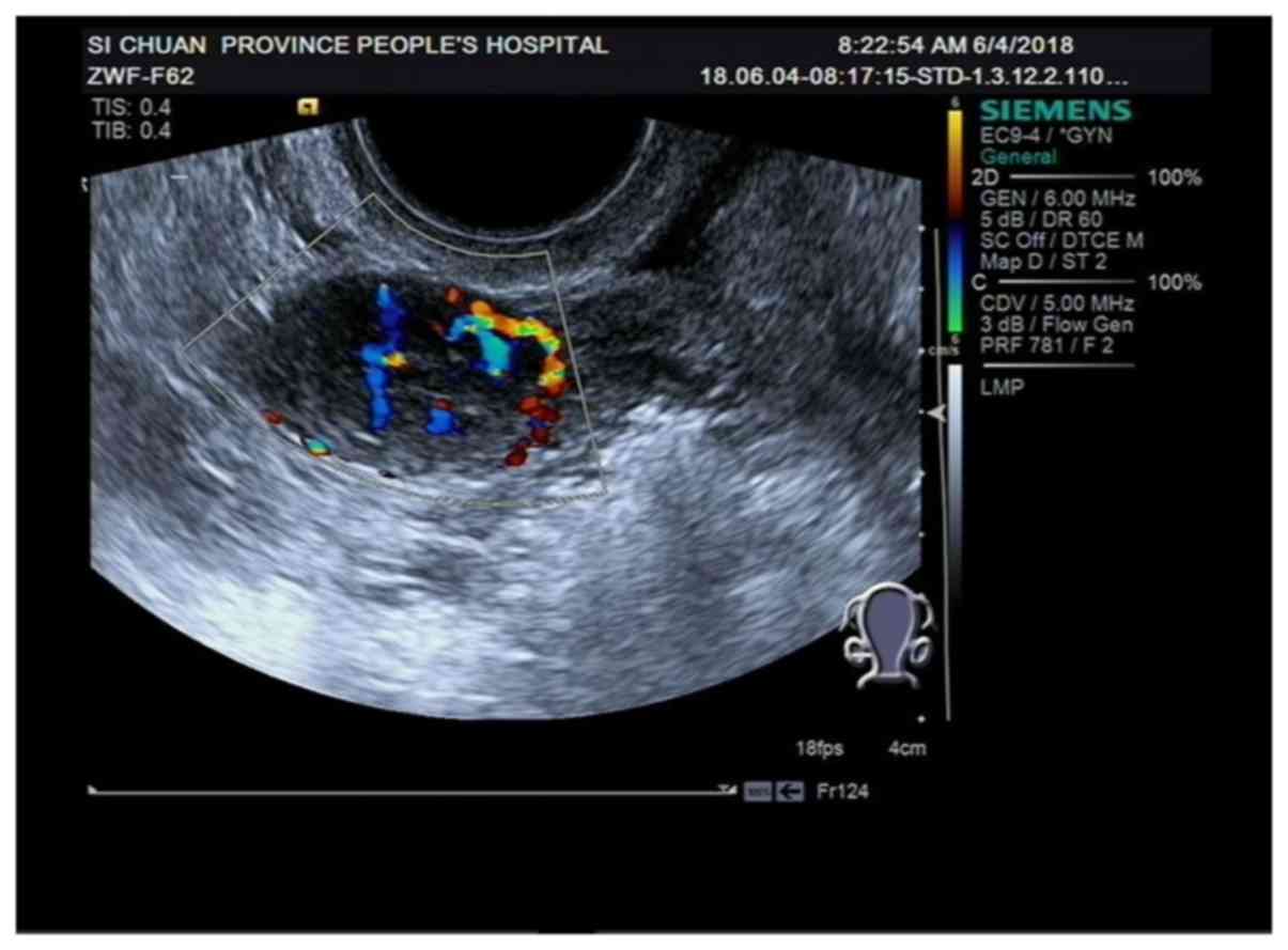
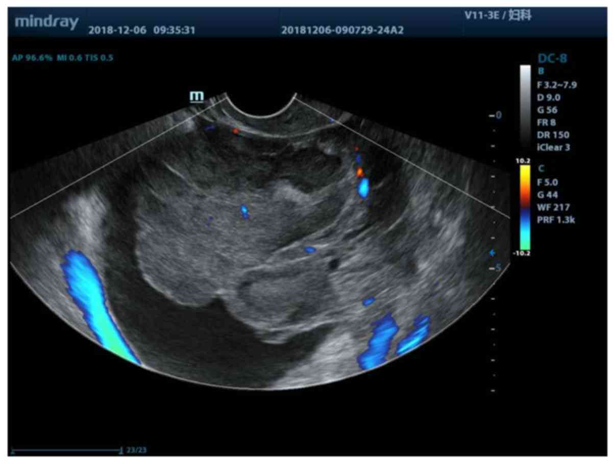
The real internal echo of color Doppler ultrasound
was significantly higher than that of two-dimensional ultrasound,
with significant statistical differences (P<0.05). The blood
flow signal of color Doppler ultrasound was significantly higher
than that of two-dimensional ultrasound, with significant
statistical differences (P<0.05). There was no statistically
significant difference in posterior echo and calcified lesion
(P>0.05) (Table III, Figs. 1 and 2).
| Table III.Comparison of results of two kinds of
ultrasound images in patients with positive ovarian sex
cord-stromal tumors. |
Table III.
Comparison of results of two kinds of
ultrasound images in patients with positive ovarian sex
cord-stromal tumors.
| Features of
imaging | Color Doppler
ultrasound group n=48 | Two-dimensional
ultrasound group n=43 | t/χ2
value | P-value |
|---|
| Internal echo |
|
| 4.135 | 0.042 |
| Solid
type | 31 (64.58) | 19 (44.19) |
|
|
|
Cystic-solid type | 17 (35.42) | 24 (55.81) |
|
|
| Posterior echo |
|
| 1.128 | 0.569 |
|
Uniformity | 10 (20.83) | 7
(16.28) |
|
|
|
Weak | 27 (56.25) | 22 (51.16) |
|
|
|
Enhanced | 11 (22.92) | 14 (32.56) |
|
|
| Blood flow
signal |
|
| 5.093 | 0.024 |
|
Obvious | 33 (68.75) | 38 (88.37) |
|
|
| Not
obvious | 15 (31.25) | 5
(11.63) |
|
|
| Calcified
lesions |
|
| 1.129 | 0.288 |
|
Yes | 16 (33.33) | 19 (44.19) |
|
|
| No | 32 (66.67) | 24 (55.81) |
|
|
Comparison of the sensitivity,
specificity and diagnostic accuracy of two kinds of ultrasound in
detecting OSCST metastasis
The diagnostic sensitivity and the coincidence rate
of color Doppler ultrasound for lymph node metastasis of OSCSTs
were significantly higher than those of two-dimensional ultrasound
(P<0.05). Differences were statistically significant (P<0.05)
(Tables IV and V).
| Table IV.Diagnostic results of color Doppler
ultrasound and two-dimensional ultrasound combined with CA125 and
CA199 detection for ovarian sex cord-stromal tumors. |
Table IV.
Diagnostic results of color Doppler
ultrasound and two-dimensional ultrasound combined with CA125 and
CA199 detection for ovarian sex cord-stromal tumors.
| Diagnostic
results | Pathology
(metastasis) | Pathology
(non-metastasis) | Total |
|---|
| Color Doppler
ultrasound |
|
Diagnosis (metastasis) | 36 | 3 | 39 |
|
Diagnosis
(non-metastasis) | 2 | 7 | 9 |
|
Total | 38 | 10 | 48 |
| Two-dimensional
ultrasound |
|
Diagnosis (metastasis) | 24 | 9 | 33 |
|
Diagnosis
(non-metastasis) | 4 | 6 | 10 |
|
Total | 28 | 15 | 43 |
| Color Doppler
ultrasound combined with CA125 and CA199 detection |
|
Diagnosis (metastasis) | 36 | 2 | 38 |
|
Diagnosis
(non-metastasis) | 2 | 8 | 10 |
|
Total | 38 | 10 | 48 |
| Two-dimensional
ultrasound combined with CA125 and CA199 detection |
|
Diagnosis (metastasis) | 24 | 6 | 30 |
|
Diagnosis
(non-metastasis) | 4 | 9 | 13 |
|
Total | 28 | 15 | 43 |
| Table V.Diagnostic efficacy analysis of
individual indicators and joint detection for ovarian sex
cord-stromal tumors. |
Table V.
Diagnostic efficacy analysis of
individual indicators and joint detection for ovarian sex
cord-stromal tumors.
| Diagnostic
value | Color Doppler
ultrasound | Two-dimensional
ultrasound | Color Doppler
ultrasound + CA125 + CA199 | Two-dimensional
ultrasound + CA125 + CA199 |
|---|
| Sensitivity | 92.31% (36/39) | 65.12%
(24/33)a | 94.74% (36/38) | 80.00%
(24/30)b |
| Specificity | 77.78% (7/9) | 60.00% (6/10) | 80.00% (8/10) | 69.23%
(9/13)b |
| Diagnostic
coincidence rate | 89.58% (43/48) | 69.77%
(30/43)a | 91.67% (44/48) | 76.74%
(33/43)b |
Discussion
OSCSTs can occur in women of any age group, and its
manifestations of abdominal pain and abdominal distension become
more and more obvious as the tumor grows larger, and even cause
pedicle torsion and rupture in severe cases (16). Most of the OSCSTs can release steroid
hormones and frequently cause corresponding clinical symptoms, such
as menstrual disorder and hemorrhage of women caused by the
increase of estrogen level, and masculinization characteristics of
a few patients caused by the increase of androgen expression
(17,18). Ultrasound is generally used to assist
the diagnosis of OSCSTs. Two-dimensional ultrasound provides early
diagnosis for patients suspected of malignant tumors by presenting
single-angle cross-sectional images of the human body. It has the
advantages of non-invasiveness, repeatability, economy and high
safety. At the same time, it is difficult to clearly express the
three-dimensional morphological structure of ovary, which affects
the accuracy of clinical differentiation of benign and malignant
ovarian tumors (19,20). Color Doppler ultrasound spatial
stereogram vascular parameters can be used to observe
neovascularization and blood cells in tumors, so as to judge the
development of the disease (21). At
present, there are few reports on the diagnosis of OSCSTs by color
Doppler ultrasound and two-dimensional ultrasound. Therefore, we
use pathological examination results as the gold standard to
explore the diagnostic value of ultrasound imaging in OSCSTs, so as
to provide a more accurate scheme for the diagnosis of OSCSTs
patients.
In our study, the application value of color Doppler
ultrasound and two-dimensional ultrasound in the diagnosis of
OSCSTs was explored. First, we investigated the differences in
size, form, boundary and location of tumors between color Doppler
ultrasound and two-dimensional ultrasound. Our results showed that
there were no significant differences in size, form, boundary and
location of tumors between patients with positive OSCSTs and those
of under two-dimensional ultrasound (P>0.05). Then we analyzed
the diagnostic efficiency of color Doppler ultrasound and
two-dimensional ultrasound for OSCSTs. The results showed that the
diagnostic sensitivity and diagnostic coincidence rate of color
Doppler ultrasound for lymph node metastasis of OSCSTs were
significantly higher than those of two-dimensional ultrasound for
lymph node metastasis of OSCSTs. The accuracy of color Doppler
ultrasound combined with CA125 and CA199 detection was higher than
that of two-dimensional ultrasound (P<0.05). Previous studies
(22–24) have suggested that the sensitivity and
diagnostic specificity of color Doppler ultrasound for ovarian
malignant tumors are higher than those of two-dimensional
ultrasound. Color Doppler ultrasound is more accurate than
two-dimensional ultrasound in displaying abnormal spots of lesions
and color blood signals. This proves our conclusion to some extent.
Then we studied the relationship between features of OSCSTs imaging
and pathological examination results of two kinds of ultrasound
examinations. The results showed that the real internal echo of
color Doppler ultrasound was significantly higher than that of
two-dimensional ultrasound, with significant statistical difference
(P<0.05). The blood flow signal of color Doppler ultrasound was
significantly higher than that of two-dimensional ultrasound, with
significant statistical difference (P<0.05). There is no
significant statistical significance in posterior echo and
calcified lesions (P>0.05), suggesting that the internal echo of
OSCSTs tumor is related to pathological type. It is often dominated
by solid hypoecho, and color Doppler has relatively high expression
of color blood flow signals. Previous studies have shown that
(25), ovarian sarcoma fibroma, as
an uncommon OSCST, has been detected by ultrasound as a solid tumor
with typical characteristics of well-defined hypoechoic masses.
However, there are few specific research documents on OSCSTs
ultrasound imaging. From this study, color Doppler ultrasound shows
more solid mass and blood flow signals than two-dimensional
ultrasound. There are no significant differences in general
clinical pathological data and tumor characteristics between
patients in the two groups. It is speculated that differences in
internal echo and blood flow signals give color Doppler ultrasound
higher diagnostic value for OSCSTs than two-dimensional ultrasound,
and the diagnostic reliability is high. From our research, it seems
that color Doppler ultrasound has more obvious diagnostic
sensitivity for internal echo and blood flow signals in features of
OSCST imaging.
In conclusion, this study shows that color Doppler
ultrasound has high diagnostic value for OSCSTs, and its diagnostic
results can be used as an important reference for diagnosis and
treatment of OSCSTs in clinical practice.
Acknowledgements
Not applicable.
Funding
This study was supported by the Science and
Technology Department of Sichuan Province (grant no.
2019YFS0439).
Availability of data and materials
The datasets used and/or analyzed during the present
study are available from the corresponding author on reasonable
request.
Authors' contributions
GH, JZ and WX conceived and designed the study. GH,
JZ, ZY, ZZ and YB were responsible for the collection, analysis and
interpretation of the data. GH drafted the manuscript. JZ and WX
revised the manuscript critically for important intellectual
content. All authors read and approved the final manuscript.
Ethics approval and consent to
participate
The study was approved by the Medical Ethics
Committee of Sichuan Provincial Hospital for Women and Children
(Chengdu, China). Signed informed consents were obtained from the
patients and/or the guardians.
Patient consent for publication
Not applicable.
Competing interests
The authors declare that they have no competing
interests.
References
|
1
|
Hanley KZ and Mosunjac MB: Practical
review of ovarian sex cord-stromal tumors. Surg Pathol Clin.
12:587–620. 2019. View Article : Google Scholar : PubMed/NCBI
|
|
2
|
Young RH: Ovarian sex cord-stromal tumours
and their mimics. Pathology. 50:5–15. 2018. View Article : Google Scholar : PubMed/NCBI
|
|
3
|
Abdullazade S, Kosemehmetoglu K, Adanir I,
Kutluay L and Usubutun A: Uterine tumors resembling ovarian sex
cord-stromal tumors: Synchronous uterine tumors resembling ovarian
sex cord-stromal tumors and ovarian sex cord tumor. Ann Diagn
Pathol. 14:432–437. 2010. View Article : Google Scholar : PubMed/NCBI
|
|
4
|
Haroon S, Zia A, Idrees R, Memon A, Fatima
S and Kayani N: Clinicopathological spectrum of ovarian sex
cord-stromal tumors; 20 years' retrospective study in a developing
country. J Ovarian Res. 6:872013. View Article : Google Scholar : PubMed/NCBI
|
|
5
|
Bairwa S, Satarkar RN, Kalhan S, Garg S,
Sangwaiya A and Singh P: Sclerosing stromal tumor: A rare ovarian
neoplasm. Iran J Pathol. 12:402–405. 2017.PubMed/NCBI
|
|
6
|
Färkkilä A, Haltia UM, Tapper J, McConechy
MK, Huntsman DG and Heikinheimo M: Pathogenesis and treatment of
adult-type granulosa cell tumor of the ovary. Ann Med. 49:435–447.
2017. View Article : Google Scholar : PubMed/NCBI
|
|
7
|
Stuart GC and Dawson LM: Update on
granulosa cell tumours of the ovary. Curr Opin Obstet Gynecol.
15:33–37. 2003. View Article : Google Scholar : PubMed/NCBI
|
|
8
|
Tamai K, Koyama T, Saga T, Kido A, Kataoka
M, Umeoka S, Fujii S and Togashi K: MR features of physiologic and
benign conditions of the ovary. Eur Radiol. 16:2700–2711. 2006.
View Article : Google Scholar : PubMed/NCBI
|
|
9
|
Kapadia SR, Ziada KM, L'Allier PL, Crowe
TD, Rincon G, Hobbs RE, Bott-Silverman C, Young JB, Nissen SE and
Tuzcu EM: Intravascular ultrasound imaging after cardiac
transplantation: Advantage of multi-vessel imaging. J Heart Lung
Transplant. 19:167–172. 2000. View Article : Google Scholar : PubMed/NCBI
|
|
10
|
Ng FC, Yam WL, Lim TYB, Teo JK, Ng KK and
Lim SK: Ultrasound-guided percutaneous nephrolithotomy: Advantages
and limitations. Investig Clin Urol. 58:346–352. 2017. View Article : Google Scholar : PubMed/NCBI
|
|
11
|
Dala-Krishna P: Ultrasound image
processing to render three-dimensional images from two-dimensional
images. Acoust Soc Am J. 131:42312004. View Article : Google Scholar : PubMed/NCBI
|
|
12
|
Franke S, Lieske H, Fischer A, Büttner L,
Czarske J, Räbiger D and Eckert S: Two-dimensional ultrasound
Doppler velocimeter for flow mapping of unsteady liquid metal
flows. Ultrasonics. 53:691–700. 2013. View Article : Google Scholar : PubMed/NCBI
|
|
13
|
Porter MB: Polycystic ovary syndrome: The
controversy of diagnosis by ultrasound. Semin Reprod Med.
26:241–251. 2008. View Article : Google Scholar : PubMed/NCBI
|
|
14
|
Chen D, Peng L and Yang P: Clinical value
of transvaginal color Doppler ultrasound in differential diagnosis
of benign and malignant ovarian tumor. Medical J Wuhan Univ.
35:893–895. 2014.(In Chinese). PubMed/NCBI
|
|
15
|
Shah D, Shah S, Parikh J, Bhatt CJ,
Vaishnav K and Bala DV: Doppler ultrasound: A good and reliable
predictor of ovarian malignancy. J Obstet Gynaecol India.
63:186–189. 2013. View Article : Google Scholar : PubMed/NCBI
|
|
16
|
Tinelli A, Pellegrino M, Malvasi A and
Lorusso V: Laparoscopical management ovarian early sex cord-stromal
tumors in postmenopausal women: A proposal method. Arch Gynecol
Obstet. 283 (Suppl 1):87–91. 2011. View Article : Google Scholar : PubMed/NCBI
|
|
17
|
Bufa A, Farkas N, Preisz Z, Poór V, Páger
C, Szukits S, Farkas B and Gőcze PM: Diagnostic relevance of
urinary steroid profiles on ovarian granulosa cell tumors: Two case
reports. J Med Case Rep. 11:1662017. View Article : Google Scholar : PubMed/NCBI
|
|
18
|
Cecchetto G, Ferrari A, Bernini G, Alaggio
R, Collini P, Virgone C, Terenziani M, Dall'igna P, Cozza R, Conte
M, et al: Sex cord stromal tumors of the ovary in children: A
clinicopathological report from the Italian TREP project. Pediatr
Blood Cancer. 56:1062–1067. 2011. View Article : Google Scholar : PubMed/NCBI
|
|
19
|
Holsbeke CV and Timmerman D: Sex
cord-stromal tumors: Clinical setting and ultrasound findings.
Ovarian Neoplasm Imaging. Springer Science + Business Media. (New
York). 2013. View Article : Google Scholar : PubMed/NCBI
|
|
20
|
Chen QN, Zhang Y and Tan JX: Diagnosis of
ovarian teratomas with two-dimensional ultrasound. Zhong Nan Da Xue
Xue Bao Yi Xue Ban. 29:319–321. 2004.(In Chinese). PubMed/NCBI
|
|
21
|
Saad AA: Vessel recognition in color
doppler ultrasound imaging. 2008. PubMed/NCBI
|
|
22
|
Zanetta G, Vergani P and Lissoni A: Color
Doppler ultrasound in the preoperative assessment of adnexal
masses. Acta Obstet Gynecol Scand. 73:637–641. 1994. View Article : Google Scholar : PubMed/NCBI
|
|
23
|
Cohen LS, Escobar PF, Scharm C, Glimco B
and Fishman DA: Three-dimensional power Doppler ultrasound improves
the diagnostic accuracy for ovarian cancer prediction. Gynecol
Oncol. 82:40–48. 2001. View Article : Google Scholar : PubMed/NCBI
|
|
24
|
Liu GA, Ye RZ and Xu M: Analysis of 50
cases of traumatic retinal detachment diagnosed by color Doppler
ultrasound and two-dimensional ultrasound. Int J Ophthalmol.
14:1263–1265. 2014.PubMed/NCBI
|
|
25
|
Chen H, Liu Y, Shen LF, Jiang MJ, Yang ZF
and Fang GP: Ovarian thecoma-fibroma groups: Clinical and
sonographic features with Res. Ovarian pathological comparison. J
Ovarian Res. 9:812016. View Article : Google Scholar : PubMed/NCBI
|