Introduction
In premenopausal women, fluctuations in circulating
estrogen and progesterone occur across the course of the menstrual
cycle. Breast tissue is highly responsive to ovarian hormones, and
the cellular and molecular changes that occur in the breast over
the course of the menstrual cycle affect breast development and
function (1,2). These hormones also affect the activity
of breast cancer cells, both directly through ligand-receptor
binding to hormone receptor positive cancer cells, and indirectly
through effects on cells within the cancer cell microenvironment
(3). An intriguing association
between the timing of surgery in relation to menstrual cycle phase
and breast cancer clinical outcomes was first proposed in the late
1980s (4,5). This concept provides a potential new
approach to improving survival outcomes for premenopausal women. If
the hormone milieu at a specific phase of the menstrual cycle
results in a more favourable outcome, then the timing of breast
cancer surgery to this phase might be a non-toxic and
cost-effective means of reducing morbidity and mortality for young
breast cancer patients. However, there is significant controversy
in the literature surrounding the impact of menstrual cycle phase
at the time of surgery on breast cancer outcomes.
Here, we review the current evidence for a
relationship between the menstrual cycle phase at the time of
surgery on breast cancer outcomes, and explore the biological
mechanisms that may contribute to a phase-specific prognosis.
Relevant articles were identified by searching the PubMed database
for clinical studies investigating the impact of menstrual cycle
stage at the time of breast cancer surgery on patient survival
outcomes, and also by reviewing the reference lists of relevant
articles. All studies with the full text available on The
University of Adelaide or SA Health Library databases were included
in the review.
Mouse studies supported by small retrospective
clinical studies suggested that changes in the characteristics of
the tumour and tumour microenvironment across the menstrual cycle
might influence the metastatic potential of tumour cells, and
affect clinical outcomes in premenopausal women. However, while
some studies suggest that surgery performed during the luteal phase
results in favourable outcomes in terms of metastatic incidence,
disease free survival, and overall survival (6–14), other
studies report the follicular phase is more favourable (5,15,16), and
other studies show no association (17–24).
We conclude that currently, there is insufficient
evidence to support a change in surgery scheduling for
premenopausal breast cancer patients. The lack of consistency in
studies is likely due to a number of differences in study design
and the small sample sizes used. There are variable approaches to
defining the menstrual cycle phase and hormone receptor status of
the tumour. Few studies controlled for prognostic factors such as
tumour size and stage, or addressed the impact of adjuvant
treatments such as chemotherapy and hormonal therapy. There are a
number of potential biological mechanisms that might affect
surgical outcomes (Fig. 1), but
currently no causal mechanisms have been demonstrated. To fully
address this lack of clear evidence, prospective, well-controlled
studies are required, supported by research on animal models that
link biological mechanisms with clinical findings.
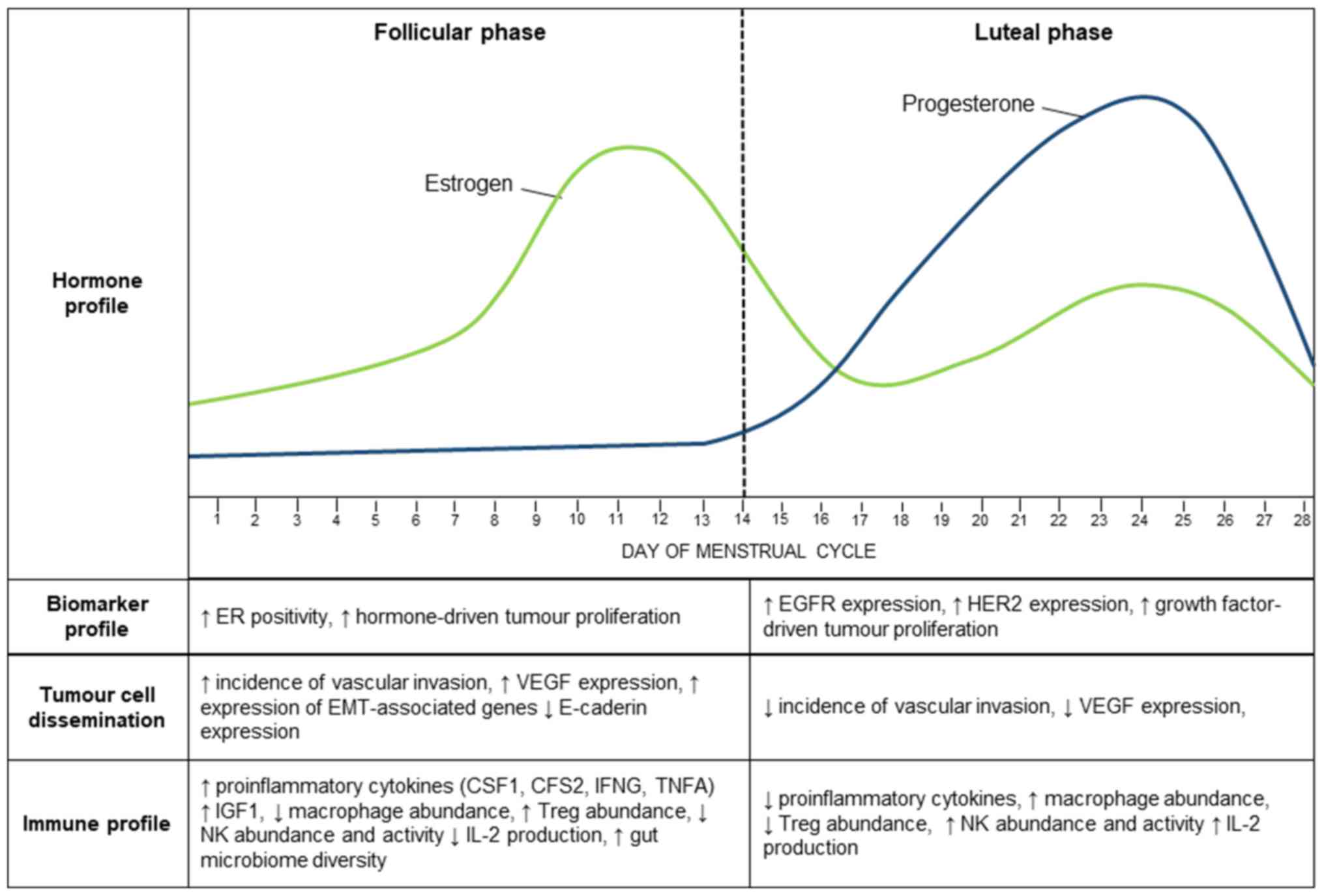 | Figure 1.Summary of the biological mechanisms
that could affect the metastatic ability of breast cancer cells and
contribute to a phase-specific prognosis. The effects of estrogen
(green) and progesterone (blue) on breast cancer biomarker
expression, tumour cell dissemination and immune function. ER,
estrogen receptor; EGFR, epidermal growth factor receptor; HER2,
human epidermal growth factor recpetor-2; VEGF, vascular
endothelial growth factor; EMT, epithelial-to-mesenchymal
transition; CSF, colony-stimulating factor; IFNG, interferon γ;
TNF-α, tumour necrosis factor α; IGF1, Insulin-like growth
factor-1. |
Impact of ovarian cycle phase at the time of
surgery on mammary cancer metastasis in rodent models
In 1988, Ratajczak et al (4) published a study showing a relationship
between the incidence of postoperative pulmonary metastasis, and
the rodent estrous cycle phase at which the mammary tumour was
removed. Using a hormone receptor-positive murine mammary
carcinoma, the authors showed that tumours resected from mice
around the time of ovulation (designated ‘near estrus’) showed
fewer incidences of pulmonary metastasis 4 weeks after surgery
compared to tumours resected at a time further away from the time
of ovulation (designated ‘post-estrus’). The study used the
cytology of vaginal smears to classify the phases of the estrous
cycle and did not assess circulating ovarian hormones in the mice.
However, this classification system would have resulted in the mice
exhibiting high circulating concentration of estrogen and low
progesterone at ‘near estrus’, and high circulating concentration
of progesterone and mid-range estrogen at ‘post-estrus’. The
authors demonstrated that the incidence of lung metastasis, as
assessed by gross morphology and bioassay, was significantly
reduced in ‘near estrus’ mice (44 of 60 mice; 73%) compared to
‘post-estrus’ mice (64 of 78 mice; 82%).
The authors proposed that the hormonal environment
at the time of surgery can influence the metastatic potential of a
cancer cell, through direct effects on the tumour, or indirect
effects on the cancer microenvironment or the host immune system.
Different hormonal environments may either facilitate or impede the
metastasis of breast cancer cells, and therefore explain the
observed differences in pulmonary metastasis with estrous cycle
phase.
However, a subsequent study by Ben-Eliyahu et
al (25) suggested that rats are
instead more susceptible to mammary carcinoma metastasis during the
proestrus phase of the estrous cycle. The authors investigated lung
metastasis in rats injected intravenously with hormone
receptor-negative cancer cells, and reported that metestrus and
diestrus stages of the cycle, which are characterised by high
circulating concentrations of progesterone and mid-range estrogen,
were protective against metastasis. Similarly, the authors
demonstrated that treatment with estrogen increased the metastatic
burden in the lung, an effect which was attenuated by progesterone
treatment (25).
The current evidence in animal models supports the
possibility that estrous cycle stage influences the risk of tumour
metastasis. However, given the conflicting results, it remains
unclear which stage of the estrous cycle may provide a more
favourable prognosis, and there is no clear understanding of the
underlying biological mechanisms which may contribute to these
phase-specific differences in outcomes.
Clinical evidence of an impact of menstrual
cycle phase at time of surgery on breast cancer metastasis
In 1989, Hrushesky et al (5) published the first retrospective review
in premenopausal women, investigating the effects of the timing of
breast cancer surgery on disease recurrence and metastasis. The
review included 44 premenopausal women, with both hormone
receptor-positive and -negative disease. The authors found that
patient outcomes varied significantly depending on the day of the
menstrual cycle that surgery was performed. In agreement with their
earlier mouse study, the authors found that women operated on close
to the time of menstruation showed poorer disease free and overall
survival outcomes, and a greater incidence of metastasis, compared
to women operated on during other phases of the cycle. This
suggests that premenopausal women might have an increased risk of
metastasis and poorer survival outcomes if surgery is performed
during the perimenstrual phase of their menstrual cycle.
However, later studies have found conflicting
results (26,27), and there is significant controversy
in the literature surrounding the effects of the menstrual cycle
stage at the time of surgery on the survival outcomes of
premenopausal breast cancers. In agreement with animal studies
published by Ben-Eliyahu et al (25), several studies in premenopausal women
have reported favourable outcomes for women when surgery is
performed during the perimenstrual phase of their menstrual cycle
(26,27). These findings are in direct
disagreement with those reported by Ratajczak et al
(4) and point to the complexities of
experimental design in affecting results.
A meta-analysis of 37 published studies (n=10,476)
suggested favourable prognosis when surgery was performed during
the luteal phase (28). Similarly, a
meta-analysis of 5353 premenopausal women demonstrated an overall
survival benefit for women operated on during the luteal phase of
the menstrual cycle (29).
Conversely, two meta-analyses of 19 published studies (30,31),
found no significant relationship between menstrual cycle stage and
patient prognosis.
The discrepancies between meta-analyses are likely
associated with differences in their methodology. Different
meta-analyses had different defining criteria for study inclusion;
restricting their analysis to studies based on only one specific
type of menstrual cycle stage classification, using one combined
prognostic outcome, or limiting analysis to cohorts of women
residing solely in Italy (30) or
the United States (29). The four
systematic reviews to date (31–34),
which examined the relationship between the menstrual cycle stage
at the time of surgery and patient outcomes, reported that there is
insufficient evidence to determine if one phase of the menstrual
cycle provides a more favourable outcome.
Confounding factors that could affect the
relationship between timing of surgery and prognosis
Despite the large number of existing studies, there
remains significant controversy in the literature surrounding how
the menstrual cycle stage at time of surgery affects breast cancer
outcomes. Disagreement between published studies could be due to a
number of confounding factors including how menstrual cycle stage
was classified for the study, variability in circulating hormone
profiles between women, tumour stage at the time of surgery, and
how psychological stress can affect ovarian hormone secretion and
menstrual cycling.
Differences in classification methods can introduce
significant variability into results, and may provide some
explanation for the differences in results between different
studies (5–24,26,27)
(Table I). Other factors include
inaccuracies in menstrual cycle data, as there can be significant
variability in cycle length (i.e., 22–36 days) between women
(35); and other factors, such as
irregular menses, use of oral contraceptives, recent pregnancies,
or differing hormonal and chemotherapy treatment regimens may
impact circulating ovarian hormones and menstrual cycle phase.
McGuire et al suggested that by changing the cut-off days
used to classify the menstrual cycle phase, a significant number of
patients can be shifted into a different phase, and this could
influence the significance and outcomes of published results
(36,37).
 | Table I.Methods for classifying the menstrual
cycle stage. |
Table I.
Methods for classifying the menstrual
cycle stage.
|
|
|
|
| Classification of
menstrual cycle using serum hormone concentrations | Classification of
menstrual cycle using patient reported last menstrual period |
|---|
|
|
|
|
|
|
|
|---|
| Author | Number of
women | Favourable
outcome | Variable
measured | Follicular | Luteal | Ovulatory | Follicular | Luteal | Midcycle | Perimenstrual |
|---|
| Hrushesky et
al (5) | 44 | Follicular | DFS, OS | – | – | – | – | – | 7-20 | 0-6, 21–32 |
| Senie et al
(6) | 283 | Luteal | DFS | – | – | – | 0-14 | >14 | 7-20 | 0-6, 21–32 |
| Badwe et al
(7) | 249 | Luteal | DFS, OS | – | – | – | 3-12 | 13-32, 0–2 | – | – |
| Wobbes et al
(17) | 89 | No
relationship | DFS | E2 100–990 pmol/l,
P4<1.3 nmol/l | E2 330- 1,500
pmol/l, P4 14–81 nmol/l | E2 86–1,700 pmol/l,
P4 1.5–9.7 nmol/l | 0-9 | 13-24 | 10-12 | – |
| Badwe et al
(18) | 271 | No
relationship | DFS, OS | P4<1.5
ng/ml | P4>1.5
ng/ml | – | 3-12 | 13-28, 0–2 | – | – |
| Corder et al
(15) | 157 | Follicular | DFS, OS | – | – | – | 3-12 | 0-2, 13–32 | – | – |
| Veronesi et
al (8) | 1,175 | Luteal | DFS | – | – | – | 0-14 | 15-36 | – | – |
| Saad et al
(10) | 84 | Luteal | DFS, OS | – | – | – | 1-12 | >12 | – | – |
| Saad et al
(9) | 96 | Luteal | DFS, OS | – | – | – | 1-12 | >12 | – | – |
| Minckwitz et
al (11) | 266 | Luteal | DFS, OS | – | – | – | 3-12 | 13-35, 0–2 | – | – |
| Holli et al
(19) | 267 | No
relationship | OS | – | – | – | 1-14 | 15-28 | – | – |
| Mohr et al
(12) | 289 | Luteal | DFS, OS | P4<4 ng/ml | P4>4 ng/ml |
| 0-12 | 13-30 | – | – |
| Vanek et al
(27) | 150 | Perimenstrual | DFS, OS | – | – | – | – | – | 7-20 | 0-6, 21–32 |
| Milella et
al (14) | 248 | Luteal | DFS, OS | – | – | – | 0-14 | 15-37 | – | – |
| Nomura et al
(23) | 721 | No
relationship | DFS, OS | – | – | – | 3–12 or 0–14 | 0-2, 13–32 or
15–36 | – | – |
| Holmburg et
al (24) | 774 | No
relationship | OS | E2<440
pmol/l | 200–700 pmol/l | 500–1300
pmol/l | – | – | – | – |
| Pujol et al
(22) | 360 | No
relationship | DFS, OS | P42.5 ng/ml,
E2<100 pmol/l and LH<10 mIU/ml | P4>2.5
ng/ml | E2100 pg/ml, 10
mIU/ml | – | – | – | – |
| Takeda et al
(26) | 28 | Perimenstrual | DFS | – | – | – | 3–12 or 0–14 | 13-28, 0–2 or 15
−36 | 7-20 | 0-6, 21–32 |
| Thorpe et al
(21) | 412 | No
relationship | DFS, OS | Not defined | Not defined | Not defined | 0–14 or 3–12 | 15–36 or 0–12,
13–12 | – | – |
| Grant et al
(20) | 834 | No
relationship | DFS, OS | P4<3 ng/ml | P4>5 ng/ml | – | 0-21 | 21-35 | – | – |
| Kucuk et al
(13) | 90 | Luteal | DFS, OS | P4<2.5 ng/ml,
E2<100 pmol/l and LH<10 mIU/ml | P4 >2.5
ng/ml | E2>100 pmol/l,
10 mIU/ml | 0-14 | 15-28 | – | – |
| Liu et al
(16) | 554 | Follicular | DFS, OS | – | – | – | 1-14 | 15-31 | – | – |
Differences in the definition of surgery could also
contribute to discordances between findings (Table II). The majority of studies that
found an association between menstrual cycle stage at the time of
surgery and patient prognosis defined surgery as the time of first
intervention. It is possible that the menstrual cycle stage when
the tumour is first manipulated, through excision or incision
biopsies or fine needle aspiration (FNA) has the greatest effect on
patient prognosis, regardless of the total number of surgeries.
Indeed, a study by Corder et al (1994) (15) reported that FNAs performed during the
follicular phase were associated with an improved patient
prognosis, but there was no association between menstrual cycle
stage at the time of first surgical intervention and patient
prognosis. On the other hand, Vanek et al (1997) (27) found that the menstrual cycle stage at
the time of both biopsy and surgery correlated with patient disease
free survival, suggesting that any time the tumour is manipulated,
through either biopsies or surgeries, might influence patient
prognosis.
| Table II.Criteria in studies investigating the
relationship between menstrual cycle stage and patient
prognosis. |
Table II.
Criteria in studies investigating the
relationship between menstrual cycle stage and patient
prognosis.
| Author | Number of
women | Favourable
outcome | Variable
measured | Surgery
definition |
|---|
| Hrushesky et
al (5) | 44 | Follicular | DFS, OS | First
intervention |
| Senie et al
(6) | 283 | Luteal | DFS | First surgical
intervention |
| Badwe et al
(7) | 249 | Luteala | DFS, OS | First
intervention |
| Wobbes et al
(17) | 89 | No
relationshipa | DFS | First surgical
intervention |
| Badwe et al
(18) | 271 | No
relationshipa | DFS, OS | First surgical
intervention |
| Corder et al
(15) | 157 | Follicular | DFS, OS | Analysed both
initial and definitive procedures |
| Veronesi et
al (8) | 1,175 | Luteal | DFS | Definitive
surgery |
| Saad et al
(10) | 84 | Luteal | DFS, OS | First surgical
intervention |
| Saad et al
(9) | 96 | Luteal | DFS, OS | Analysed both
initial and definitive procedures |
| Minckwitz et
al (11) | 266 | Luteal | DFS, OS | First surgical
intervention |
| Holli et al
(19) | 267 | No
relationship | OS | Undefined |
| Mohr et al
(12) | 289 | Luteala | DFS, OS | First
intervention |
| Vanek et al
(27) | 150 | Perimenstrual | DFS, OS | Analysed both
initial and definitive procedures |
| Milella et
al (14) | 248 | Luteal | DFS, OS | Definitive
surgery |
| Nomura et al
(23) | 721 | No
relationship | DFS, OS | Definitive
surgery |
| Holmburg et
al (24) | 774 | No
relationshipa | OS | Definitive
surgery |
| Pujol et al
(22) | 360 | No
relationship | DFS, OS | First
intervention |
| Takeda et al
(26) | 28 |
Perimenstruala | DFS | First surgical
intervention |
| Thorpe et al
(21) | 412 | No
relationship | DFS, OS | First surgical
intervention |
| Grant et al
(20) | 834 | No
relationship | DFS, OS | First surgical
intervention |
| Kucuk et al
(13) | 90 | Luteal | DFS, OS | First surgical
intervention |
| Liu et al
(16) | 554 | Follicular | DFS, OS | Undefined |
To date, the majority of human studies have
suggested that menstrual cycle stage at the time of surgery does
indeed affect breast cancer outcomes; however, have disagreed on
what stage of the cycle is optimal. It is unclear whether these
observed effects of menstrual cycling are due to menstrual cycle
phase per se, or due to biological effects of circulating hormones
on breast cancer metastasis. Serum concentrations of estrogen and
progesterone vary significantly between women of the same menstrual
cycle stage. There is evidence that it is the elevated
concentration of circulating progesterone during the luteal phase
that exerts a protective effect against metastatic incidence
(12,18). If favourable outcomes are associated
with higher concentration of circulating progesterone, then
treatment with progesterone prior to surgery may be a feasible
approach to improving breast cancer outcomes. Indeed, it has been
reported that the injection of hydroxyprogesterone prior to surgery
is associated with improved disease free survival for node positive
breast cancer patients (38).
However, there is controversy in the literature on the beneficial
effects of progesterone on prognosis, and not all studies found a
beneficial relationship between progesterone concentrations and
survival outcomes (22).
Alternatively, it may be that high luteinsing
hormone (LH) or follicle-stimulating hormone (FSH) concentrations,
which peak prior to ovulation, are responsible for poorer rates of
disease free and overall survival independent of estrogen and
progesterone concentrations. FSH and LH can increase the invasive
ability of breast cancer cells in vitro and in vivo
(39,40); and in breast cancer patients LH
expression is increased in breast tumour tissue compared to normal
breast tissue (41). However, the
roles of LH and FSH in breast cancer initiation and progression are
not well defined, and how they may contribute to metastasis
warrants further investigation.
Several studies have shown that the effects of the
menstrual cycle phase at the time of surgery on prognosis is more
pronounced in lymph node positive patients (Table III). Lymph node positive tumours
operated on during the luteal phase (6–8,11), or when circulating concentrations of
progesterone were high (12,18), showed improved survival outcomes;
however, these differences were less pronounced, or not observed,
in node negative tumours. The more pronounced effect may be due to
lymph node positive tumours already showing the potential for
metastasis, and the hormonal environment at the time of surgery may
further facilitate tumour cell metastasis in lymph node positive
disease. However, not all studies have found a relationship between
menstrual cycle phase and outcomes in lymph node positive patients
(17,42).
| Table III.Nodal status of patients involved in
studies which examined the relationship between menstrual cycle
stage and patient prognosis. |
Table III.
Nodal status of patients involved in
studies which examined the relationship between menstrual cycle
stage and patient prognosis.
|
|
|
| Nodal status |
|---|
|
|
|
|
|
|---|
| Author | Number | Favourable
outcome | Pos | Neg | Ukn |
|---|
| Hrushesky et
al (5) | 44 | Follicular | 16 | 28 | 0 |
| Senie et al
(6) | 283 | Luteala | 117 | 166 | 0 |
| Badwe et al
(7) | 249 | Luteala | 126 | 123 | 0 |
| Wobbes et al
(17) | 89 | No
relationship | 46 | 39 | 4 |
| Badwe et al
(18) | 271 | No
relationship | 119 | 151 | 1 |
| Corder et al
(15) | 157 | Follicular | 66 | 91 | 0 |
| Veronesi et
al (8) | 1,175 | Luteala | 436 | 739 | 0 |
| Saad et al
(10) | 84 | Luteal | 45 | 39 | 0 |
| Saad et al
(9) | 96 | Lutealb | 50 | 46 | 0 |
| Minckwitz et
al (11) | 266 | Luteala | 146 | 120 | 0 |
| Holli et al
(19) | 267 | No
relationship | 78 | 89 | 100 |
| Mohr et al
(12) | 289 | Lutealb | 140 | 149 | 0 |
| Vanek et al
(27) | 150 | Perimenstrual | 59 | 80 | 11 |
| Milella et
al (14) | 248 | Luteal | 155 | 93 | 0 |
| Nomura et al
(23) | 721 | No
relationship | 329 | 392 | 0 |
| Holmburg et
al (24) | 774 | No
relationship | – | – | – |
| Pujol et al
(22) | 360 | No
relationship | 137 | 220 | 3 |
| Takeda et al
(26) | 28 | Perimenstrual | 15 | 13 | 0 |
| Thorpe et al
(21) | 412 | No
relationship | 208 | 193 | 11 |
| Grant et al
(20) | 834 | No
relationship | 328 | 500 | 6 |
| Kucuk et al
(13) | 90 | Luteal | 44 | 46 | 0 |
| Liu et al
(16) | 554 | Follicular | 214 | 340 | 0 |
Another confounding factor in these studies may be
the acute psychological impact of a breast cancer diagnosis on
ovarian hormones and menstrual cycle length. Stressful life events
affect the hypothalamo-pituitary-ovarian axis through
catecholamine-induced inhibition of gonadotropin-releasing hormone,
suppressing ovulation and progesterone secretion (43). The impact of stress on circulating
estrogen, progesterone and menstrual cycle length (44) is difficult to address in
retrospective studies on timing of surgery with menstrual cycle
phase.
Impact of menstrual cycle phase at time of
surgery on adjuvant therapy
Hormone receptor expression in breast cancer directs
decision-making around use of adjuvant therapies, and influences
the extent to which a tumour responds to treatment. The majority of
studies investigating the effect of cycle phase on breast cancer
outcomes did not take into account the percent positivity of
hormone receptors (Table IV), nor
the treatment regimen given to patients (Table V). However, as hormone receptor
expression and adjuvant therapy use are independent predictors of
improved survival, differences in treatment regimens and treatment
responses between menstrual cycle phases could confound results if
not accounted for.
| Table IV.ER, PR and HER2 expression. |
Table IV.
ER, PR and HER2 expression.
|
|
|
| Receptor
expression |
|---|
|
|
|
|
|
|---|
| Author | Number | Favourable
outcome | ER+ | ER‒ | Ukn | PR+ | PR‒ | Ukn | HER2+ | HER2‒ | Ukn |
|---|
| Hrushesky et
al (5) | 44 | Follicular | 27c | 17c | 0 | 27c | 17c | 0 | – | – | – |
| Senie et al
(6) | 283 | Luteal | 126 | 88 | 69 | – | – | – | – | – | – |
| Badwe et al
(7) | 249 | Luteal | 145c | 65c | 39c | 119c | 84c | 46c | – | – | – |
| Wobbes et al
(17) | 89 | No
relationship | 52 | 26 | 11 | 59 | 23 | 7 | – | – | – |
| Badwe et al
(18) | 271 | No
relationship | – | – | – | – | – | – | – | – | – |
| Corder et al
(15) | 157 | Follicular | – | – | – | – | – | – | – | – | – |
| Veronesi et
al (8) | 1,175 | Luteal | 926c | 249c | 0c | 905e | 270e | 0e | – | – | – |
| Saad et al
(10) | 84 | Luteal | 36c | 48c | 0c | 48c | 34c | 2c | – | – | – |
| Saad et al
(9) | 96 | Luteal | 36c | 68c | 12c | 48c | 34c | 14c | – | – | – |
| Minckwitz et
al (11) | 266 | Luteal | 120d | 115d | 31d | 126d | 96d | 44d | – | – | – |
| Holli et al
(19) | 267 | No
relationship | 126c | 107c | 34c | 172c | 61c | 34c | – | – | – |
| Mohr et al
(12) | 289 | Luteal | – | – | – | – | – | – | – | – | – |
| Vanek et al
(27) | 150 | Perimenstrual | 77 | 52 | 21 | 67 | 51 | 32 | – | – | – |
| Milella et
al (14) | 248 | Luteal | 127 a | 121a | 0a | – | – | – | – | – | – |
| Nomura et al
(23) | 721 | No
relationship | 400 | 284 | 37 | – | – | – | – | – | – |
| Holmburg et
al (24) | 774 | No
relationship | – | – | – | – | – | – | – | – | – |
| Pujol et al
(22) | 360 | No
relationship | 222c | 138c | 0c | 264c | 96c | 0c | – | – | – |
| Takeda et al
(26) | 28 | Perimenstrual | 4 | 16 | 8 | – | – | – | – | – | – |
| Thorpe et al
(21) | 412 | No
relationship | – | – | – | – | – | – | – | – | – |
| Grant et al
(20) | 834 | No
relationship | 591 | 237 | 6 | – | – | – | – | – | – |
| Kucuk et al
(13) | 90 | Luteal | 66a | 24a | 0a | – | – | – | – | – | – |
| Liu et al
(16) | 554 | Follicular | 341b | 213b | 0b | 238 | 256 | 60 | 318b | 168b | 68b |
| Table V.Distribution of treatments within
studies examining the relationship between menstrual cycle stage
and prognosis. |
Table V.
Distribution of treatments within
studies examining the relationship between menstrual cycle stage
and prognosis.
|
|
|
| Treatment |
|
|---|
|
|
|
|
|
|
|---|
| Author | Number | Favourable
outcome | Chemo-therapy | Hormonal
therapy | Radiation | No therapy | Not defined in
methods | Adjusted for
treatment |
|---|
| Hrushesky et
al (5) | 44 | Follicular | 31 | – | 28 | 13 | – | No |
| Senie et al
(6) | 283 | Luteal | – | – | – | – | 283 | No |
| Badwe et al
(7) | 249 | Luteal | 60 | – | 1 | 188 | 126 (N0
patients) | Noa |
| Wobbes et al
(17) | 89 | No
relationship | 46 | – | – | – | 43 (N0
patients) | No |
| Badwe et al
(18) | 271 | No
relationship | 54 | – | – | 66 | 151 (N0
patients) | No |
| Corder et al
(15) | 157 | Follicular | – | – | – | – | 157 | No |
| Veronesi et
al (8) | 1,175 | Luteal | 385 | – | – | 51 | 739 (N0
patients) | No |
| Saad et al
(10) | 84 | Luteal | 41 | – | – | 43 | – | Yesb |
| Saad et al
(9) | 96 | Luteal | 43 | – | – | 53 | 50 (N0
patients) | Noa |
| Minckwitz et
al (11) | 266 | Luteal | 151 | – | – | 115 | – | Noa |
| Holli et al
(19) | 267 | No
relationship | – | – | – | – | 267 | No |
| Mohr et al
(12) | 289 | Luteal | 35 | – | – | 99 | 149 (N0
patients) | No |
| Vanek et al
(27) | 150 | Perimenstrual | – | – | – | – | 150 | No |
| Milella et
al (14) | 248 | Luteal | 248 | – | – | – | – | Noa |
| Nomura et al
(23) | 721 | No
relationship | 582 | 429 | – | – | – | Noa |
| Holmburg et
al (24) | 774 | No
relationship | – | – | – | – | 774 | No |
| Pujol et al
(22) | 360 | No
relationship | – | – | – | – | 360 | Noa |
| Takeda et al
(26) | 28 | Perimenstrual | – | – | – | – | 28 | No |
| Thorpe et al
(21) | 412 | No
relationship | 278 | 278 | 230 | – | – | Yes |
| Grant et al
(20) | 834 | No
relationship | 624 | 564 | 490 | – | – | Yesb |
| Kucuk et al
(13) | 90 | Luteal | 60 | – | – | 30 | – | Yes |
| Liu et al
(16) | 554 | Follicular | – | – | – | – | 554 | No |
Breast cancer hormone receptor expression fluctuates
across the menstrual cycle. Breast cancer tissue samples are more
likely to be estrogen receptor (ER) positive, and exhibit greater
ER positivity when taken during the follicular phase compared to
the luteal phase (22,45). Furthermore, breast cancer samples
exhibit greater progesterone receptor (PR) positivity during the
ovulatory phase, compared to either follicular or luteal phases
(22). The percentage of ER and PR
positive cells in a tumour is a predictor of the response to
therapy, where increasing hormone receptor expression is associated
with an increased benefit to endocrine therapy (46,47).
Changes in hormone receptor expression with menstrual cycle phase
might therefore affect the extent to which the tumour responds to
treatment.
Similarly, growth factor receptor expression also
fluctuates across the course of the menstrual cycle, and could
contribute to a phase-specific prognosis. Increased expression of
the epidermal growth factor receptor (EGFR) and human epidermal
growth factor recpetor-2 (HER2) is observed during the follicular
phase of the menstrual cycle (48,49), and
has been associated with increased metastasis and poorer survival
outcomes (50,51). Increased signalling through growth
factor receptors during the follicular phase could promote breast
cancer cell survival, facilitate metastasis, and contribute to the
poorer outcomes observed during the follicular phase. However,
other studies have instead suggested that EGFR and HER2 expression
is highest during the luteal phase in the normal breast (52), and that its expression is inversely
related to ER expression which peaks during the follicular phase
(53). Furthermore, the in
vitro treatment of breast cancer cells with estrogen and
progesterone results in the switching from hormone-driven to growth
factor-driven cell growth (54).
Together, this suggests that the increasing concentrations of
progesterone during the luteal phase may increase growth
factor-dependent cancer cell function, and contribute to a poorer
prognosis, as opposed to estrogen-dependent cancer cell function
during the follicular phase. To date, only one study that examines
the relationship between the timing of surgery and patient outcomes
has assessed HER2 expression (Table
IV). Liu et al (16) took
into account HER2 expression, and found that HER2 expression did
not fluctuate across the menstrual cycle, nor was it a prognostic
factor for disease free survival. However, the authors did not
consider the intensity of HER2 expression.
Several studies (11,14) have
suggested that the effects of menstrual cycle phase are more
pronounced in ER positive tumours, however the influence of PR and
HER2 positivity on prognosis remains unclear. Expression of ER, PR
and HER2 may be influenced by fluctuating concentrations of
estrogen and progesterone, affecting cancer cell function and risk
of metastasis. Changes in expression of hormone and growth factor
receptors may also affect clinical decision-making around use of
adjuvant therapies in some premenopausal women (55,56),
which could influence use of adjuvant treatments and explain why
one stage of the menstrual cycle is associated with poorer survival
outcomes. Therefore, hormone receptor and growth factor receptor
expression may be a confounding factor on menstrual cycle
phase-specific prognosis, or there may be alterations in tumour
cell biology across the menstrual cycle that affect metastatic
potential.
Biological mechanisms that link menstrual
cycle phase to increased breast cancer cell dissemination
Several studies provide preclinical evidence that
the manipulation of breast tumours during surgery or biopsy can
increase the number of circulating tumour cells in the blood
(57–60). The hormonal environment at the time
of surgery may have effects on these circulating tumour cells and
their microenvironment, to facilitate the establishment and
survival of tumour cell metastases and contribute to phase-specific
prognoses (61).
Estrogen and progesterone can modulate angiogenesis,
vascular invasion, and the immune system, to promote a
proangiogenic and immunosuppressive environment supportive of
metastasis. In premenopausal women, breast tumours resected during
the follicular phase of the menstrual cycle show increased
incidence of vascular invasion (62). Preclinical studies have shown that
expression of vascular endothelial growth factor (VEGF), a growth
factor that plays key roles in angiogenesis and vascular invasion,
is positively associated with estrogen concentration and its
expression is blocked by estrogen antagonists in vivo
(63,64). VEGF expression is highest during the
follicular phase of the menstrual cycle, and expression is reduced
with increasing concentrations of progesterone during the luteal
phase (65). Any relationship
between the timing of surgery and patient outcomes may be
influenced by increasing concentrations of estrogen during the
follicular phase promoting a proangiogenic environment favourable
for breast cancer metastasis.
Metastasis involves the migration of cells from the
primary tumour in the breast to a distant site at which they must
be able to establish. During the follicular phase, unopposed
estrogen may facilitate metastasis by increasing the risk of
dissemination of malignant cells during tumour handling during
surgery. In addition to stimulating angiogenesis and vascular
invasion, estrogen promotes the expression of genes involved in
epithelial-to-mesenchymal transition (EMT), and allows for cells to
detach and gain access to lymph and blood vessels (66). In vitro and in vivo
stimulation with estrogen promotes proliferation of breast cancer
cells and induces protease production. Simultaneously, estrogen
downregulates E-cadherin expression, an effect which can be
reversed with anti-estrogenic treatment, consequently increasing
the invasive ability of tumour cells (67,68).
Biological mechanisms that link menstrual
cycle phase to suboptimal immune response to breast cancer
The immune system plays a key role in removing
cancer cells and preventing metastasis, and therefore an
immunosuppressive environment at the time of surgery may increase
the metastatic potential of cancer cells. Hormonal fluctuations
during the menstrual cycle have direct and indirect effects on the
immune system. Circulating estrogen during the follicular phase of
the menstrual cycle can reduce immune activity, phagocytic
activity, and alter expression of cytokines, which may promote
tumour metastasis, establishment and survival. Conversely,
progesterone can supress the effects of estrogen.
Macrophages and regulatory T cells (Tregs) play
critical roles in the immune evasion abilities of breast cancer
cells. The abundance of Tregs correlates with serum concentrations
of estrogen; Tregs are most abundant during the follicular phase of
the menstrual cycle, and their abundance decreases during the
luteal phase (69). Furthermore,
treatment with estradiol promotes the proliferation of Tregs and
enhances their immunosuppressive functions (70). Similarly, progesterone is known to
have immunosuppressive activity, and regulates Treg abundance and
phenotype (71). The abundance and
function of macrophages also fluctuates across the ovarian cycle of
mice, where lowest macrophage abundance is observed in the mouse
mammary gland during the estrus phase, when concentrations of
estrogen are highest (72,73).
Reduced natural killer (NK) cell abundance and
activity is associated with increased metastatic incidence. Breast
cancer patients with low NK activity are at a greater risk of
developing metastatic recurrence (74). Furthermore, in mice, the metestrus
phase of the estrous cycle shows lowest NK cell activity and
interleukin-2 production, and is associated with the highest
incidence of pulmonary metastasis (75). The effects of cycle phase on the
abundance and activity of NK cells may be mediated by estrogen.
Treatment of mice with estrogen results in inhibition of NK cell
activity, and is associated with an increased incidence of
pulmonary metastasis (76).
Similarly, tamoxifen treatment of postmenopausal women resulted in
enhanced NK cell activity (77). It
is possible that high concentrations of estrogen during the
follicular phase reduce NK activity, resulting in an
immunosuppressive and pro-metastatic environment; conversely, high
progesterone concentrations during the luteal phase promote an
environment more resistant to tumour metastasis.
Estrogen also influences the expression of
pro-inflammatory cytokines, including CSF1, CSF2, IFNG and TNFA. In
mice, expression of pro-inflammatory cytokines is greatest at the
estrus phase of the ovarian cycle, when concentrations of estrogen
peak, and their increased expression is mitigated by progesterone
during different ovarian cycle stages (78). Furthermore, estrogen treatment alone,
or in combination with progesterone, can stimulate insulin-like
growth factor 1 (IGF1) which can increase breast cancer cell
proliferation and inhibit apoptosis (79,80).
Conversely, concentrations of IGF1 in serum are reduced following
progesterone treatment alone (79,81).
A relationship between the gut microbiome and the
immune system has been described, where disturbance in diversity
and alterations in relative abundance of different bacterial phyla
and genera can influence the local and systemic immune environment
(82) and increase breast cancer
metastasis in mice (83). An
association has been suggested between circulating concentrations
of estrogen in blood and gut microbiota diversity, whereby
increased circulating concentrations of estrogen contribute to a
more diverse microbiome (84,85). If
the stage of the menstrual cycle influences gut microbiota
diversity, then cross-talk between the altered microbiome and the
immune system may result in an environment that favours tumour cell
metastasis, and thus the timing of surgery could influence survival
outcomes. However, this phenomenon has not yet been explored.
Fluctuations in estrogen and progesterone across the
menstrual cycle can influence immune cell abundance and activity,
and change the cytokine environment. It is possible that altered
immune function at a specific menstrual cycle phase may affect the
metastatic ability of breast cancer cells; allowing for tumour
cells to evade the immune system, and facilitate the spread,
survival, and establishment of metastatic cells following
surgery.
Conclusion
The current evidence from clinical studies and
animal models supports the possibility that menstrual cycle phase
at the time of surgery influences risk of tumour metastasis.
However, given the conflicting results, it remains unclear whether
there is an optimal time of the month to perform surgery.
Currently, there is insufficient evidence to support a change in
surgery scheduling for premenopausal breast cancer patients. This
issue has dogged breast cancer surgery for decades; knowledge of an
optimal time of the month to conduct surgery would be a simple,
non-toxic, and cost-effective approach to improve patient outcomes.
Key considerations for further studies are clear definitions for
the different phases of the menstrual cycle based on both last
menstrual period and circulating hormone concentrations,
stratification by tumour subtype and nodal status, as well as
consideration of confounding factors, including irregular menses,
the use of oral contraceptives, and neoadjuvant and adjuvant
therapy. The impact of tumour manipulation during both diagnosis
and excision on patient prognosis should also be assessed. A
significant problem with the current clinical studies is the lack
of insight from mechanistic research that would elucidate the
important variables to control for. While there are a number of
plausible biological mechanisms that could collectively lead to
altered survival (Fig. 1),
supporting evidence is limited. Elucidation of the specific
confounding factors, as well as biological mechanistic pathways
that may explain the potential relationship between timing of
surgery and survival will greatly assist in designing robust
well-controlled clinical studies to evaluate this paradigm.
Acknowledgements
Not applicable.
Funding
This work is supported by The Hospital Research
Foundation and The Queen Elizabeth Hospital Haem/Onc Scheme A. WVI
is a THRF Fellow.
Availability of data and materials
Not applicable.
Authors contributions
SB, PD, DW, AT, TP and WI wrote the manuscript. All
authors read and approved the final manuscript.
Ethics approval and consent to
participate
Not applicable.
Patients consent for publication
Not applicable.
Competing interests
The authors declare that they have no competing
interests.
References
|
1
|
Vogel PM, Georgiade NG, Fetter BF, Vogel
FS and McCarty KS Jr: The correlation of histologic changes in the
human breast with the menstrual cycle. Am J Pathol. 104:23–34.
1981.PubMed/NCBI
|
|
2
|
Ramakrishnan R, Khan SA and Badve S:
Morphological changes in breast tissue with menstrual cycle. Mod
Pathol. 15:1348–1356. 2002. View Article : Google Scholar : PubMed/NCBI
|
|
3
|
Brisken C: Progesterone signalling in
breast cancer: A neglected hormone coming into the limelight. Nat
Rev Cancer. 13:385–396. 2013. View
Article : Google Scholar : PubMed/NCBI
|
|
4
|
Ratajczak HV, Sothern RB and Hrushesky WJ:
Estrous influence on surgical cure of a mouse breast cancer. J Exp
Med. 168:73–83. 1988. View Article : Google Scholar : PubMed/NCBI
|
|
5
|
Hrushesky WJ, Bluming AZ, Gruber SA and
Sothern RB: Menstrual influence on surgical cure of breast cancer.
Lancet. 2:949–952. 1989. View Article : Google Scholar : PubMed/NCBI
|
|
6
|
Senie RT, Rosen PP, Rhodes P and Lesser
ML: Timing of breast cancer excision during the menstrual cycle
influences duration of disease-free survival. Ann Intern Med.
115:337–342. 1991. View Article : Google Scholar : PubMed/NCBI
|
|
7
|
Badwe RA, Gregory WM, Chaudary MA,
Richards MA, Bentley AE, Rubens RD and Fentiman IS: Timing of
surgery during menstrual cycle and survival of premenopausal women
with operable breast cancer. Lancet. 337:1261–1264. 1991.
View Article : Google Scholar : PubMed/NCBI
|
|
8
|
Veronesi U, Luini A, Mariani L, Del
Vecchio M, Alvez D, Andreoli C, Giacobone A, Merson M, Pacetti G,
Raselli R, et al: Effect of menstrual phase on surgical treatment
of breast cancer. Lancet. 343:1545–1547. 1994. View Article : Google Scholar : PubMed/NCBI
|
|
9
|
Saad Z, Bramwell V, Duff J, Girotti M,
Jory T, Heathcote G, Turnbull I, Garcia B and Stit L: Timing of
surgery in relation to the menstrual cycle in premenopausal women
with operable breast cancer. Br J Surg. 81:217–220. 1994.
View Article : Google Scholar : PubMed/NCBI
|
|
10
|
Saad Z, Vincent M, Bramwell V, Stitt L,
Duff J, Girotti M, Jory T, Heathcote G, Turnbull I and Garcia B:
Timing of surgery influences survival in receptor-negative as well
as receptor-positive breast cancer. Eur J Cancer. 30A:1348–1352.
1994. View Article : Google Scholar : PubMed/NCBI
|
|
11
|
von Minckwitz G, Kaufmann M, Dobberstein
S, Grischke EM and Diel IJ: Surgical procedure can explain varying
influence of menstrual cycle on prognosis of premenopausal breast
cancer patients. Breast. 4:29–32. 1995. View Article : Google Scholar
|
|
12
|
Mohr PE, Wang DY, Gregory WM, Richards MA
and Fentiman IS: Serum progesterone and prognosis in operable
breast cancer. Br J Cancer. 73:1552–1555. 1996. View Article : Google Scholar : PubMed/NCBI
|
|
13
|
Kucuk AI and Atalay C: The relationship
between surgery and phase of the menstrual cycle affects survival
in breast cancer. J Breast Cancer. 15:434–440. 2012. View Article : Google Scholar : PubMed/NCBI
|
|
14
|
Milella M, Nisticò C, Ferraresi V, Vaccaro
A, Fabi A, D'Ottavio AM, Botti C, Giannarelli D, Lopez M, Cortesi
E, et al: Breast cancer and timing of surgery during menstrual
cycle: A 5-year analysis of 248 premenopausal women. Breast Cancer
Res Treat. 55:259–266. 1999. View Article : Google Scholar : PubMed/NCBI
|
|
15
|
Corder AP, Cross M, Julious SA, Mullee MA
and Taylor I: The timing of breast cancer surgery within the
menstrual cycle. Postgrad Med J. 70:281–284. 1994. View Article : Google Scholar : PubMed/NCBI
|
|
16
|
Liu Y, Wang Y, Zhou L, Yin K, Yin W and Lu
J: Prognostic effect of menstrual cycle on timing of surgery in
premenopausal breast cancer patients. Am J Surg. 210:506–511. 2015.
View Article : Google Scholar : PubMed/NCBI
|
|
17
|
Wobbes T, Thomas CM, Segers MF, Peer PG,
Bruggink ED and Beex LV: The phase of the menstrual cycle has no
influence on the disease-free survival of patients with mammary
carcinoma. Br J Cancer. 69:599–600. 1994. View Article : Google Scholar : PubMed/NCBI
|
|
18
|
Badwe RA, Wang DY, Gregory WM, Fentiman
IS, Chaudary MA, Smith P, Richards MA and Rubens RD: Serum
progesterone at the time of surgery and survival in women with
premenopausal operable breast cancer. Eur J Cancer. 30A:445–448.
1994. View Article : Google Scholar : PubMed/NCBI
|
|
19
|
Holli K, Isola J and Hakama M: Prognostic
effect of timing of operation in relation to menstrual phase of
breast cancer patient-fact or fallacy. Br J Cancer. 71:124–127.
1995. View Article : Google Scholar : PubMed/NCBI
|
|
20
|
Grant CS, Ingle JN, Suman VJ, Dumesic DA,
Wickerham DL, Gelber RD, Flynn PJ, Weir LM, Intra M, Jones WO, et
al: Menstrual cycle and surgical treatment of breast cancer:
Findings from the NCCTG N9431 study. J Clin Oncol. 27:3620–3626.
2009. View Article : Google Scholar : PubMed/NCBI
|
|
21
|
Thorpe H, Brown SR, Sainsbury JR, Perren
TJ, Hiley V, Dowsett M, Nejim A and Brown JM: Timing of breast
cancer surgery in relation to menstrual cycle phase: no effect on
3-year prognosis: The ITS study. Br J Cancer. 98:39–44. 2008.
View Article : Google Scholar : PubMed/NCBI
|
|
22
|
Pujol P, Daures JP, Brouillet JP, Chang S,
Rouanet P, Bringer J, Grenier J and Maudelonde T: A prospective
prognostic study of the hormonal milieu at the time of surgery in
premenopausal breast carcinoma. Cancer. 91:1854–1861. 2001.
View Article : Google Scholar : PubMed/NCBI
|
|
23
|
Nomura Y, Kataoka A, Tsutsui S, Murakami S
and Takenaka Y: Lack of correlation between timing of surgery in
relation to the menstrual cycle and prognosis of premenopausal
patients with early breast cancer. Eur J Cancer. 35:1326–1330.
1999. View Article : Google Scholar : PubMed/NCBI
|
|
24
|
Holmberg L, Nordén T, Lindgren A, Wide L,
Degerman M and Adami HO: Pre-operative oestradiol levels-relation
to survival in breast cancer. Eur J Surg Oncol. 27:152–156. 2001.
View Article : Google Scholar : PubMed/NCBI
|
|
25
|
Ben-Eliyahu S, Page GG, Shakhar G and
Taylor AN: Increased susceptibility to metastasis during
pro-oestrus/oestrus in rats: Possible role of oestradiol and
natural killer cells. Br J Cancer. 74:1900–1907. 1996. View Article : Google Scholar : PubMed/NCBI
|
|
26
|
Takeda Y, Nonaka Y, Yanagie H, Yoshizaki I
and Eriguchi M: Correlation between timing of surgery in relation
to the menstrual cycle and prognosis of premenopausal breast cancer
patients. Biomed Pharmacother. 55 (Suppl 1):133s–137s. 2001.
View Article : Google Scholar : PubMed/NCBI
|
|
27
|
Vanek VW, Kadivar TF and Bourguet CC:
Correlation of menstrual cycle at time of breast cancer surgery to
disease-free and overall survival. South Med J. 90:780–788. 1997.
View Article : Google Scholar : PubMed/NCBI
|
|
28
|
Badwe RA, Mittra I and Havaldar R: Timing
of surgery during the menstrual cycle and prognosis of breast
cancer. J Biosci. 25:113–120. 2000. View Article : Google Scholar : PubMed/NCBI
|
|
29
|
Lemon HM and Rodriguez-Sierra JF: Timing
of breast cancer surgery during the luteal menstrual phase may
improve prognosis. Nebr Med J. 81:110–115. 1996.PubMed/NCBI
|
|
30
|
Mondini G, Decian F, Sorice G, Friedman D,
Spirito C, Costantini M, Sormani MP and Civalleri D: Timing of
surgery related to menstrual cycle and prognosis of premenopausal
women with breast cancer. Anticancer Res. 17:787–790.
1997.PubMed/NCBI
|
|
31
|
Klonoff-Cohen H, An R, Fries T, Le J and
Matt GE: Timing of breast cancer surgery, menstrual phase, and
prognosis: Systematic review and meta-analysis. Crit Rev Oncol
Hematol. 102:1–14. 2016. View Article : Google Scholar : PubMed/NCBI
|
|
32
|
Chaudhry A, Puntis ML, Gikas P and Mokbel
K: Does the timing of breast cancer surgery in pre-menopausal women
affect clinical outcome? An update. Int Semin Surg Oncol. 3:372006.
View Article : Google Scholar : PubMed/NCBI
|
|
33
|
Kroman N: Timing of breast cancer surgery
in relation to the menstrual cycle-the rise and fall of a
hypothesis. Acta Oncol. 47:576–579. 2008. View Article : Google Scholar : PubMed/NCBI
|
|
34
|
Samuel M, Wai KL, Brennan VK and Yong WS:
Timing of breast surgery in premenopausal breast cancer patients.
Cochrane Database Syst Rev. CD0037202011.PubMed/NCBI
|
|
35
|
Sherman BM and Korenman SG: Hormonal
characteristics of the human menstrual cycle throughout
reproductive life. J Clin Invest. 55:699–706. 1975. View Article : Google Scholar : PubMed/NCBI
|
|
36
|
McGuire WL, Hilsenbeck S and Clark GM:
Optimal mastectomy timing. J Natl Cancer Inst. 84:346–348. 1992.
View Article : Google Scholar : PubMed/NCBI
|
|
37
|
McGuire WL: The optimal timing of
mastectomy: Low tide or high tide? Ann Intern Med. 115:401–403.
1991. View Article : Google Scholar : PubMed/NCBI
|
|
38
|
Badwe R, Hawaldar R, Parmar V, Nadkarni M,
Shet T, Desai S, Gupta S, Jalali R, Vanmali V, Dikshit R and Mittra
I: Single-injection depot progesterone before surgery and survival
in women with operable breast cancer: A randomized controlled
trial. J Clin Oncol. 29:2845–2851. 2011. View Article : Google Scholar : PubMed/NCBI
|
|
39
|
Sanchez AM, Flamini MI, Zullino S, Russo
E, Giannini A, Mannella P, Naccarato AG, Genazzani AR and Simoncini
T: Regulatory actions of LH and follicle-stimulating hormone on
breast cancer cells and mammary tumors in rats. Front Endocrinol
(Lausanne). 9:2392018. View Article : Google Scholar : PubMed/NCBI
|
|
40
|
Sanchez AM, Flamini MI, Russo E, Casarosa
E, Pacini S, Petrini M, Genazzani AR and Simoncini T: LH and FSH
promote migration and invasion properties of a breast cancer cell
line through regulatory actions on the actin cytoskeleton. Mol Cell
Endocrinol. 437:22–234. 2016. View Article : Google Scholar : PubMed/NCBI
|
|
41
|
Silva EG, Mistry D, Li D, Kuerer HM,
Atkinson EN, Lopez AN, Shannon R and Hortobagyi GN: Elevated
luteinizing hormone in serum, breast cancer tissue, and normal
breast tissue from breast cancer patients. Breast Cancer Res Treat.
76:125–130. 2002. View Article : Google Scholar : PubMed/NCBI
|
|
42
|
Gnant MF, Seifert M, Jakesz R, Adler A,
Mittlboeck M and Sevelda P: Breast cancer and timing of surgery
during menstrual cycle. A 5-year analysis of 385 pre-menopausal
women. Int J Cancer. 52:707–12. 1992. View Article : Google Scholar : PubMed/NCBI
|
|
43
|
Schenker JG, Meirow D and Schenker E:
Stress and human reproduction. Eur J Obstet Gynecol Reprod Biol.
45:1–8. 1992. View Article : Google Scholar : PubMed/NCBI
|
|
44
|
Nagma S, Kapoor G, Bharti R, Batra A,
Batra A, Aggarwal A and Sablok A: To evaluate the effect of
perceived stress on menstrual function. J Clin Diagn Res.
9:QC01–QC03. 2015.PubMed/NCBI
|
|
45
|
Atalay C, Kanliöz M and Altinok M:
Menstrual cycle and hormone receptor status in breast cancer
patients. Neoplasma. 49:2782002.PubMed/NCBI
|
|
46
|
Dowsett M, Allred C, Knox J, Quinn E,
Salter J, Wale C, Cuzick J, Houghton J, Williams N, Mallon E, et
al: Relationship between quantitative estrogen and progesterone
receptor expression and human epidermal growth factor receptor 2
(HER-2) status with recurrence in the arimidex, tamoxifen, alone or
in combination trial. J Clin Oncol. 26:1059–1065. 2008. View Article : Google Scholar : PubMed/NCBI
|
|
47
|
Viale G, Regan MM, Maiorano E,
Mastropasqua MG, Golouh R, Perin T, Brown RW, Kovács A, Pillay K,
Ohlschlegel C, et al: Chemoendocrine compared with endocrine
adjuvant therapies for node-negative breast cancer: Predictive
value of centrally reviewed expression of estrogen and progesterone
receptors-international breast cancer study group. J Clin Oncol.
26:1404–1410. 2008. View Article : Google Scholar : PubMed/NCBI
|
|
48
|
Oliver DJ and Ingram DM: Timing of surgery
during the menstrual cycle for breast cancer: Possible role of
growth factors. Eur J Cancer. 31A:325–328. 1995. View Article : Google Scholar : PubMed/NCBI
|
|
49
|
Balsari A, Casalini P, Tagliabue E, Greco
M, Pilotti S, Agresti R, Giovanazzi R, Alasio L, Rumio C,
Cascinelli N, et al: Fluctuation of HER2 expression in breast
carcinomas during the menstrual cycle. Am J Pathol. 155:1543–1547.
1999. View Article : Google Scholar : PubMed/NCBI
|
|
50
|
Chia S, Norris B, Speers C, Cheang M,
Gilks B, Gown AM, Huntsman D, Olivotto IA, Nielsen TO and Gelmon K:
Human epidermal growth factor receptor 2 overexpression as a
prognostic factor in a large tissue microarray series of
node-negative breast cancers. J Clin Oncol. 26:5697–5704. 2008.
View Article : Google Scholar : PubMed/NCBI
|
|
51
|
Lee HJ, Seo AN, Kim EJ, Jang MH, Kim YJ,
Kim JH, Kim SW, Ryu HS, Park IA, Im SA, et al: Prognostic and
predictive values of EGFR overexpression and EGFR copy number
alteration in HER2-positive breast cancer. Br J Cancer.
112:103–111. 2015. View Article : Google Scholar : PubMed/NCBI
|
|
52
|
Gompel A, Martin A, Simon P, Schoevaert D,
Plu-Bureau G, Hugol D, Audouin J, Leygue E, Truc JB and Poitout P:
Epidermal growth factor receptor and c-erbB-2 expression in normal
breast tissue during the menstrual cycle. Breast Cancer Res Treat.
38:227–235. 1996. View Article : Google Scholar : PubMed/NCBI
|
|
53
|
Sainsbury JR, Farndon JR, Needham GK,
Malcolm AJ and Harris AL: Epidermal-growth-factor receptor status
as predictor of early recurrence of and death from breast cancer.
Lancet. 1:1398–1402. 1987.PubMed/NCBI
|
|
54
|
Need EF, Selth LA, Trotta AP, Leach DA,
Giorgio L, O'Loughlin MA, Smith E, Gill PG, Ingman WV, Graham JD
and Buchanan G: The unique transcriptional response produced by
concurrent estrogen and progesterone treatment in breast cancer
cells results in upregulation of growth factor pathways and
switching from a Luminal A to a Basal-like subtype. BMC Cancer.
15:7912015. View Article : Google Scholar : PubMed/NCBI
|
|
55
|
Bernhardt SM, Dasari P, Walsh D, Townsend
AR, Price TJ and Ingman WV: Hormonal modulation of breast cancer
gene expression: Implications for intrinsic subtyping in
premenopausal women. Front Oncol. 6:2412016. View Article : Google Scholar : PubMed/NCBI
|
|
56
|
Bernhardt SM, Dasari P, Glynn DJ, Woolford
L, Raymond W, Moldenhauer LM, Walsh D, Townsend AR, Price TJ and
Ingman WV: Abstract P1-10-12: Menstrual cycling critically affects
the Oncotype DX 21-gene signature: implications for predictive
biomarker assays in premenopausal women. Cancer Res. 80:P1-10-12.
2020.
|
|
57
|
McCulloch P, Choy A and Martin L:
Association between tumour angiogenesis and tumour cell shedding
into effluent venous blood during breast cancer surgery. Lancet.
346:1334–1335. 1995. View Article : Google Scholar : PubMed/NCBI
|
|
58
|
Hu XC and Chow LW: Fine needle aspiration
may shed breast cells into peripheral blood as determined by
RT-PCR. Oncology. 59:217–222. 2000. View Article : Google Scholar : PubMed/NCBI
|
|
59
|
Juratli MA, Siegel ER, Nedosekin DA,
Sarimollaoglu M, Jamshidi-Parsian A, Cai C, Menyaev YA, Suen JY,
Galanzha EI and Zharov VP: In vivo long-term monitoring of
circulating tumor cells fluctuation during medical interventions.
PLoS One. 10:e01376132015. View Article : Google Scholar : PubMed/NCBI
|
|
60
|
Mathenge EG, Dean CA, Clements D,
Vaghar-Kashani A, Photopoulos S, Coyle KM, Giacomantonio M, Malueth
B, Nunokawa A, Jordan J, et al: Core needle biopsy of breast cancer
tumors increases distant metastases in a mouse model. Neoplasia.
16:950–960. 2014. View Article : Google Scholar : PubMed/NCBI
|
|
61
|
Horowitz M, Neeman E, Sharon E and
Ben-Eliyahu S: Exploiting the critical perioperative period to
improve long-term cancer outcomes. Nat Rev Clin Oncol. 12:213–226.
2015. View Article : Google Scholar : PubMed/NCBI
|
|
62
|
Fentiman IS: 12. Timing of surgery for
breast cancer. Int J Clin Pract. 56:188–190. 2002.PubMed/NCBI
|
|
63
|
Garvin S and Dabrosin C: Tamoxifen
inhibits secretion of vascular endothelial growth factor in breast
cancer in vivo. Cancer Res. 63:8742–8748. 2003.PubMed/NCBI
|
|
64
|
Garvin S, Nilsson UW, Huss FR, Kratz G and
Dabrosin C: Estradiol increases VEGF in human breast studied by
whole-tissue culture. Cell Tissue Res. 325:245–251. 2006.
View Article : Google Scholar : PubMed/NCBI
|
|
65
|
Heer K, Kumar H, Speirs V, Greenman J,
Drew PJ, Fox JN, Carleton PJ, Monson JR and Kerin MJ: Vascular
endothelial growth factor in premenopausal women-indicator of the
best time for breast cancer surgery? Br J Cancer. 78:1203–1207.
1998. View Article : Google Scholar : PubMed/NCBI
|
|
66
|
Saad Z, Bramwell VH, Wilson SM, O'Malley
FP, Jeacock J and Chambers AF: Expression of genes that contribute
to proliferative and metastatic ability in breast cancer resected
during various menstrual phases. Lancet. 351:1170–1173. 1998.
View Article : Google Scholar : PubMed/NCBI
|
|
67
|
Oesterreich S, Deng W, Jiang S, Cui X,
Ivanova M, Schiff R, Kang K, Hadsell DL, Behrens J and Lee AV:
Estrogen-mediated down-regulation of E-cadherin in breast cancer
cells. Cancer Res. 63:5203–5208. 2003.PubMed/NCBI
|
|
68
|
Rochefort H, Augereau P, Briozzo P, Capony
F, Cavailles V, Freiss G, Garcia M, Maudelonde T, Morisset M and
Vignon F: Structure, function, regulation and clinical significance
of the 52K pro-cathepsin D secreted by breast cancer cells.
Biochimie. 70:943–949. 1988. View Article : Google Scholar : PubMed/NCBI
|
|
69
|
Arruvito L, Sanz M, Banham AH and Fainboim
L: Expansion of CD4+ CD25+ and
FOXP3+ regulatory T cells during the follicular phase of
the menstrual cycle: Implications for human reproduction. J
Immunol. 178:2572–2578. 2007. View Article : Google Scholar : PubMed/NCBI
|
|
70
|
Prieto GA and Rosenstein Y: Oestradiol
potentiates the suppressive function of human CD4 CD25 regulatory T
cells by promoting their proliferation. Immunology. 118:58–65.
2006. View Article : Google Scholar : PubMed/NCBI
|
|
71
|
Lee JH, Ulrich B, Cho J, Park J and Kim
CH: Progesterone promotes differentiation of human cord blood fetal
T cells into T regulatory cells but suppresses their
differentiation into Th17 cells. J Immunol. 187:1778–1787. 2011.
View Article : Google Scholar : PubMed/NCBI
|
|
72
|
Chua AC, Hodson LJ, Moldenhauer LM,
Robertson SA and Ingman WV: Dual roles for macrophages in ovarian
cycle-associated development and remodelling of the mammary gland
epithelium. Development. 137:4229–4238. 2010. View Article : Google Scholar : PubMed/NCBI
|
|
73
|
Hodson LJ, Chua AC, Evdokiou A, Robertson
SA and Ingman WV: Macrophage phenotype in the mammary gland
fluctuates over the course of the estrous cycle and is regulated by
ovarian steroid hormones. Biol Reprod. 89:652013. View Article : Google Scholar : PubMed/NCBI
|
|
74
|
Mackay IR, Goodyear MD, Riglar C and
Penschow J: Effect on natural killer and antibody-dependent
cellular cytotoxicity of adjuvant cytotoxic chemotherapy including
melphalan in breast cancer. Cancer Immunol Immunother. 16:98–100.
1983. View Article : Google Scholar : PubMed/NCBI
|
|
75
|
Hrushesky WJ, Gruber SA, Sothern RB,
Hoffman RA, Lakatua D, Carlson A, Cerra F and Simmons RL: Natural
killer cell activity: Age, estrous- and circadian-stage dependence
and inverse correlation with metastatic potential. J Natl Cancer
Inst. 80:1232–1237. 1988. View Article : Google Scholar : PubMed/NCBI
|
|
76
|
Hanna N and Schneider M: Enhancement of
tumor metastasis and suppression of natural killer cell activity by
beta-estradiol treatment. J Immunol. 130:974–980. 1983.PubMed/NCBI
|
|
77
|
Berry J, Green BJ and Matheson DS:
Modulation of natural killer cell activity by tamoxifen in stage I
post-menopausal breast cancer. Eur J Cancer Clin Oncol. 23:517–520.
1987. View Article : Google Scholar : PubMed/NCBI
|
|
78
|
Dasari P, Sharkey DJ, Noordin E, Glynn DJ,
Hodson LJ, Chin PY, Evdokiou A, Robertson SA and Ingman WV:
Hormonal regulation of the cytokine microenvironment in the mammary
gland. J Reprod Immunol. 106:58–66. 2014. View Article : Google Scholar : PubMed/NCBI
|
|
79
|
Clarke RB, Howell A and Anderson E: Type I
insulin-like growth factor receptor gene expression in normal human
breast tissue treated with oestrogen and progesterone. Br J Cancer.
75:251–257. 1997. View Article : Google Scholar : PubMed/NCBI
|
|
80
|
Dabrosin C: Increase of free insulin-like
growth factor-1 in normal human breast in vivo late in the
menstrual cycle. Breast Cancer Res Treat. 80:193–198. 2003.
View Article : Google Scholar : PubMed/NCBI
|
|
81
|
Papa V, Hartmann KK, Rosenthal SM, Maddux
BA, Siiteri PK and Goldfine ID: Progestins induce down-regulation
of insulin-like growth factor-I (IGF-I) receptors in human breast
cancer cells: Potential autocrine role of IGF-II. Mol Endocrinol.
5:709–717. 1991. View Article : Google Scholar : PubMed/NCBI
|
|
82
|
Belkaid Y and Hand TW: Role of the
microbiota in immunity and inflammation. Cell. 157:121–141. 2014.
View Article : Google Scholar : PubMed/NCBI
|
|
83
|
Rosean CB, Bostic RR, Ferey JCM, Feng T,
Azar FN, Tung KS, Dozmorov MG, Smirnova E, Bos PD and Rutkowski MR:
Preexisting commensal dysbiosis is a host-intrinsic regulator of
tissue inflammation and tumor cell dissemination in hormone
receptor-positive breast cancer. Cancer Res. 79:3662–3675. 2019.
View Article : Google Scholar : PubMed/NCBI
|
|
84
|
Flores R, Shi J, Fuhrman B, Xu X, Veenstra
TD, Gail MH, Gajer P, Ravel J and Goedert JJ: Fecal microbial
determinants of fecal and systemic estrogens and estrogen
metabolites: A cross-sectional study. J Transl Med. 10:2532012.
View Article : Google Scholar : PubMed/NCBI
|
|
85
|
Fuhrman BJ, Feigelson HS, Flores R, Gail
MH, Xu X, Ravel J and Goedert JJ: Associations of the fecal
microbiome with urinary estrogens and estrogen metabolites in
postmenopausal women. J Clin Endocrinol Metab. 99:4632–4640. 2014.
View Article : Google Scholar : PubMed/NCBI
|


