Introduction
Angiogenesis, the formation of new vascular
structures, is essential for tumor growth and metastasis (1,2). The
degree of angiogenesis is a marker of diagnosis and prognosis in
several human solid tumors (3–5).
Endothelial progenitor cells (EPCs) have the potential to migrate,
proliferate and differentiate into vascular or lymphatic
endothelial cells, contributing to angiogenesis in ischemic
diseases and tumor progression (6,7).
Hepatocellular carcinoma (HCC) is one of the most
prevalent tumor types and the second leading cause of
cancer-related mortality in China (8). HCC is a highly angiogenic tumor, and
the degree of angiogenesis correlates directly with prognosis
(9,10). Due to vigorous angiogenesis, HCC has
a high incidence of early postoperative in situ or distant
site recurrence, leading to a poor long-term prognosis (11). Previous studies have reported that
EPCs contribute to angiogenesis in HCC (12–14). The
number of EPCs is also known to be positively correlated with the
invasive stages of HCC (15).
Furthermore, HCC metastasis progression is inhibited by reducing
the EPC population in an orthotopic liver cancer model (16). Pro-angiogenesis factors, such as
vascular endothelial growth factor (VEGF) and platelet-derived
growth factor, are highly expressed in human HCC tissues, and EPCs
are considered to participate in this process (17). Therefore, elucidating the
interactions between HCC pathophysiological changes and EPCs is
critical for developing new therapeutic options.
Bile acid levels are increased in most patients with
HCC, due to tumor progression (18,19).
Previous studies have revealed that disruption of the bile acid
balance is associated with the development of liver diseases
(18,20). Moreover, bile salt may function as a
survival agonist and potential carcinogen, inducing chemoresistance
in HCC (21,22). Another study reported that
tauroursodeoxycholic acid, a type of bile acid, promoted blood
vessel repair by recruiting vasculogenic progenitor cells
(CD34+) (23)
Additionally, it has been demonstrated that EPCs are activated in
the bone marrow by VEGF and migrate to the site of tumor
angiogenesis (24,25). The effects of VEGF are primarily
mediated via the VEGF receptor-2 (VEGFR-2) in endothelial cells
(26–28). However, the mechanisms underlying the
contribution of EPC recruitment to tumor cell metastasis are
complicated, and the relationship between bile acid levels and
EPC-induced angiogenesis remains unknown. Thus, the present study
detected VEGFR-2 and CD34 expression levels in HCC tumors, and the
relationship between the bile acid levels and EPC number was
subsequently analyzed.
Materials and methods
Patients, tissue samples and
peripheral blood
The present study obtained 72 pairs of tumor and
matched adjacent tissues all from patients (median age, 54; age
range, 33–72 years; 60 males and 12 females) with HCC who were
undergoing curative resection at the Liver Surgery Department of
Eastern Hepatobiliary Surgery Hospital (EHBH; Shanghai, China)
between June 2013 and September 2013. According to the
pathologist's judgment criteria, <3 cm was classed as ‘adjacent’
to cancer tissue, 3–5 cm was classed as ‘near’ cancer tissue and
>5 cm was defined as ‘distant’ to cancer tissue. These tissues
were used to construct tissue microarrays (TMAs) for
immunohistochemical (IHC) analysis. All the patients were followed
up for ≥5 years, with a median follow-up period of 42 months
(range, 1–76 months). Additionally, 500 µl peripheral blood samples
from 64 other patients with HCC were obtained before they had
undergone curative resection. Clinicopathological characteristics,
such as gender, age, tumor size, and bile acid, α-fetoprotein (AFP)
and total bilirubin levels, were obtained from the patient's
medical records. The study was approved by the Committee of
Research Ethical Research at EHBH (approval no. EHBHKY2018-1-019),
and prior patient written informed consent was obtained from all
enrolled patients.
TMA-IHC staining
All the samples were frozen at −196°C in liquid
nitrogen until further examination. The samples were fixed in 4%
paraformaldehyde at room temperature for 24 h and were then
embedded in paraffin. The samples embedded in paraffin for TMA were
collected using needles (diameter, 1.5 mm) from a section of tumors
and matched adjacent tissues. TMAs were cut at 3 µm using a manual
rotary microtome (RM2235; Leica Microsystems, Inc). In total, one
set of TMA was evaluated for VEGFR-2 expression, and another set
was evaluated for CD34 expression. TMAs were deparaffinized in
xylene, rehydrated via a graded alcohol series (100, 85 and 75%)
and then peroxidase activity was blocked using 3%
H2O2 in methanol for 15 min at room
temperature. For antigen retrieval, TMAs were boiled twice at 96°C
under atmospheric pressure in citrate buffer (pH 6.0; cat. no.
E673000; Sangon Biotech Co., Ltd.) for 5 min. Non-specific binding
was blocked by 5% goat serum (cat. no. E661003; Sangon Biotech Co.,
Ltd.) in PBS for 15 min at room temperature. After washing with
PBS, the sections were incubated with primary antibodies (VEGFR-2:
1:1,000; cat. no. ab9530; Abcam; CD34: 1:100; cat. no. sc-74499;
Santa Cruz Biotechnology, Inc.) for 1 h at room temperature and
then at 4°C overnight. After washing, TMAs were incubated with a
horseradish peroxidase-conjugated secondary antibody (1:1,000; cat.
no. ab6789; Abcam) for 30 min at 37°C, the color was developed by a
15 min incubation at room temperature with DAB solution (cat. no.
E670033; Sangon Biotech Co., Ltd.) and then the sections were
weakly counterstained with hematoxylin for 10 min at room
temperature. Negative controls were included using the replacement
of the primary antibody with PBS.
A single-blind method was set to evaluate the IHC
results independently by a pathologist. The counting protocol was
performed according to a previous report (29). CD34 and VEGFR-2 were counted in a
microscopic field using a light microscope at ×400 magnification
(0.0156 mm2/field of view). In total, three different
random areas were selected, the percentage score and intensity
score were calculated and then the average score was calculated.
The percentages of TMA-IHC positive cells were scored into four
categories according to the staining intensity: 1 for 0–10%, 2 for
10–40%, 3 for 40–70% and 4 for 70–100%. The TMA-IHC staining
intensities were also scored into four grades: 1, 2, 3 and 4, as
aforementioned. The sum of the percentages and intensity scores was
used as the final TMA-IHC staining score and was defined as
follows: 1–3, low expression; and 4–8, high expression. The sample
was assigned to each group according to the sum of the percentage
score and the intensity score.
Flow cytometry
For flow cytometry analysis, a total of 500 µl
peripheral blood was collected from each patient with HCC. The
blood samples were hemolyzed using 2 ml ammonium chloride solution
(STEMCELL Technologies, Inc.), vortexed and incubated at room
temperature for 30 min. Subsequently, the samples were washed in
PBS and centrifuged at 500 × g for 5 min at 4°C, the supernatant
was discarded and the remaining cells (peripheral blood mononuclear
cells) were resuspended in PBS. The samples were then stained with
monoclonal antibodies against CD34 (1:50; cat. no. 11-0349), CD133
(1:100; cat. no. 17-1338) and VEGFR-2 (1:50; cat. no. 12-5821) (all
from Affymetrix; Thermo Fisher Scientific, Inc.) at 4°C for 1 h.
Next, the samples were subjected to two-dimensional side
scatter-fluorescence histogram analysis using a FACS instrument
(Beckman Coulter, Inc.). Data analyses were performed using Flow-Jo
(version X) flow cytometry analysis software (FlowJo LLC).
Statistical analysis
All experiments were repeated three times. A
χ2 test was used to compare the CD34 and VEGFR-2
immunostaining results in different clinical feature groups. Cox
regression was used to conduct univariate and multivariate analyses
of prognostic factors for survival. Overall survival (OS) curves
were estimated using the Kaplan-Meier method and the log-rank test.
Student's t-test was used to compare two sets of data. Linear
regression was used to analyze the relationship between the bile
acid level and EPC and CPC numbers. Data were analyzed using Stata
Statistical Software (Release 13; StataCorp LLC). P<0.05 was
considered to indicate a statistically significant difference.
Results
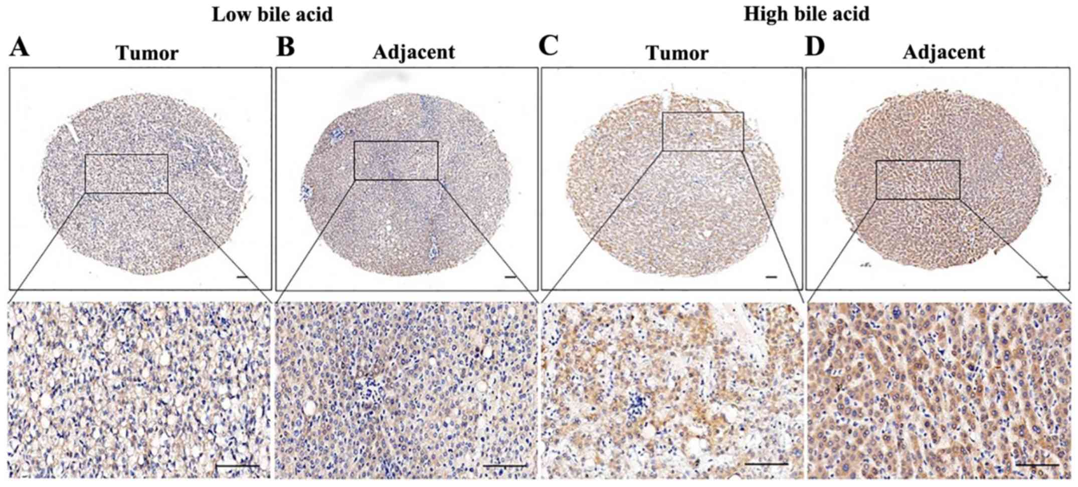
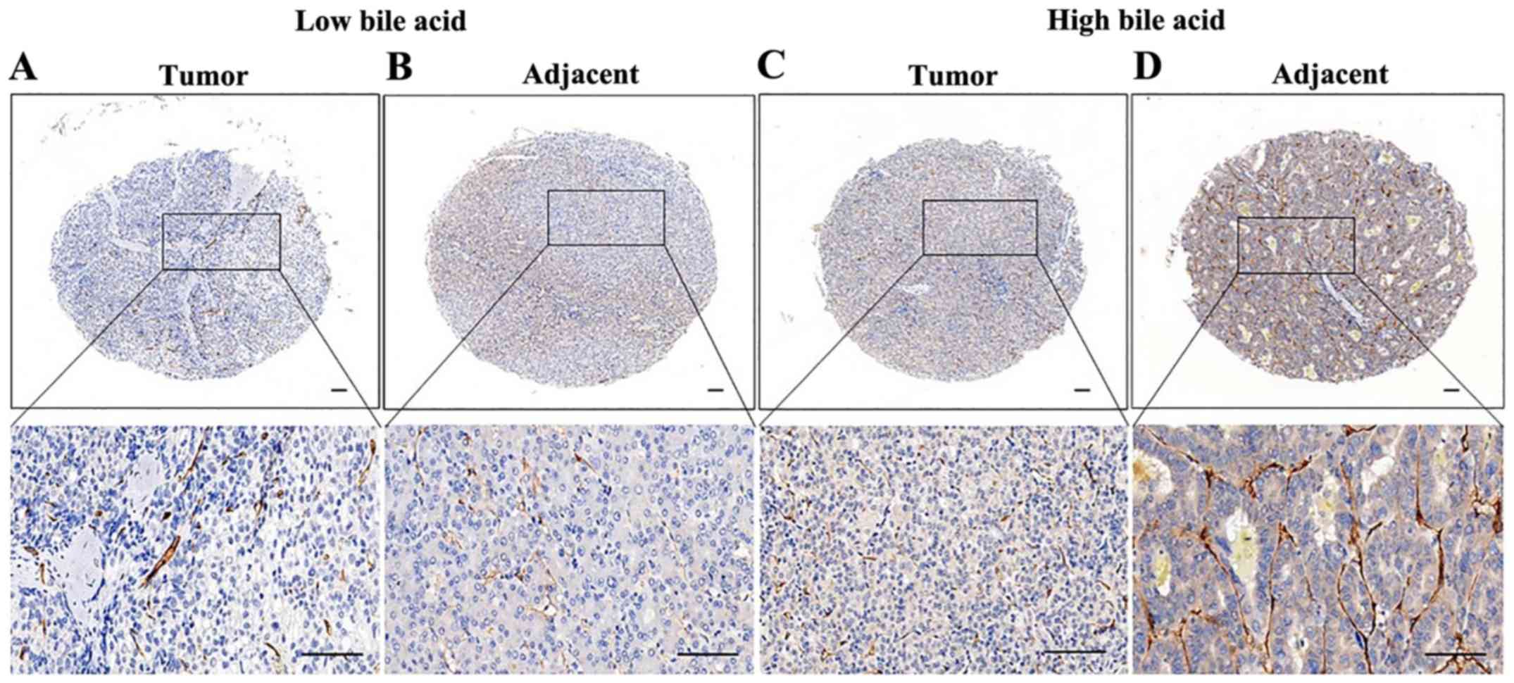
VEGFR-2 and CD34 expression levels and
clinicopathological characteristics
Among the 72 patients, 41 (56.94%) and 44 (61.11%)
demonstrated high expression levels of VEGFR-2 and CD34,
respectively. Representative IHC staining images of VEGFR-2 and
CD34 are presented in Figs. 1 and
2. VEGFR-2 expression was found to
be significantly associated with the tumor size (P<0.001), bile
acid level (P=0.014) and AFP level (P=0.011; Table I). The other clinical
characteristics, including sex, age, HBV infection, TNM stage,
pathological grade, recrudescence, liver cirrhosis, satellite
nodules, venous invasion, Child-Pugh stage (30) and total bilirubin level, were not
directly associated with the expression of VEGFR-2 (Table I). However, CD34 expression was
significantly associated with the tumor size (P=0.009),
recrudescence (P<0.001) and bile acid level (P=0.009). Other
clinical characteristics, including sex, age, HBV infection, TNM
stage, pathological grade, liver cirrhosis, satellite nodules,
venous invasion, Child-Pugh stage, AFP level and total bilirubin
level were not directly related to the expression of CD34 (Table II).
 | Table I.Association between VEGFR-2
expression in tumor tissues and clinicopathological characteristics
in patients with HCC (n=72). |
Table I.
Association between VEGFR-2
expression in tumor tissues and clinicopathological characteristics
in patients with HCC (n=72).
|
|
| VEGFR-2 |
|
|
|---|
|
|
|
|
|
|
|---|
| Clinicopathological
parameters | Number of cases
(%) | Low (%) | High (%) | χ2 | P-value |
|---|
| Sex |
|
|
|
|
|
|
Male | 60 (83) | 24 (77) | 36 (88) | 0.012 | 0.91 |
|
Female | 12 (23) | 7 (23) | 5 (12) |
|
|
| Age, years |
|
|
|
|
|
|
≤50 | 24 (33) | 10 (32) | 14 (34) | 0.028 | 0.87 |
|
>50 | 48 (67) | 21 (68) | 27 (66) |
|
|
| HBV infection |
|
|
|
|
|
|
Positive | 56 (78) | 22 (71) | 34 (83) | 1.46 | 0.23 |
|
Negative | 16 (22) | 9 (29) | 7 (17) |
|
|
| Tumor, cm |
|
|
|
|
|
| ≤5 | 35 (47) | 24 (77) | 11 (27) | 18.09 |
<0.001b |
|
>5 | 37 (53) | 7 (23) | 30 (73) |
|
|
| TNM stage |
|
|
|
|
|
|
I–II | 25 (35) | 11 (35) | 14 (34) | 1.06 | 0.30 |
|
III–IV | 47 (65) | 20 (65) | 27 (66) |
|
|
| Recrudescence |
|
|
|
|
|
|
Yes | 38 (53) | 15 (48) | 23 (56) | 0.42 | 0.52 |
| No | 34 (47) | 16 (52) | 18 (44) |
|
|
| Liver
cirrhosis |
|
|
|
|
|
|
Yes | 49 (68) | 20 (65) | 29 (71) | 0.31 | 0.58 |
| No | 23 (32) | 11 (35) | 12 (29) |
|
|
| Satellites
nodules |
|
|
|
|
|
|
Yes | 43 (60) | 16 (52) | 27 (66) | 1.49 | 0.22 |
| No | 29 (40) | 15 (48) | 14 (34) |
|
|
| Venous
invasion |
|
|
|
|
|
|
Yes | 52 (72) | 23 (74) | 29 (71) | 0.11 | 0.75 |
| No | 20 (28) | 8 (26) | 12 (29) |
|
|
| Differentiation
grade |
|
|
|
|
|
|
Well | 21 (29) | 13 (42) | 8 (20) | 4.79 | 0.091 |
|
Moderate | 29 (40) | 9 (29) | 20 (49) |
|
|
|
Poor | 22 (31) | 9 (29) | 13 (31) |
|
|
| Child-Pugh
stage |
|
|
|
|
|
| A | 25 (35) | 10 (32) | 15 (37) | 0.15 | 0.70 |
| B and
C | 47 (65) | 21 (68) | 26 (63) |
|
|
| Bile acid,
µmol/l |
|
|
|
|
|
|
≤10 | 30 (42) | 18 (58) | 12 (29) | 6.023 | 0.014a |
|
>10 | 42 (58) | 13 (42) | 29 (71) |
|
|
| AFP, ng/ml |
|
|
|
|
|
|
≤20 | 34 (47) | 20 (65) | 14 (34) | 6.53 | 0.011a |
|
>20 | 38 (53) | 11 (35) | 27 (66) |
|
|
| Total bilirubin,
µmol/l |
|
|
|
|
|
|
≤21 | 39 (54) | 14 (45) | 25 (61) | 1.78 | 0.18 |
|
>21 | 33 (46) | 17 (55) | 16 (39) |
|
|
| Table II.Correlation between CD34 expression
and clinicopathological characteristics in patients with HCC
(n=72). |
Table II.
Correlation between CD34 expression
and clinicopathological characteristics in patients with HCC
(n=72).
|
|
| CD34 |
|
|
|---|
|
|
|
|
|
|
|---|
| Clinicopathological
parameters | Number of cases
(%) | Low (%) | High (%) | χ2 | P-value |
|---|
| Sex |
|
|
|
|
|
|
Male | 60 (83) | 22 (79) | 38 (86) | 0.75 | 0.39 |
|
Female | 12 (17) | 6 (21) | 6 (14) |
|
|
| Age, years |
|
|
|
|
|
|
≤50 | 24 (33) | 10 (36) | 14 (32) | 0.12 | 0.73 |
|
>50 | 48 (67) | 18 (64) | 30 (68) |
|
|
| HBV infection |
|
|
|
|
|
|
Positive | 56 (78) | 21 (75) | 35 (80) | 0.21 | 0.65 |
|
Negative | 16 (22) | 7 (25) | 9 (20) |
|
|
| Tumor, cm |
|
|
|
|
|
| ≤5 | 35 (47) | 19 (68) | 16 (36) | 6.79 | 0.009a |
|
>5 | 37 (53) | 9 (32) | 28 (64) |
|
|
| TNM stage |
|
|
|
|
|
|
I–II | 25 (35) | 12 (43) | 13 (30) | 1.34 | 0.25 |
|
III–IV | 47 (65) | 16 (57) | 31 (70) |
|
|
| Recrudescence |
|
|
|
|
|
|
Yes | 38 (53) | 6 (21) | 32 (73) | 18.07 |
<0.001b |
| No | 34 (47) | 22 (79) | 12 (27) |
|
|
| Liver
cirrhosis |
|
|
|
|
|
|
Yes | 49 (68) | 19 (68) | 30 (68) | 0.001 | 0.98 |
| No | 23 (32) | 9 (32) | 14 (32) |
|
|
| Satellites
nodules |
|
|
|
|
|
|
Yes | 43 (60) | 14 (50) | 29 (66) | 1.80 | 0.18 |
| No | 29 (40) | 14 (50) | 15 (34) |
|
|
| Venous
invasion |
|
|
|
|
|
|
Yes | 52 (72) | 18(64) | 34 (77) | 1.44 | 0.23 |
| No | 20 (28) | 10 (36) | 10 (23) |
|
|
| Differentiation
grade |
|
|
|
|
|
|
Well | 21 (29) | 8 (29) | 13 (30) | 0.055 | 0.97 |
|
Moderate | 29 (40) | 11 (39) | 18 (40) |
|
|
|
Poor | 22 (31) | 9 (32) | 13 (30) |
|
|
| Child-Pugh
stage |
|
|
|
|
|
| A | 25 (35) | 8 (29) | 17 (39) | 0.77 | 0.38 |
| B and
C | 47 (65) | 20 (71) | 27 (61) |
|
|
| Bile acid,
µmol/l |
|
|
|
|
|
|
≤10 | 30 (42) | 17 (61) | 13 (30) | 6.84 | 0.009a |
|
>10 | 42 (58) | 11 (39) | 31 (70) |
|
|
| AFP, ng/ml |
|
|
|
|
|
|
≤20 | 34 (47) | 15 (54) | 19 (43) | 0.74 | 0.39 |
|
>20 | 38 (53) | 13 (46) | 25 (57) |
|
|
| Total bilirubin,
µmol/l |
|
|
|
|
|
|
≤21 | 39 (54) | 13 (36) | 26 (57) | 0.041 | 0.84 |
|
>21 | 33 (46) | 10 (64) | 18 (43) |
|
|
Comparison of VEGFR-2 and CD34
expression in tumor and matched adjacent tissue according to the
bile acid level
Previous analyses have revealed that both VEGFR-2
and CD34 expression levels are associated with the bile acid level
(24,31). Therefore, the expression levels of
these two biomarkers in tumor and matched adjacent tissues were
compared with the bile acid level. The expression levels of VEGFR-2
and CD34 were higher in tumors compared with matched adjacent
tissues, but there was no difference between the high and low bile
acid groups (VEGFR-2, P=0.96, Table
III; CD34, P=0.78, Table IV).
The score of VEGFR-2 (P=0.044; Table
SI) was higher in the high bile acid group compared with that
in the low bile acid group in the non-tumor adjacent tissues, while
the score of CD34 (P=0.020; Table
SII) was higher in the high bile acid group compared with that
in the low bile acid group in both the tumor and matched adjacent
tissues.
| Table III.High expression of vascular
endothelial growth factor receptor 2 in hepatocellular carcinoma
patient tumor and matched adjacent tissues in low and high bile
acid groups. |
Table III.
High expression of vascular
endothelial growth factor receptor 2 in hepatocellular carcinoma
patient tumor and matched adjacent tissues in low and high bile
acid groups.
| Classification | Tumor | Adjacent | χ2 | P-value |
|---|
| Low bile acid | 12 | 14 |
|
|
| High bile acid | 29 | 33 | 0.03 | 0.96 |
| Table IV.High expression of CD34 in
hepatocellular carcinoma patient tumor and matched adjacent tissues
in low and high bile acid groups. |
Table IV.
High expression of CD34 in
hepatocellular carcinoma patient tumor and matched adjacent tissues
in low and high bile acid groups.
| Classification | Tumor | Adjacent | χ2 | P-value |
|---|
| Low bile acid | 13 | 11 |
|
|
| High bile acid | 31 | 30 | 0.077 | 0.78 |
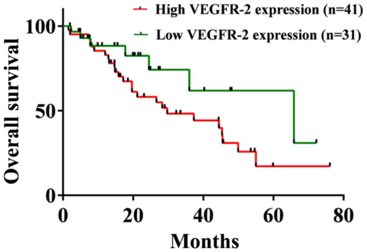
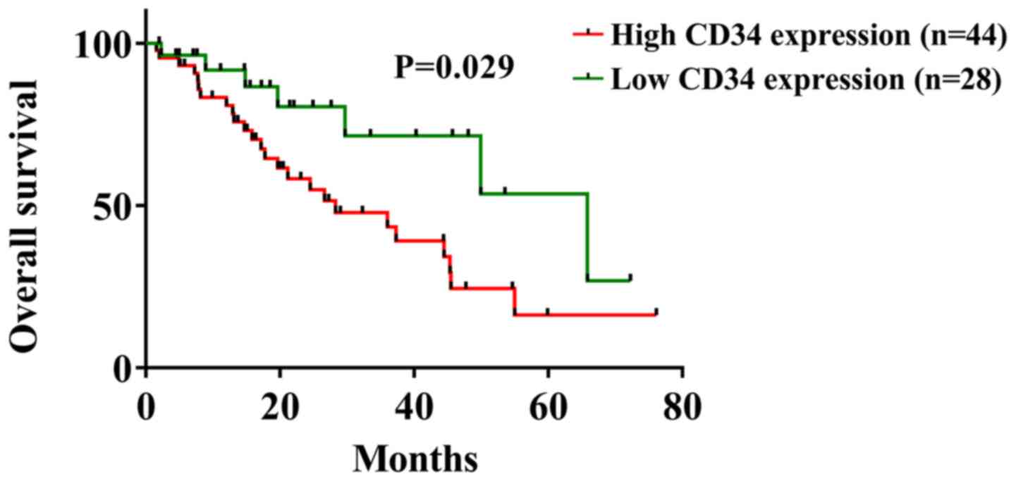
Comparison of the prognostic impact of
VEGFR-2 and CD34 expression in tumor tissues
At the last follow up, 52.78% of all cases presented
recurrence and mortality occurred in 44.44% cases. The OS of
patients with high VEGFR-2 expression was decreased compared with
patients with low VEGFR-2 expression, but this difference was not
significant (29.73 months vs. 65.83 months, respectively; P=0.094;
Fig. 3). Additionally, patients with
high CD34 expression had a significant difference in OS compared
with patients with low CD34 expression in Kaplan-Meier curve
analysis (28.30 months vs. 65.38 months, respectively; P=0.029;
Fig. 4).
Univariate and multivariate analyses of OS were
performed in patients with HCC. Univariate analysis indicated that
the TNM stage (P=0.030), recrudescence (P=0.048), VEGFR-2
(P<0.001) and CD34 (P=0.005) were significantly associated with
OS (Table V). Moreover, multivariate
Cox regression analysis suggested that VEGFR-2 (P=0.020) and CD34
(P=0.035) were independent prognostic factors for OS, while the TNM
stage and recrudescence were not (Table
V).
| Table V.Univariate and Multivariate analyses
of prognostic factors for survival in hepatocellular carcinoma. |
Table V.
Univariate and Multivariate analyses
of prognostic factors for survival in hepatocellular carcinoma.
|
| Univariable | Multivariable |
|---|
|
|
|
|
|---|
| Variables | P-value | HR (95% CI) | P-value | HR (95% CI) |
|---|
| Sex |
|
|
|
|
| Male
and female | 0.24 | 0.53
(0.18–1.54) |
|
|
| Age, years |
|
|
|
|
| ≤50 and
>50 | 0.55 | 0.80
(0.38–1.68) |
|
|
| HBV infection |
|
|
|
|
|
Positive and negative | 0.098 | 1.93
(0.89–4.20) |
|
|
| Tumor, cm |
|
|
|
|
| ≤5 and
>5 | 0.34 | 0.70
(0.34–1.43) |
|
|
| TNM stage |
|
|
|
|
| I–II
and III–IV | 0.030a | 0.42
(0.19–0.92) | 0.55 | 0.78
(0.35–1.76) |
| Recrudescence |
|
|
|
|
| Yes and
no | 0.048a | 0.47
(0.22–0.99) | 0.61 | 0.82
(0.37–1.79) |
| Liver
cirrhosis |
|
|
|
|
| Yes and
no | 0.37 | 0.72
(0.35–1.48) |
|
|
| Satellites
nodules |
|
|
|
|
| Yes and
no | 0.11 | 0.54
(0.25–1.16) |
|
|
| Venous
invasion |
|
|
|
|
| Yes and
no | 0.077 | 0.50
(0.23–1.08) |
|
|
| Differentiation
grade |
|
|
|
|
| Well,
Moderate and Poor | 0.065 | 0.51
(0.24–1.04) |
|
|
| Child-Pugh
stage |
|
|
|
|
| A, B
and C | 0.90 | 1.05
(0.52–2.11) |
|
|
| Bile acid
(µmol/l) |
|
|
|
|
| ≤10 and
>10 | 0.58 | 1.22
(0.61–2.42) |
|
|
| AFP (ng/ml) |
|
|
|
|
| ≤20 and
>20 | 0.14 | 1.77
(0.84–3.72) |
|
|
| Total bilirubin
(µmol/l) |
|
|
|
|
| ≤21 and
>21 | 0.20 | 1.59
(0.79–3.21) |
|
|
| VEGFR-2 |
|
|
|
|
| High
and low expression |
<0.001c | 0.15
(0.051–0.42) | 0.020a | 0.27
(0.089–0.81) |
| CD34 |
|
|
|
|
| High
and low expression | 0.005b | 0.057
(0.008–0.42) | 0.035a | 0.11
(0.015–0.87) |
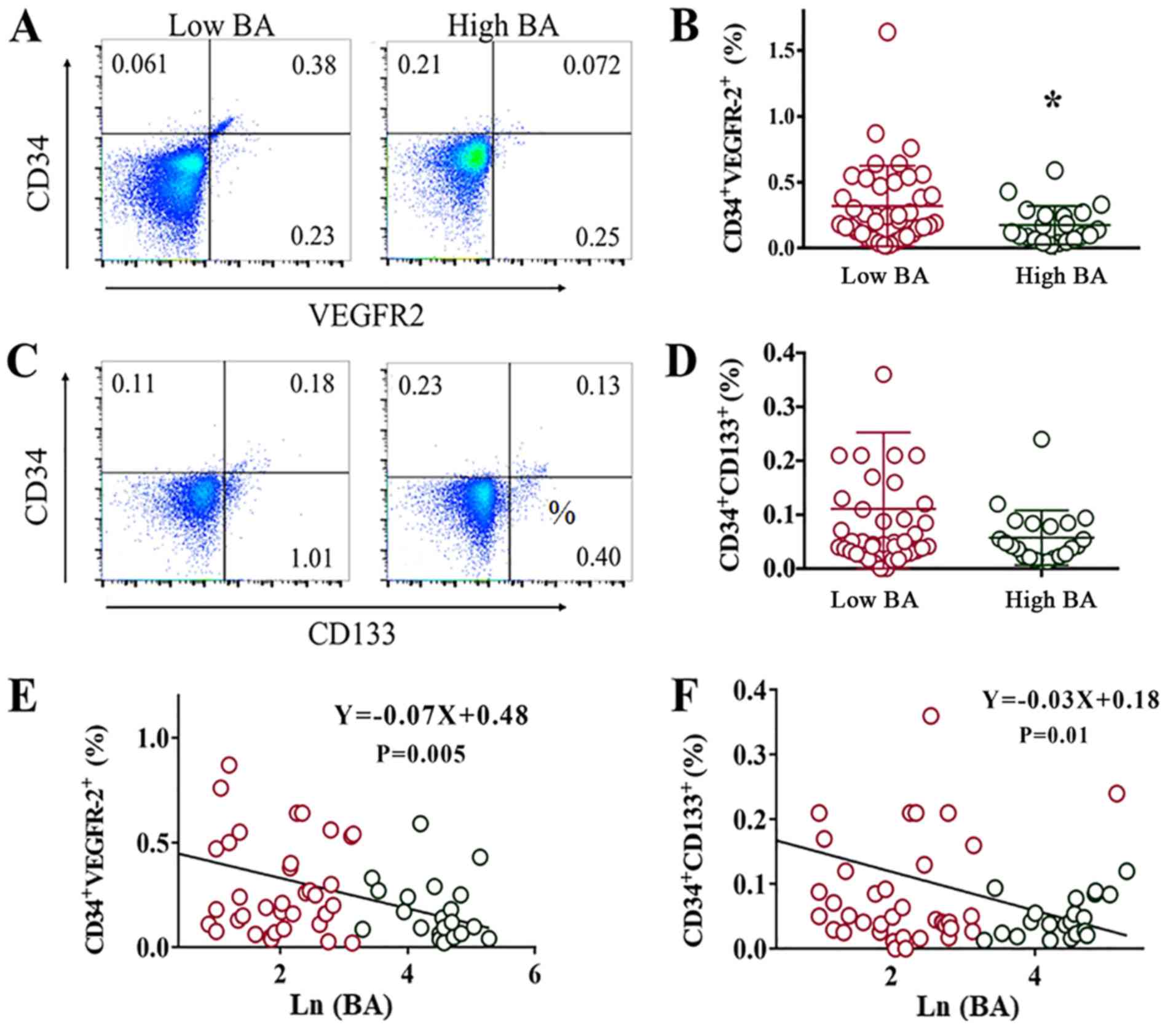
Comparison of the number of EPCs and
circulating progenitor cells (CPCs) in peripheral blood cells of
patients with HCC patients before surgery
As aforementioned, the expression levels of VEGFR-2
and CD34 were related to the bile acid level. Additionally, VEGFR-2
and CD34 expression levels were closely associated to the number of
EPCs and CPCs. To determine the relationship between the bile acid
level and the number of EPCs and CPCs, flow cytometry was performed
on the peripheral blood of patients with HCC before curative
resection. These data indicated that the number of EPCs and CPCs
was reduced in the high bile acid group compared with the low bile
acid group (Fig. 5A-D). However,
only EPCs had a statistically significant difference (P=0.044;
Fig. 5B). Furthermore, EPC (P=0.005;
Fig. 5E) and CPC (P=0.01; Fig. 5F) numbers were negatively related to
the bile acid levels, and the EPCs and CPCs demonstrated an
undifferentiated bilirubin state in different groups (Fig. S1).
Discussion
Pathological angiogenesis is a crucial hallmark of
cancer progression, including in HCC. Although the relationship
between VEGFR-2 expression in tumors and clinicopathological
characteristics has been extensively studied (32–34), the
expression levels of VEGFR-2 and CD34 in both tumor tissues and
adjacent tissues from patients with HCC remains elusive. The
present results demonstrated that VEGFR-2 and CD34 expression
levels were negatively related to OS in patients with HCC.
Additionally, both expression markers were independent prognosis
factors according to univariate and multivariate analyses. Huang
et al (35) reported that
high expression of VEGFR-2 in HCC was related to a large tumor
diameter, poor differentiation, high serum AFP levels, multifocal
gross classification and <5 years of survival. Moreover, it was
revealed that high VEGFR-2 expression and stage grouping with TNM
classification were independent prognostic factors (35); these findings were consistent with
the present results. VEGFR-2 is known to be a key target in
anti-angiogenesis during anti-tumor treatment (36–38).
Tsuji et al (39) revealed
that CD34 expression in the capillaries and sinusoids of
non-cancerous hepatic tissue was a risk factor for the multicentric
recurrence of HCC. In the present study, it was found that CD34
expression in HCC tumor tissue was a risk factor for recurrence.
Furthermore, the morphology of VEGFR-2- and CD34-positive cells in
HCC tumors reported in previous studies were consistent with those
in the current study (39–41).
VEGFR-2 and CD34 co-expression has previously been
used as a characteristic marker of EPCs (42–44).
Zahran et al (44) reported
that EPCs were higher in patients with HCC compared with healthy
controls, and increased levels of EPCs were associated with worse
OS in HCC. EPCs present an undifferentiated bilirubin state in
different groups (44), a finding
that was corroborated by the present study. However, in the current
study, the number of EPCs in the peripheral blood of patients with
HCC was negatively related with the bile acid level. In addition,
VEGFR-2 and CD34 expression levels were increased in the high bile
acid group. The cause of this contradiction may be that EPCs were
activated into tumor cells from the peripheral blood in the high
bile acid group. Zhu et al (42) revealed that various subgroups of bone
marrow-derived cells exhibit synergistic effects of BM-derived
CD45+CD133+ and
VEGFR2+CD133+ cells on HCC tumor progression
at different stages, facilitate the recovery of bone marrow
function and promote tumor growth.
In the present study, bile acid was suggested to
promote EPC-induced angiogenesis in HCC tumors. The bile acid level
was positively related to the VEGFR-2 and CD34 expression levels in
tumor and matched adjacent tissues. Moreover, the number of EPCs
was decreased in the high bile acid group in the peripheral blood
of patients with HCC. A possible explanation for this phenomenon is
that EPCs and CPCs may be activated by bile acid, thus
translocating to adjacent tissues and stimulating tumor progression
(24). However, different types of
bile acid may have various roles in HCC. For instance, Wang et
al (22) showed that bile acid
induced tumor cell survival and chemoresistance in HCC.
Additionally, the interaction between the enterohepatic circulation
of bile acid and intestinal flora complicates the role of bile acid
in HCC. For example, Yamada et al (18) demonstrated that bile acid was
regulated by gut microbiota and promoted HCC development. This
should be further investigated in future studies.
The present study has some limitations. EPC-induced
angiogenesis should be confirmed via the merged imaging of
proliferation, stem cell and endothelial markers, such as BrdU,
CD34 and VEGFR-2 using confocal immunofluorescence. Furthermore,
the results would be more reliable if tissue and blood samples were
derived from the same group of patients. The effects and mechanism
of bile acid on EPCs and angiogenesis also require further
investigation using primary cells in vitro and in
vivo.
In conclusion, the present study demonstrated that
bile acid promoted VEGFR-2 and CD34 expression induced by EPCs in
HCC, and that these two markers were expressed at higher levels in
tumor tissues compared with in matched adjacent tissues. Moreover,
it was indicated that VEGFR-2 and CD34 expression levels were both
negatively related to OS.
Supplementary Material
Supporting Data
Acknowledgements
Not applicable.
Funding
The present study was supported by the Shanghai
Municipal Education Commission (grant no. 2019 Technology
Education-01-8), the Shanghai Pudong New Area Municipal Commission
of Health and Family Planning Funding (grant nos. PW2015D-3 and
PWZXQ2017-06) and in part by the National Nature Science Foundation
of China (grant no. 81971223).
Availability of data and materials
The datasets used and/or analyzed during the present
study are available from the corresponding author on reasonable
request.
Authors' contributions
WFY, JLC and YFJ developed the experimental concept
and design of the study. LW analyzed the clinicopathological
characteristic factors and conducted the flow cytometry. Tissue
microarray, immunohistochemical staining and statistical analyses
were performed by JLC. RL conducted the immunohistochemical
staining evaluation. JLC, WFY and YFJ drafted and revised the
manuscript. All authors have read and approved the final
manuscript.
Ethics approval and consent to
participate
The present study was approved by the Committee of
Research Ethical Research at EHBH (approval no. EHBHKY2018- 1-019),
and prior patient written informed consent was obtained from all
enrolled patients.
Patient consent for publication
Not applicable.
Competing interests
The authors declare that they have no competing
interests.
References
|
1
|
St Croix BD, Rak JW and Kerbel RS:
Consequences of angiogenesis for tumor progression, metastasis and
cancer therapy. Anticancer Drugs. 6:3–18. 1995. View Article : Google Scholar : PubMed/NCBI
|
|
2
|
Hervey-Jumper SL, Garton HJL, Lau D,
Altshuler D, Quint DJ, Robertson PL, Muraszko KM and Maher CO:
Differences in vascular endothelial growth factor receptor
expression and correlation with the degree of enhancement in
medulloblastoma. J Neurosurg Pediatr. 14:121–128. 2014. View Article : Google Scholar : PubMed/NCBI
|
|
3
|
Li S, Xu HX, Wu CT, Wang WQ, Jin W, Gao
HL, Li H, Zhang SR, Xu JZ, Qi ZH, et al: Angiogenesis in pancreatic
cancer: Current research status and clinical implications.
Angiogenesis. 22:15–36. 2019. View Article : Google Scholar : PubMed/NCBI
|
|
4
|
Folkman J: What is the evidence that
tumors are angiogenesis dependent. J Natl Cancer Inst. 82:4–6.
1990. View Article : Google Scholar : PubMed/NCBI
|
|
5
|
Viallard C and Larrivee B: Tumor
angiogenesis and vascular normalization: Alternative therapeutic
targets. Angiogenesis. 20:409–426. 2017. View Article : Google Scholar : PubMed/NCBI
|
|
6
|
Peters EB: Endothelial progenitor cells
for the vascularization of engineered tissues. Tissue Eng Part B
Rev. 24:1–24. 2018. View Article : Google Scholar : PubMed/NCBI
|
|
7
|
Mandraffino G and Saitta A: Endothelial
and circulating progenitor cells: Between diseases and therapies.
Curr Med Chem. 25:4476–4477. 2018. View Article : Google Scholar : PubMed/NCBI
|
|
8
|
Zhou M, Wang H, Zeng X, Yin P, Zhu J, Chen
W, Li X, Wang L, Wang L, Liu Y, et al: Mortality, morbidity, and
risk factors in China and its provinces, 1990-2017: A systematic
analysis for the global burden of disease study 2017. Lancet.
394:1145–1158. 2019. View Article : Google Scholar : PubMed/NCBI
|
|
9
|
Zhu AX, Duda DG, Sahani DV and Jain RK:
HCC and angiogenesis: Possible targets and future directions. Nat
Rev Clin Oncol. 8:292–301. 2011. View Article : Google Scholar : PubMed/NCBI
|
|
10
|
Rafii S and Lyden D: Cancer. A few to flip
the angiogenic switch. Science. 319:163–164. 2008. View Article : Google Scholar : PubMed/NCBI
|
|
11
|
Poon RT, Fan ST, Lo CM, Liu CL, Lam CM,
Yuen WK, Yeung C and Wong J: Extended hepatic resection for
hepatocellular carcinoma in patients with cirrhosis: Is it
justified? Ann Surg. 236:602–611. 2002. View Article : Google Scholar : PubMed/NCBI
|
|
12
|
Ho JW, Pang RW, Lau C, Sun CK, Yu WC, Fan
ST and Poon RT: Significance of circulating endothelial progenitor
cells in hepatocellular carcinoma. Hepatology. 44:836–843. 2006.
View Article : Google Scholar : PubMed/NCBI
|
|
13
|
Sieghart W, Fellner S, Reiberger T,
Ulbrich G, Ferlitsch A, Wacheck V and Peck-Radosavljevic M:
Differential role of circulating endothelial progenitor cells in
cirrhotic patients with or without hepatocellular carcinoma. Dig
Liver Dis. 41:902–906. 2009. View Article : Google Scholar : PubMed/NCBI
|
|
14
|
Yu D, Sun X, Qiu Y, Zhou J, Wu Y, Zhuang
L, Chen J and Ding Y: Identification and clinical significance of
mobilized endothelial progenitor cells in tumor vasculogenesis of
hepatocellular carcinoma. Clin Cancer Res. 13:3814–3824. 2007.
View Article : Google Scholar : PubMed/NCBI
|
|
15
|
Shaked Y, Henke E, Roodhart JM, Mancuso P,
Langenberg MH, Colleoni M, Daenen LG, Man S, Xu P, Emmenegger U, et
al: Rapid chemotherapy-induced acute endothelial progenitor cell
mobilization: Implications for antiangiogenic drugs as
chemosensitizing agents. Cancer Cell. 14:263–273. 2008. View Article : Google Scholar : PubMed/NCBI
|
|
16
|
Li CX, Shao Y, Ng KT, Liu XB, Ling CC, Ma
YY, Geng W, Fan ST, Lo CM and Man K: FTY720 suppresses liver tumor
metastasis by reducing the population of circulating endothelial
progenitor cells. PLoS One. 7:e323802012. View Article : Google Scholar : PubMed/NCBI
|
|
17
|
Lee ES, Han EM, Kim YS, Shin BK, Kim CH,
Kim HK, Won NH, Yeom BW, Kim I and Leong ASY: Occurrence of c-kit+
tumor cells in hepatitis B virus-associated hepatocellular
carcinoma. Am J Clin Pathol. 124:31–36. 2005. View Article : Google Scholar : PubMed/NCBI
|
|
18
|
Yamada S, Takashina Y, Watanabe M,
Nagamine R, Saito Y, Kamada N and Saito H: Bile acid metabolism
regulated by the gut microbiota promotes non-alcoholic
steatohepatitis-associated hepatocellular carcinoma in mice.
Oncotarget. 9:9925–9939. 2018. View Article : Google Scholar : PubMed/NCBI
|
|
19
|
Liu N, Feng H, Lv Y, Liu Q, Deng J, Xia Y,
Guo C and Zhou Y: Role of bile acids in the diagnosis and
progression of liver cirrhosis: A prospective observational study.
Exp Ther Med. 18:4058–4066. 2019.PubMed/NCBI
|
|
20
|
Wu WB, Chen YY, Zhu B, Peng XM, Zhang SW
and Zhou ML: Excessive bile acid activated NF-kappa B and promoted
the development of alcoholic steatohepatitis in farnesoid X
receptor deficient mice. Biochimie. 115:86–92. 2015. View Article : Google Scholar : PubMed/NCBI
|
|
21
|
Liao M, Zhao J, Wang T, Duan J, Zhang Y
and Deng X: Role of bile salt in regulating Mcl-1 phosphorylation
and chemoresistance in hepatocellular carcinoma cells. Mol Cancer.
10:442011. View Article : Google Scholar : PubMed/NCBI
|
|
22
|
Wang C, Yang M, Zhao J, Li X, Xiao X,
Zhang Y, Jin X and Liao M: Bile salt (glycochenodeoxycholate acid)
induces cell survival and chemoresistance in hepatocellular
carcinoma. J Cell Physiol. 234:10899–10906. 2019. View Article : Google Scholar : PubMed/NCBI
|
|
23
|
Cho JG, Lee JH, Hong SH, Lee HN, Kim CM,
Kim SY, Yoon KJ, Oh BJ, Kim JH, Jung SY, et al:
Tauroursodeoxycholic acid, a bile acid, promotes blood vessel
repair by recruiting vasculogenic progenitor cells. Stem Cells.
33:792–805. 2015. View Article : Google Scholar : PubMed/NCBI
|
|
24
|
Takahashi T, Kalka C, Masuda H, Chen D,
Silver M, Kearney M, Magner M, Isner JM and Asahara T: Ischemia-
and cytokine-induced mobilization of bone marrow-derived
endothelial progenitor cells for neovascularization. Nat Med.
5:434–438. 1999. View
Article : Google Scholar : PubMed/NCBI
|
|
25
|
Rafii S: Circulating endothelial
precursors: Mystery, reality, and promise. J Clin Invest.
105:17–19. 2000. View
Article : Google Scholar : PubMed/NCBI
|
|
26
|
Carmeliet P and Jain R: Angiogenesis in
cancer and other diseases. Nature. 407:249–257. 2000. View Article : Google Scholar : PubMed/NCBI
|
|
27
|
Dvorak HF: Vascular permeability
factor/vascular endothelial growth factor: A critical cytokine in
tumor angiogenesis and a potential target for diagnosis and
therapy. J Clin Oncol. 20:4368–4380. 2002. View Article : Google Scholar : PubMed/NCBI
|
|
28
|
Ferrara N, Hillan KJ, Gerber HP and
Novotny W: Discovery and development of bevacizumab, an anti-VEGF
antibody for treating cancer. Nat Rev Drug Discov. 3:391–400. 2004.
View Article : Google Scholar : PubMed/NCBI
|
|
29
|
Hamanishi J, Mandai M, Iwasaki M, Okazaki
T, Tanaka Y, Yamaguchi K, Higuchi T, Yagi H, Takakura K, Minato N,
et al: Programmed cell death 1 ligand 1 and tumor-infiltrating CD8+
T lymphocytes are prognostic factors of human ovarian cancer. Proc
Natl Acad Sci USA. 104:3360–3365. 2007. View Article : Google Scholar : PubMed/NCBI
|
|
30
|
Zhao S, Wang M, Yang Z, Tan K, Zheng D, Du
X and Liu L: Comparison between child-pugh score and
albumin-bilirubin grade in the prognosis of patients with HCC after
liver resection using time-dependent ROC. Ann Transl Med.
8:5392020. View Article : Google Scholar : PubMed/NCBI
|
|
31
|
Glaser S, Onori P, Gaudio E, Ueno Y,
Pannarale L, Franchitto A, Francis H, Mancinelli R, Carpino G,
Venter J, et al: Taurocholic acid prevents biliary damage induced
by hepatic artery ligation in cholestatic rats. Dig Liver Dis.
42:709–717. 2010. View Article : Google Scholar : PubMed/NCBI
|
|
32
|
Nakamura K, Zen Y, Sato Y, Kozaka K,
Matsui O, Harada K and Nakanuma Y: Vascular endothelial growth
factor, its receptor Flk-1, and hypoxia inducible factor-1alpha are
involved in malignant transformation in dysplastic nodules of the
liver. Hum Pathol. 38:1532–1546. 2007. View Article : Google Scholar : PubMed/NCBI
|
|
33
|
Iavarone M, Lampertico P, Iannuzzi F,
Manenti E, Donato MF, Arosio E, Bertolini F, Primignani M,
Sangiovanni A and Colombo M: Increased expression of vascular
endothelial growth factor in small hepatocellular carcinoma. J
Viral Hepat. 14:133–139. 2007. View Article : Google Scholar : PubMed/NCBI
|
|
34
|
Jia JB, Zhuang PY, Sun HC, Zhang JB, Zhang
W, Zhu XD, Xiong YQ, Xu HX and Tang ZY: Protein expression
profiling of vascular endothelial growth factor and its receptors
identifies subclasses of hepatocellular carcinoma and predicts
survival. J Cancer Res Clin Oncol. 135:847–854. 2009. View Article : Google Scholar : PubMed/NCBI
|
|
35
|
Huang J, Zhang X, Tang Q, Zhang F, Li Y,
Feng Z and Zhu J: Prognostic significance and potential therapeutic
target of VEGFR2 in hepatocellular carcinoma. J Clin Pathol.
64:343–348. 2011. View Article : Google Scholar : PubMed/NCBI
|
|
36
|
Llovet JM, Ricci S, Mazzaferro V, Hilgard
P, Gane E, Blanc JF, de Oliveira AC, Santoro A, Raoul JL, Forner A,
et al: Sorafenib in advanced hepatocellular carcinoma. N Engl J
Med. 359:378–390. 2008. View Article : Google Scholar : PubMed/NCBI
|
|
37
|
Wang H, Chen K, Niu G and Chen X:
Site-specifically biotinylated VEGF(121) for nearinfrared
fluorescence imaging of tumor angiogenesis. Mol Pharm. 6:285–294.
2009. View Article : Google Scholar : PubMed/NCBI
|
|
38
|
Giannelli G, Sgarra C, Porcelli L,
Azzariti A, Antonaci S and Paradiso A: EGFR and VEGFR as potential
target for biological therapies in HCC cells. Cancer Lett.
262:257–264. 2008. View Article : Google Scholar : PubMed/NCBI
|
|
39
|
Tsuji N, Ishiguro S, Sasaki Y and Kudo M:
CD34 expression in noncancerous liver tissue predicts multicentric
recurrence of hepatocellular carcinoma. Dig Dis. 31:467–471. 2013.
View Article : Google Scholar : PubMed/NCBI
|
|
40
|
Yang ZF and Poon RT: Vascular changes in
hepatocellular carcinoma. Anat Rec (Hoboken). 291:721–734. 2008.
View Article : Google Scholar : PubMed/NCBI
|
|
41
|
Segatelli V, de Oliveira EC, Boin IF,
Ataide EC and Escanhoela CA: Evaluation and comparison of
microvessel density using the markers CD34 and CD105 in
regenerative nodules, dysplastic nodules and hepatocellular
carcinoma. Hepatol Int. 8:260–265. 2014.PubMed/NCBI
|
|
42
|
Zhu X, Zhou H, Luo J, Cui Y, Li H, Zhang
W, Fang F, Li Q and Zhang T: Different but synergistic effects of
bone marrow-derived VEGFR2+ and VEGFR2−CD45+ cells during
hepatocellular carcinoma progression. Oncol Lett. 13:63–68. 2017.
View Article : Google Scholar : PubMed/NCBI
|
|
43
|
Fürstenberger G, von Moos R, Lucas R,
Thürlimann B, Senn HJ, Hamacher J and Boneberg EM: Circulating
endothelial cells and angiogenic serum factors during neoadjuvant
chemotherapy of primary breast cancer. Br J Cancer. 94:524–531.
2006. View Article : Google Scholar : PubMed/NCBI
|
|
44
|
Zahran AM, Abdel-Rahim MH, Refaat A, Sayed
M, Othman MM, Khalak LMR and Hetta HF: Circulating hematopoietic
stem cells, endothelial progenitor cells and cancer stem cells in
hepatocellular carcinoma patients: Contribution to diagnosis and
prognosis. Acta Oncol. 59:33–39. 2020. View Article : Google Scholar : PubMed/NCBI
|