Introduction
Nasopharyngeal carcinoma (NPC) is a highly malignant
tumor arising from epithelial cells (1). In 2015, there were 60,600 newly
diagnosed NPC and 34,100 deaths in China (2). Despite the low morbidity and mortality,
the treatment of NPC has always been difficult in clinical
practice. NPC is occult and adjacent to optic nerve, trigeminal
nerve, and internal carotid artery, so it is difficult to be
treated by surgery (3). With the
exception of a small number of patients with early NPC who can be
cured by radical surgery, most patients are treated with palliative
chemoradiotherapy to improve the condition (4). At present, there are few efficacy
assessment indicators for NPC, and imaging examinations are mainly
applied to assess the efficacy of chemoradiotherapy (5). However, serological test is less
harmful, faster and more convenient. Therefore, it is of great
significance to find a specific serological index (6).
Patients with NPC always suffer from cognitive
decline after chemoradiotherapy. Currently, Montreal Cognitive
Assessment (MoCA) score and Das-Naglieri cognitive assessment
system are commonly used to assess the cognitive function of
patients (7). However, subjective
bias may occur in artificial scoring, so it is very important to
find a new observation index for cognitive function assessment
(8). MicroRNAs (miRs) is a class of
newly discovered, highly conserved, endogenous, non-coding, hairpin
nucleotide transcripts that widely exist in eukaryotic cells and
are approximately 19–25 bases in length and 18–25 nucleotides in
size (9). Some miRNAs are reported
to be closely related to the occurrence and progression of NPC
(10). As an important member of the
miR-34 family, miR-34a is located at 1p36.23 and mainly distributed
in brain tissue under normal circumstances. A study found that it
has a significantly reduced expression in NPC and was expected to
become a potential diagnostic indicator for NPC (11). Another study suggested that miR-34a
was closely associated to cognitive function, that is, cognitive
function could be improved by downregulating the expression of
miR-34a (12). Therefore, we
speculate that there may be a correlation between miR-34a and
cognitive function, but whether miR-34a is related to the cognitive
impairment in patients with NPC has not been confirmed yet.
Therefore, this study explored the potential value
of miR-34a in assessing cognitive function and radiotherapy
efficacy in patients with NPC by determining its expression before
and after radiotherapy, so as to provide reference for
clinicians.
Patients and methods
Clinical data
A total of 89 patients with NPC admitted to Shunde
Hospital (Foshan, China) from July 2015 to February 2017 were
enrolled, 39 patients who failed to complete treatment were
excluded, and the remaining 50 served as a treated group. Moreover,
50 healthy individuals who underwent physical examination in the
hospital during the same period were collected as a control group.
Laboratory test indexes and imaging tests of patients in the
control group were normal. The study was approved by the Ethics
Committee of Shunde Hospital. Signed informed consents were
obtained from the patients and/or guardians. Inclusion criteria:
Patients aged over 18 years; patients conforming to AJCC 7th
version (13) and staged III–IVB;
patients diagnosed with NPC through imaging and pathological
examinations; Eastern Cooperative Oncology Group (ECOG) score ≤1.
Exclusion criteria: Patients complicated with other tumors;
patients receiving corresponding anti-cancer treatment before this
treatment; patients intolerant to chemoradiotherapy; breast-feeding
women and patients with cognitive dysfunction.
Main reagents and drugs
Cisplatin sodium chloride injection (Guizhou Hanfang
Pharmaceutical Co., Ltd.); total RNA extraction kit EasyPure miRNA
kit - TransScript Green miRNA Two-Step qRT-PCR SuperMix (Beijing
TransGen Biotech Company; AQ 202-01, ER601-01); PCR instrument (ABI
7500; Applied Biosystems; Thermo Fisher Scientific, Inc.).
Treatment
All the patients were treated with concurrent
chemoradiotherapy (CCRT), and 6 MV–X-ray, 9-field three-dimensional
conformal radiotherapy (3D-CRT) and intensity modulated
radiotherapy (IMRT) were performed. The specific steps were as
follows: The tumor volume was measured by MRI and CT localization
images, prescription dose: PGTVnx, 70.4–72.3 Gy/32 f; PGTVnd,
66–70.4 Gy/32 f; PCTV 1, 60–62 Gy/30 f; PCTV 2, 54–55.8 Gy/30 f,
once a day, five times a week. The radiation dose to temporal lobes
was obtained using dose-volume histogram (DVH). Chemotherapy was
performed with cisplatin at the same time. The specific protocol
was as follows: One cycle of cisplatin (100 mg/m2) was
given on the 1st, 22nd and 43rd days, respectively, 21 days each
cycle (14).
Expression of miR-34a in serum of
patients
Samples of peripheral blood (5 ml) were collected
from patients before and after 3 cycles of CCRT (after treatment)
and from subjects in the control group, placed at room temperature
for 30 min, and centrifuged at 1,500 × g for 10 min to collect the
serum. Total RNAs were extracted using a EasyPure miRNA kit, and
the purity, concentration and integrity were detected using an
ultraviolet spectrophotometer and agarose gel electrophoresis. The
RNAs were reverse transcribed into cDNAs by 2X TS miRNA reaction
mix in transScript Green miRNA two-step qRT-PCR supermix kit, and
the specific procedure was performed according to the
manufacturer's kit instructions. Then PCR amplification was carried
out. PCR reaction system: 1 µl of cDNA, 0.4 µl of each upstream and
downstream primers, 10 µl of 2X TransTaq® Tip Green qPCR
SuperMix, 0.4 µl of Passive Reference Dye (50X), finally made up to
20 µl with ddH2O. PCR reaction conditions:
Pre-denaturation at 94°C for 30 sec, denaturation at 94°C for 5
sec, annealing at 60°C for 15 sec, extension for 10 sec, for a
total of 40 cycles. Each sample was tested in 3 repeat wells, and
the experiment was carried out 3 times. In this study, U6 was used
as an internal reference and the 2−∆∆Cq was used to
analyze the data (15). miR-34a
upstream primer 5′-GTGCAGGGTCCGAGGT-3′, downstream primer
5′-GCCGCTGGCAGTGTCTTAGCTG-3′, U6 upstream primer
5′-CTCGCTTCGGCAGCACA-3′, downstream primer
5′-AACGCTTCACGAATTTGCGT-3′.
Efficacy evaluation criteria
According to Response Evaluation Criteria in Solid
Tumors, the short-term efficacy was evaluated and divided into 4
grades: Complete remission (CR), partial remission (PR), stable
disease (SD) and progressive disease (PD (Table I).
 | Table I.Efficacy evaluation. |
Table I.
Efficacy evaluation.
| Efficacy grade | Evaluation
criteria |
|---|
| CR | After treatment, the
lesions disappeared completely for >4 weeks |
| PR | After treatment, the
total maximum diameter of the lesions decreased by ≥50% |
| SD | After treatment, the
total maximum diameter of the lesions decreased by <50% |
| PD | After treatment, the
total maximum diameter of the lesions decreased by ≥25% or new
lesions appeared |
Outcome measures
Main outcome measures: The expression of miR-34a in
the control and the treated group before treatment was compared.
The diagnostic value of miR-34a was assessed using a receiver
operating characteristic (ROC) curve, and the changes of miR-34a
before and after treatment were compared. The changes of MoCA score
(all the subjects were scored by a senior psychiatrist in the
hospital) before and after treatment were compared. A higher score
indicated a better cognitive function of the patients (30 points in
total).
Secondary outcome measures: CR patients were
classified as significant group and PR patients as general group.
The expression of miR-34a before treatment was compared between the
two groups, and the area under the ROC curve (AUC) was obtained.
The correlation between miR-34a and MoCA score after treatment was
analyzed.
Statistical analysis
In this study, SPSS 20.0 software package (Guangzhou
Pomine Information Technology Co., Ltd.) was used to carry out
statistical analysis on the collected data. GraphPad Prism 7
(Shanghai Cabit Information Technology Co., Ltd.) was used to draw
the figures. Kolmogorov-Smirnov (K-S) test was used to analyze the
data distribution. Counting data expressed as percentage (%) were
analyzed using the Chi-square test (denoted by χ2).
Measurement data conforming to normal distribution and variance
homogeneity were expressed as the mean ± SD. Independent t-test was
used for analysis between the two groups, while paired t-test was
used for comparison before and after treatment within the group
(denoted by t). ROC was used to assess the predictive and
diagnostic values of miR-34a for NPC. Pearson test was used for
determining the correlation between miR-34a and MoCA score after
treatment. P<0.05 indicates a statistically significant
difference.
Results
Comparison of baseline data
There was no significant difference in sex, age and
body mass index (BMI) (P>0.05) between the two groups, as shown
in Table II.
| Table II.Comparison of baseline data. |
Table II.
Comparison of baseline data.
| Factor | Observation group
(n=50) | Control group
(n=50) | t/χ2 | P-value |
|---|
| Sex |
| Male | 39 (78.00) | 42 (84.00) | 0.585 | 0.444 |
|
Female | 11 (22.00) | 8
(16.00) |
|
|
| Age (years) |
52.1±7.2 |
53.4±6.3 | 0.961 | 0.339 |
| BMI
(kg/m2) | 21.25±1.47 | 21.48±1.55 | 0.761 | 0.448 |
| Clinical staging |
| III | 5
(10.00) |
|
|
|
| IVa | 36 (72.00) |
|
|
|
| IVb | 9
(18.00) |
|
|
|
| T staging |
| T2 | 3
(6.00) |
|
|
|
| T3 | 4
(8.00) |
|
|
|
| T4 | 43 (86.00) |
|
|
|
| N staging |
| N0 | 4
(8.00) |
|
|
|
| N1 | 8
(16.00) |
|
|
|
| N2 | 27 (54.00) |
|
|
|
| N3 | 11 (22.00) |
|
|
|
| Pathological
type |
|
Keratinizing squamous cell
carcinoma | 25 (50.00) |
|
|
|
|
Non-keratinizing squamous cell
carcinoma | 25 (50.00) |
|
|
|
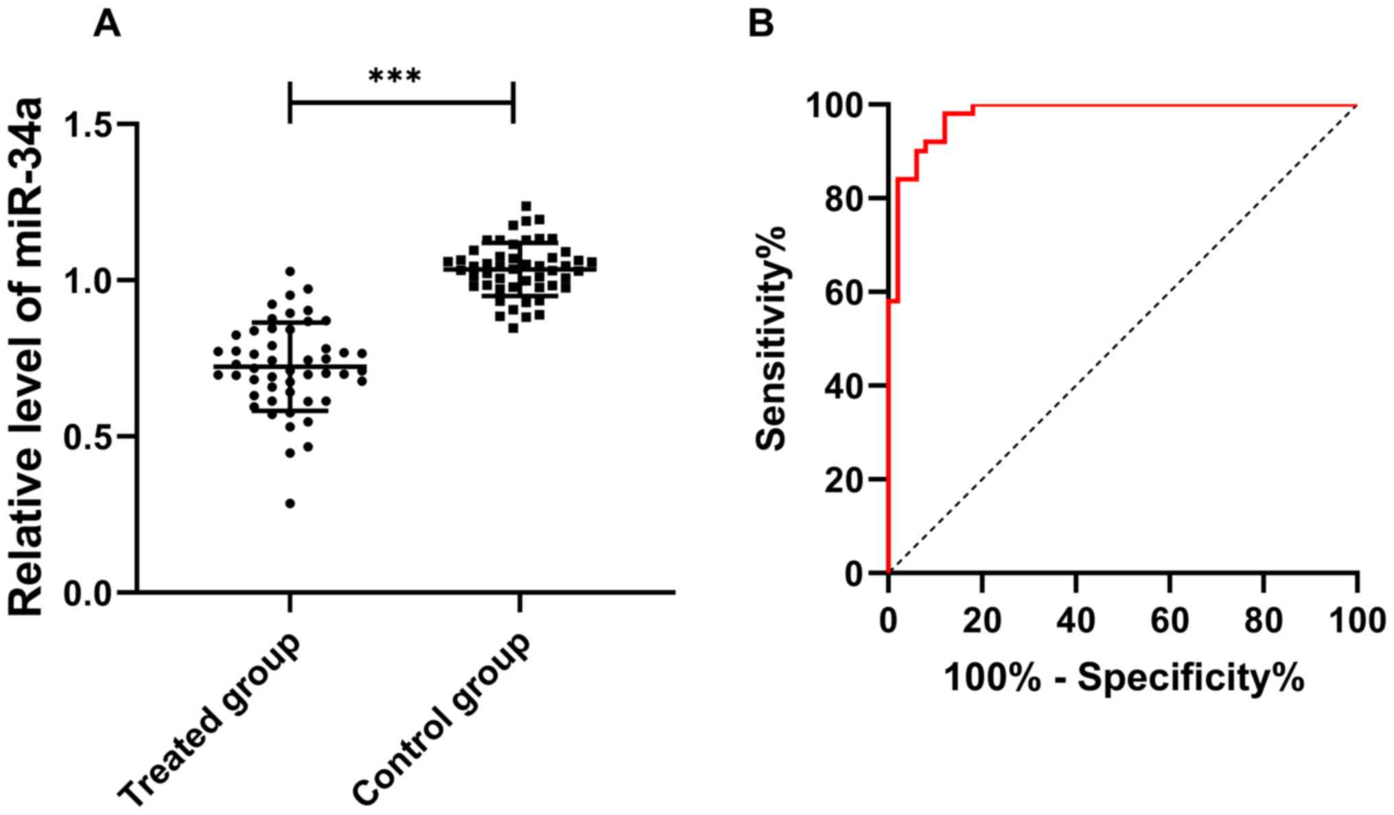
Expression and diagnostic value of
miR-34a in NPC
The expression of miR-34a in the control group
(1.035±0.085) was significantly higher than that in the treated
group (0.723±0.141) (t=13.364, P<0.001). ROC curve showed that
the AUC of miR-34 was 0.979, showing a high diagnostic value
(Fig. 1).
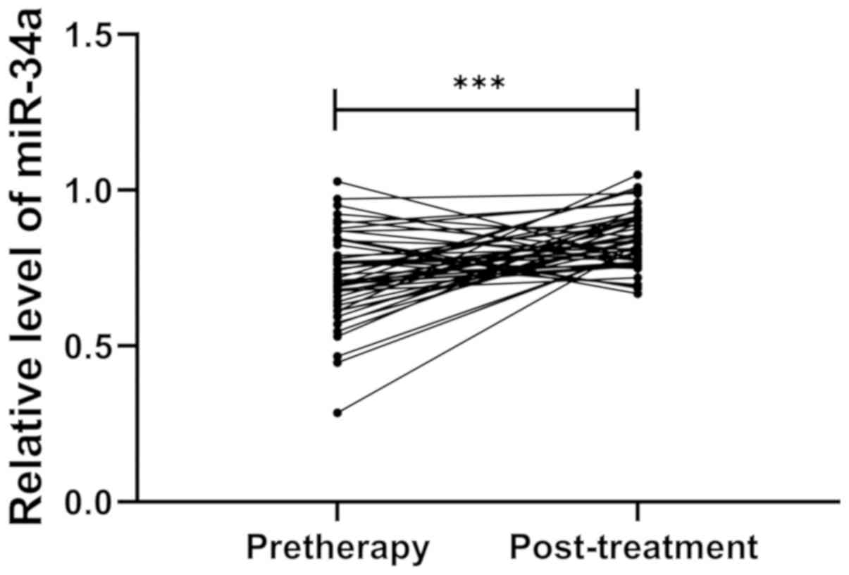
miR-34a expression before and after
treatment
The expression of miR-34a in the treated group
increased significantly after treatment compared with that before
treatment (t=4.559, P<0.001), as shown in Fig. 2.
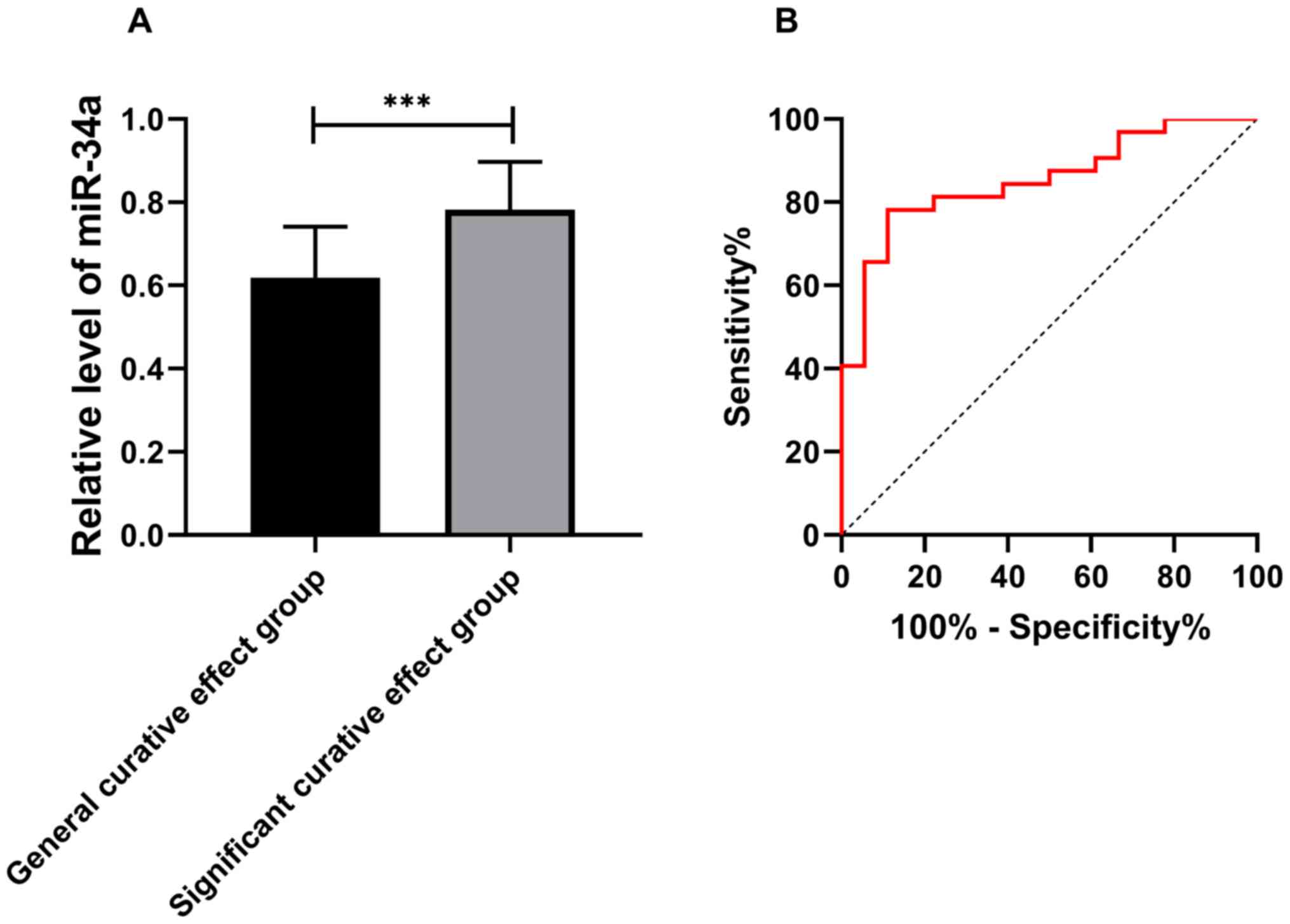
Predictive value of miR-34a in
NPC
Evaluation of the clinical efficacy of patients in
the treated group showed that there were 32 CR patients and 18 PR
patients after treatment. The CR patients were classified as the
significant group and PR patients as the general group. The
expression of miR-34a in the significant group was higher than that
in the general group before treatment (t=4.704, P<0.001). The
ROC curve showed that the AUC was 0.852 (Fig. 3).
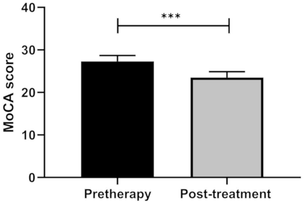
MoCA score changes before and after
treatment
The MoCA score of patients after treatment
(23.52±1.37) was significantly lower than that before treatment
(27.32±1.36) (t=13.042, P<0.001) (Fig. 4).
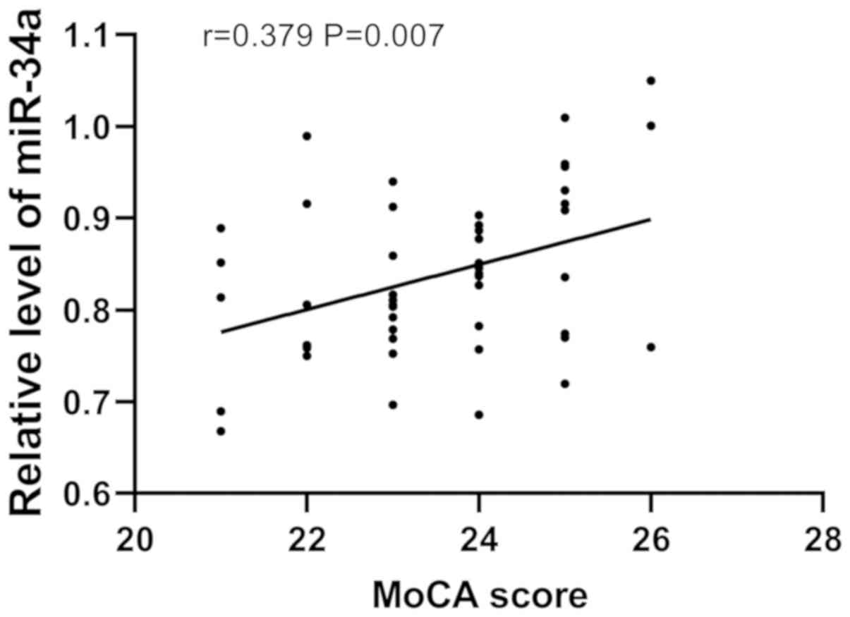
Correlation between miR-34a expression
and MoCA score after treatment
Pearson correlation analysis showed that the
expression of miR-34a was positively correlated with the MoCA score
after treatment, that is, MoCA score increased gradually with the
increase of miR-34a expression (r=0.379, P=0.006), as shown in
Fig. 5.
Discussion
Due to the concealed onset of NPC, majority of
patients are in advanced stage when diagnosed, missing the optimal
treatment timing. At present, early NPC patients are mainly treated
with radiotherapy alone, and patients in advanced stage are treated
with platinum plus radiotherapy (16,17).
Efficacy evaluation is mainly based on imaging tests after
treatment that determine the size of solid tumors. However, the
tests have a certain impact on the human body, and the imaging
results take time (18). Therefore,
we questioned whether it is possible to predict the efficacy by
performing tests on the patient before treatment, in order to
assess the clinical effect in advance and to improve the treatment
efficiency. However, there are few relevant indicators for clinical
prediction of clinical efficacy, so it is very important to find
one.
miRs are a class of highly conserved non-coding
small RNAs widely distributed in eukaryotic cells, which play an
important role in the development and progression of malignant
tumors (19). Studies have shown
that (20,21) a variety of miRs are capable of
regulating the occurrence and development of NPC. Previous studies
have found that miR-34a, a member of the miRs family, regulates
biological function in prostate cancer and renal cell cancer
(22,23). Recent studies have shown that miR-34a
has a low expression in NPC (24).
However, whether it can be used as a efficacy predictor has not
been studied. Therefore, in this study, the predictive value of
miR-34a in efficacy evaluation of NPC patients after CCRT was
explored to provide indicators for the clinic. The expression of
serum miR-34a was first detected, and the results showed that the
expression in the treated group was significantly lower than that
in the control group. Moreover, the ROC curve showed that miR-34a
had extremely high diagnostic value in patients with advanced NPC.
In the study of Long et al (25), it was found that hyperfration
radiotherapy enhanced cell apoptosis and upregulated miR-34a
expression. Moreover, the expression of miR-34a before and after
treatment through clinical trials was compared, and it was found
that the expression was significantly increased after treatment,
which was consistent with the results of Long et al
(25), indicating that the
expression of miR-34a increased after CCRT. Furthermore, the
patients were divided into the significant group and the general
group according to the curative effect, the expression of miR-34a
before treatment between the two groups was compared. The results
indicated that the expression in significant group was
significantly higher than that in general group. ROC curve showed
that the AUC was >0.8, suggesting that observing the expression
of miR-34a before treatment was a potential predictor for assessing
the efficacy of CCRT in patients with NPC. The higher the
expression of miR-34a before treatment, the more significant
improvement of the clinical curative effect.
Although the condition of the patient and survival
are improved, the cognitive function of patients may be declined
after radiotherapy, which reduces the quality of life of patients.
At present, various scores and scale are preferred means to assess
cognitive function, subjective errors are inevitable. A study
(26) found that upregulation of
miR-34a alleviated total abdominal irradiation (TAI)-mediated
cognitive impairment by restoring BFNF expression in hippocampus.
Therefore, we speculated whether the expression of miR-34a is
related to the cognitive function of patients. Therefore, the
changes of cognitive function of patients before and after
treatment were compared, and it was found that the MoCA score after
treatment was significantly lower than that before treatment. Qiu
et al (27) stated that the
MoCA score of NPC patients after radiotherapy was significantly
lower than that before treatment, which was consistent with our
research results. Moreover, Pearson correlation analysis showed
that there was a positive correlation between miR-34a and MoCA
score, indicating that the expression of miR-34a can be used as a
potential observation indicator for patients with cognitive
dysfunction. However, MoCA score may be affected by cancer staging,
side effects, nutritional status and other factors, interfering
with our findings. Thus, we will supplement more cognitive function
related scales to investigate the association between miR-34a and
cognitive function.
This study found that miR-34a can be used as a
predictor of clinical efficacy and an observation indicator of
cognitive function in NPC patients after CCRT. However, there are
still limitations. The survival of NPC patients was not followed
up, so whether miR-34a is related to the survival remains unknown.
This is a clinical experiment, the relevant mechanism between
miR-34a and patients' cognitive function is unclear. Only samples
of patients with advanced NPC were collected, the expression of
miR-34a in patients with early NPC and those without CCRT is not
clear.
In conclusion, there is a positive correlation
between miR-34a and cognitive function of patients. Moreover,
observing the expression of miR-34a before treatment can be used as
a potential observation predictor of the efficacy of CCRT in
patients with NPC.
Acknowledgements
Not applicable.
Funding
No funding was received.
Availability of data and materials
The datasets used and/or analyzed during the present
study are available from the corresponding author on reasonable
request.
Authors' contributions
WD conceived and designed the study, and drafted the
manuscript. WD, AL and JY collected, analyzed and interpreted the
experimental data, and revised the manuscript critically for
important intellectual content. All authors read and approved the
final manuscript.
Ethics approval and consent to
participate
The study was approved by the Ethics Committee of
Shunde Hospital (Foshan, China). Signed informed consents were
obtained from the patients and/or guardians.
Patient consent for publication
Not applicable.
Competing interests
The authors declare that they have no competing
interests.
References
|
1
|
Chua MLK, Wee JTS, Hui EP and Chan ATC:
Nasopharyngeal carcinoma. Lancet. 387:1012–1024. 2016. View Article : Google Scholar : PubMed/NCBI
|
|
2
|
Chen W, Zheng R, Baade PD, Zhang S, Zeng
H, Bray F, Jemal A, Yu XQ and He J: Cancer statistics in China,
2015. CA Cancer J Clin. 66:115–132. 2016. View Article : Google Scholar : PubMed/NCBI
|
|
3
|
Sun Y, Li WF, Chen NY, Zhang N, Hu GQ, Xie
FY, Sun Y, Chen XZ, Li JG, Zhu XD, et al: Induction chemotherapy
plus concurrent chemoradiotherapy versus concurrent
chemoradiotherapy alone in locoregionally advanced nasopharyngeal
carcinoma: A phase 3, multicentre, randomised controlled trial.
Lancet Oncol. 17:1509–1520. 2016. View Article : Google Scholar : PubMed/NCBI
|
|
4
|
Ribassin-Majed L, Marguet S, Lee AWM, Ng
WT, Ma J, Chan ATC, Huang PY, Zhu G, Chua DTT, Chen Y, et al: What
is the best treatment of locally advanced nasopharyngeal carcinoma?
An individual patient data network meta-analysis. J Clin Oncol.
35:498–505. 2017. View Article : Google Scholar : PubMed/NCBI
|
|
5
|
Chen YZ, Li WF, Wang JY, Wang JM, Ou RY,
Zheng XW, Xu YS and Zhao L: Evaluation of time-phase effect on
18F-FDG PET/CT delineation methods for treatment
planning of nasopharyngeal carcinoma. Clin Nucl Med. 41:354–361.
2016. View Article : Google Scholar : PubMed/NCBI
|
|
6
|
Chen Z, Xu L, Xu X and Yuan C: The
clinical value of detecting circulating tumour cells in the
peripheral blood of nasopharyngeal carcinoma patients. Oncol Lett.
15:6283–6290. 2018.PubMed/NCBI
|
|
7
|
McDowell LJ, Ringash J, Xu W, Chan B, Lu
L, Waldron J, Rock K, So N, Huang SH, Giuliani M, et al: A cross
sectional study in cognitive and neurobehavioral impairment in
long-term nasopharyngeal cancer survivors treated with
intensity-modulated radiotherapy. Radiother Oncol. 131:179–185.
2019. View Article : Google Scholar : PubMed/NCBI
|
|
8
|
Fasfous AF, Al-Joudi HF, Puente AE and
Pérez-García M: Neuropsychological measures in the Arab World: A
systematic review. Neuropsychol Rev. 27:158–173. 2017. View Article : Google Scholar : PubMed/NCBI
|
|
9
|
Kozomara A, Birgaoanu M and
Griffiths-Jones S: miRBase: From microRNA sequences to function.
Nucleic Acids Res. 47D:D155–D162. 2019. View Article : Google Scholar
|
|
10
|
Gao W, Lam JW, Li JZ, Chen SQ, Tsang RK,
Chan JY and Wong TS: MicroRNA-138-5p controls sensitivity of
nasopharyngeal carcinoma to radiation by targeting EIF4EBP1. Oncol
Rep. 37:913–920. 2017. View Article : Google Scholar : PubMed/NCBI
|
|
11
|
Wu R, Qiu E, Lin R, Wang J and Lin H:
Regulation of nasopharyngeal carcinoma cell proliferation by
targeting Notch1 with miR-34a. Int J Clin Exp Pathol. 9:8811–8816.
2016.
|
|
12
|
Xu Y, Chen P, Wang X, Yao J and Zhuang S:
miR-34a deficiency in APP/PS1 mice promotes cognitive function by
increasing synaptic plasticity via AMPA and NMDA receptors.
Neurosci Lett. 670:94–104. 2018. View Article : Google Scholar : PubMed/NCBI
|
|
13
|
Edge SB and Compton CC: The American Joint
Committee on Cancer: the 7th edition of the AJCC cancer staging
manual and the future of TNM. Ann Surg Oncol. 17:1471–1474. 2010.
View Article : Google Scholar : PubMed/NCBI
|
|
14
|
Dechaphunkul A, Danchaivijitr P,
Jiratrachu R, Dechaphunkul T, Sookthon C, Jiarpinitnun C, Paoin C,
Setakornnukul J, Niyomnaitham S, Suktitipat B, et al: 1076P
Comparison of 3-weekly cisplatin versus 3-weekly carboplatin in
patients with locally advanced nasopharyngeal carcinoma (LA-NPC)
receiving concurrent chemoradiotherapy (CCRT): A multicenter
retrospective study. Ann Oncol. 29 (Suppl. 8):viii3842018.
View Article : Google Scholar
|
|
15
|
Livak KJ and Schmittgen TD: Analysis of
relative gene expression data using real-time quantitative PCR and
the 2(-Delta Delta C(T)) method. Methods. 25:402–408. 2001.
View Article : Google Scholar : PubMed/NCBI
|
|
16
|
Ai QY, King AD, Mo FKF, Law BKH, Bhatia
KS, Ma BB, Poon DMC and Kam MKM: Prediction of distant metastases
from nasopharyngeal carcinoma: Improved diagnostic performance of
MRI using nodal volume in N1 and N2 stage disease. Oral Oncol.
69:74–79. 2017. View Article : Google Scholar : PubMed/NCBI
|
|
17
|
Tan T, Lim WT, Fong KW, Cheah SL, Soong
YL, Ang MK, Ng QS, Tan D, Ong WS, Tan SH, et al: Concurrent
chemo-radiation with or without induction gemcitabine, Carboplatin,
and Paclitaxel: A randomized, phase 2/3 trial in locally advanced
nasopharyngeal carcinoma. Int J Radiat Oncol Biol Phys. 91:952–960.
2015. View Article : Google Scholar : PubMed/NCBI
|
|
18
|
Chan SC, Yeh CH, Yen TC, Ng SH, Chang JT,
Lin CY, Yen-Ming T, Fan KH, Huang BS, Hsu CL, et al: Clinical
utility of simultaneous whole-body 18F-FDG PET/MRI as a single-step
imaging modality in the staging of primary nasopharyngeal
carcinoma. Eur J Nucl Med Mol Imaging. 45:1297–1308. 2018.
View Article : Google Scholar : PubMed/NCBI
|
|
19
|
Agarwal V, Bell GW, Nam JW and Bartel DP:
Predicting effective microRNA target sites in mammalian mRNAs.
eLife. 4:e050052015. View Article : Google Scholar
|
|
20
|
Spence T, Bruce J, Yip KW and Liu FF:
MicroRNAs in nasopharyngeal carcinoma. Linchuang Zhongliuxue Zazhi.
5:172016.(In Chinese).
|
|
21
|
Lee KT, Tan JK, Lam AK and Gan SY:
MicroRNAs serving as potential biomarkers and therapeutic targets
in nasopharyngeal carcinoma: A critical review. Crit Rev Oncol
Hematol. 103:1–9. 2016. View Article : Google Scholar : PubMed/NCBI
|
|
22
|
Chen WY, Liu SY, Chang YS, Yin JJ, Yeh HL,
Mouhieddine TH, Hadadeh O, Abou-Kheir W and Liu YN: MicroRNA-34a
regulates WNT/TCF7 signaling and inhibits bone metastasis in
Ras-activated prostate cancer. Oncotarget. 6:441–457. 2015.
View Article : Google Scholar : PubMed/NCBI
|
|
23
|
Toraih EA, Ibrahiem AT, Fawzy MS, Hussein
MH, Al-Qahtani SAM and Shaalan AAM: MicroRNA-34a: A key regulator
in the hallmarks of renal cell carcinoma. Oxid Med Cell Longev.
2017:32693792017. View Article : Google Scholar : PubMed/NCBI
|
|
24
|
Huang G, Du MY, Zhu H, Zhang N, Lu ZW,
Qian LX, Zhang W, Tian X, He X and Yin L: miRNA-34a reversed
TGF-β-induced epithelial-mesenchymal transition via suppression of
SMAD4 in NPC cells. Biomed Pharmacother. 106:217–224. 2018.
View Article : Google Scholar : PubMed/NCBI
|
|
25
|
Long Z, Wang B, Tao D, Huang Y and Tao Z:
Hypofractionated radiotherapy induces miR-34a expression and
enhances apoptosis in human nasopharyngeal carcinoma cells. Int J
Mol Med. 34:1388–1394. 2014. View Article : Google Scholar : PubMed/NCBI
|
|
26
|
Cui M, Xiao H, Li Y, Dong J, Luo D, Li H,
Feng G, Wang H and Fan S: Total abdominal irradiation exposure
impairs cognitive function involving miR-34a-5p/BDNF axis. Biochim
Biophys Acta Mol Basis Dis. 1863:2333–2341. 2017. View Article : Google Scholar : PubMed/NCBI
|
|
27
|
Qiu Y, Guo Z, Han L, Yang Y, Li J, Liu S
and Lv X: Network-level dysconnectivity in patients with
nasopharyngeal carcinoma (NPC) early post-radiotherapy:
Longitudinal resting state fMRI study. Brain Imaging Behav.
12:1279–1289. 2018. View Article : Google Scholar : PubMed/NCBI
|