Introduction
Uterine corpus endometrial cancer (UCEC) is the
second most prevalent type of malignancy among women in the United
States of America (1). Despite the
rapid development of the modern medical industry, the mortality of
UCEC has been continuously increasing (2). Due to a lack of effective therapeutic
strategies, the 5-year survival rate of patients with
advanced-stage disease is only 16%. However, patients diagnosed at
an early stage have a favorable prognosis (3,4).
Recently, cancer antigen 125 (CA125) and human epididymis protein 4
(HE4) have been utilized as serum biomarkers in UCEC; however, they
only have modest effects due to relatively low predictive accuracy
(5–7). Therefore, it is necessary to identify
reliable molecular biomarkers to predict prognosis, guide
treatments and monitor recurrence.
Mammaglobin B, also referred to as secretoglobin
family 2A member 1 (SCGB2A1), is a member of the uteroglobin
superfamily which is localized on chromosome 11q12.2 and includes
nine human secretoglobins (8,9).
SCGB2A1 was first isolated from the human endometrium, and
it is highly homologous to mammaglobin A (secretoglobin family 2A
member 2) (10). Although its
biological function has not been clarified, the differential
expression and specific significance of SCGB2A1 in various
malignancies have been reported (11). SCGB2A1 has been identified as
a candidate biomarker for the detection of lymph node
micrometastases in breast cancer (12,13) and
abdominal cancer types (14). In
addition, SCGB2A1 has been considered as a promising
diagnostic marker for occult tumor cells in effusions of several
malignancies (15,16) and as a potential immunotherapeutic
target in ovarian cancer (17).
However, to the best of our knowledge, the prognostic value of
SCGB2A1 in UCEC has not been reported, although Tassi et
al (18) observed the
overexpression of SCGB2A1 in endometrioid endometrial
cancer.
The present study assessed the prognostic
significance of SCGB2A1 in UCEC using bioinformatics.
Additionally, gene set enrichment analysis (GSEA) was performed to
further explore the function of SCGB2A1. A number of other
databases were utilized to explore the significance of
SCGB2A1 in transcriptomics, proteomics, and the immune
microenvironment. In conclusion, the present study may provide
further insights into potential therapeutic targets in UCEC.
Materials and methods
Oncomine database analysis
The Oncomine database (http://www.oncomine.com) (19) was utilized to compare the
differential expression levels of SCGB2A1 between tumor and
normal tissues in various tumor types. The threshold was set
according to the following values: P<0.0001; fold change >2;
and gene ranking of all.
Clinical Proteomic Tumor Analysis
Consortium (CPTAC) database analysis
The CPTAC database enables large-scale proteome and
genome analyses, in order to understand the molecular basis of
cancer (20). UALCAN (http://ualcan.path.uab.edu) (21), a comprehensive web resource for
analyzing cancer-omics data, includes CPTAC analysis for various
tumor types. The analysis of protein expression levels of
SCGB2A1 in UCEC was performed by UALCAN based on the CPTAC
database. UALCAN performed the comparison of differential
expression between each two groups by using t-tests (22), and similar results from the UALCAN
using the same statistical methods have been published previously
(23–25). Differential protein expression of
SCGB2A1 between UCEC and normal tissues, and the association
between clinical characteristics and protein expression levels of
SCGB2A1, were analyzed. Additionally, all P-values from the
UALCAN were adjusted using Bonferroni's correction.
Tumor Immune Estimation Resource
(TIMER) analysis
TIMER (https://cistrome.shinyapps.io/timer/) (26) is a tool for the systematic analysis
of tumor-infiltrating immune cells (TIICs) across diverse types of
cancer in The Cancer Genome Atlas (TCGA) database (https://cancergenome.nih.gov/) (27). TIMER consists of several modules: The
‘DiffExp’ module provides the differential expression between tumor
and adjacent normal tissues for genes in TCGA; the ‘Gene’ module
provides visualization of the association between gene expression
and tumor purity and immune infiltration levels in tumors; the
‘Survival’ module provides survival curves of TIICs at high and low
levels and genes in specific tumors; and the ‘SCNA’ module provides
the comparison of tumor infiltration levels among tumors with
different somatic copy number alterations (SCNAs) for a given gene.
Defined by Genomic Identification of Significant Targets in Cancer
2.0 (28,29), SCNAs include deep deletion (−2),
arm-level deletion (−1), diploid/normal (0), arm-level gain
(1) and high amplification (2). The infiltration level for each SCNA
category in UCEC was compared with that in normal tissues using a
Wilcoxon rank-sum test. SCGB2A1 was analyzed using the
‘DiffExp’, ‘Gene’, ‘Survival’, and ‘SCNA’ modules.
Downloaded data
RNA-sequencing (RNA-seq) expression data of UCEC and
corresponding clinical data were downloaded from TCGA. The details
of RNA-seq data were as follows: Project, TCGA-UCEC; data category,
transcriptome profiling; data type, gene expression quantification;
workflow type, HTSeq-FPKM. Furthermore, data of normal samples were
excluded.
Statistical analysis and nomogram
construction
Statistical analysis was performed using R software
(v.3.6.2) (30). Expression
differences for discrete variables were visualized using boxplots
and the survival curve was drawn using the survival package
(https://cran.r-project.org/web/views/Survival.html).
The association between clinical characteristics and SCGB2A1
expression was determined by logistic regression analysis. Notably,
the median value of SCGB2A1 expression was set as the
cut-off value. Furthermore, univariate Cox analysis was used to
estimate the prognostic value of certain clinicopathologic
variables, including age, BMI, grade, stage, peritoneal cytology,
pelvic lymph node status, para-aortic lymph node status,
histological subtype, myometrial invasion, residual tumor and tumor
status. Additionally, multivariate Cox analysis was performed to
identify the independent prognostic value of SCGB2A1 with
stage, peritoneal cytology, pelvic lymph node status, myometrial
invasion, and tumor status.
Following integration of the results of univariate
and multivariate Cox analysis, 6 variables (stage, tumor status,
peritoneal cytology, pelvic lymph node status, myometrial invasion,
and SCGB2A1 expression) were selected for nomogram
construction. The rms package (https://cran.r-project.org/web/packages/rms/index.html)
in R was used to construct the nomogram.
GSEA
The present study performed GSEA (31), which determines whether an a
priori defined set of genes indicates statistically significant
differences between 2 biological states, to identify the potential
mechanism of SCGB2A1 in UCEC. In the present study, GSEA
software v3.0 was used to analyze the ‘h.all.v6.2.symbols.gmt’ and
‘c2.cp.biocarta.v6.2.symbols.gmt’ gene sets from the Molecular
Signatures Database (32). Based on
the expression levels of SCGB2A1, ‘high’ and ‘low’ were
applied as phenotype labels. For each analysis, 1,000 gene set
permutations were run to obtain the normalized enrichment score
(NES). False discovery rate <0.25 and normal P<0.05, were
used as the cut-off to identify the significantly enriched gene
sets.
Results
Pan-cancer analysis of SCGB2A1 mRNA
expression in different databases
The Oncomine and TCGA databases were utilized to
determine the mRNA expression levels of SCGB2A1 in tumor and
normal tissues in different tumor types. According to the Oncomine
database, SCGB2A1 was expressed at low levels in breast,
colorectal, gastric, and kidney cancer, melanoma, ovarian and
prostate cancer, and sarcoma, whereas overexpression of
SCGB2A1 was identified in breast, esophageal, kidney, and
ovarian cancer in some analyses (P<0.0001; Fig. 1A). Detailed information of
SCGB2A1 expression in various cancer types based on the
Oncomine database is shown in Table
SI. In addition, all tumor and adjacent normal tissues in TCGA
were analyzed to further comprehend the differential expression of
SCGB2A1 (Fig. 1B). The
results revealed that SCGB2A1 expression was markedly
decreased in breast invasive carcinoma, colon adenocarcinoma,
esophageal carcinoma, head and neck cancer, kidney renal clear cell
carcinoma, lung adenocarcinoma, lung squamous cell carcinoma,
prostate, rectum, and stomach adenocarcinoma, and thyroid carcinoma
compared with in adjacent normal tissues. However, SCGB2A1
expression was markedly increased in cholangiocarcinoma, liver
hepatocellular carcinoma and UCEC tissues compared with in adjacent
normal tissues.
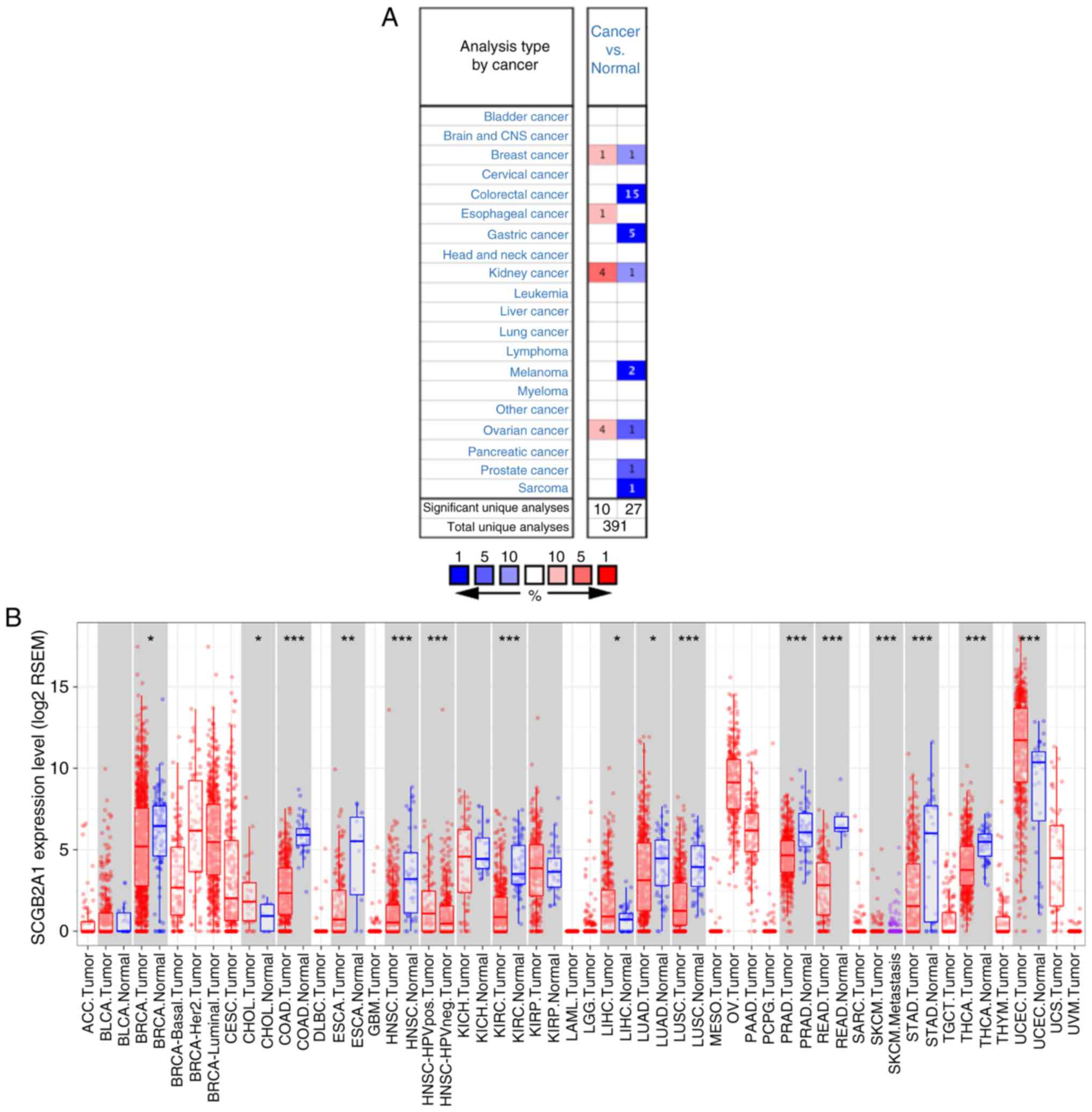 | Figure 1.Pan-cancer analysis of SCGB2A1 mRNA
expression in different databases. (A) SCGB2A1 mRNA expression in
different tumor types compared with normal samples according to
different analyses of the Oncomine database. The number represents
the count of significant unique analyses. Red represents
overexpression of the gene and blue represents low expression of
the gene. All P<0.0001 cancer vs. normal. (B) mRNA expression
levels of SCGB2A1 in different tumor types analyzed by TIMER based
on TCGA. Red indicates tumor tissues and blue indicates normal
tissues. *P<0.05, **P<0.01 and ***P<0.001. SCGB2A1,
secretoglobin family 2A member 1; TIMER, Tumor Immune Estimation
Resource; TCGA, The Cancer Genome Atlas; BRCA, breast invasive
carcinoma; CHOL, cholangiocarcinoma; COAD, colon adenocarcinoma;
ESCA, esophageal carcinoma; HNSC, head and neck cancer; KIRC,
kidney renal clear cell carcinoma; LIHC, liver hepatocellular
carcinoma; LUAD, lung adenocarcinoma; LUSC, lung squamous cell
carcinoma; PRAD, prostate adenocarcinoma; READ, rectum
adenocarcinoma; SKCM, skin cutaneous melanoma; STAD, stomach
adenocarcinoma; THCA, thyroid carcinoma; UCEC, uterine corpus
endometrial carcinoma. |
Patient characteristics
Gene expression and clinical data of 545 primary
tumors from the TCGA-UCEC project were downloaded in June 2019.
After discarding unqualified samples with apparently abnormal data
or gene expression data missing (Table
SII), the data of 540 patients were retained for further
analysis. Notably, a 64-year-old female patient was identified in
the database with a weight of 93 kg, but her height was recorded as
only 66 cm. As the accuracy of these data could not be verified,
the data of this patient was excluded in a previous study (33). Therefore, this data was defined as
apparently abnormal data in the present study. The
clinicopathological characteristics of these patients, including
age, BMI, grade, stage, peritoneal cytology status, lymph node
status, histology, myometrial invasion, tumor status, residual
tumor, and surgery approach, are shown in Table I. The median age of these patients
was 64 years old, ranging between 31 and 90 years old, while the
median BMI was 32.2, ranging between 17.4 and 81.6.
 | Table I.Clinical characteristics of patients
with uterine corpus endometrial cancer (n=540) downloaded from The
Cancer Genome Atlas database. |
Table I.
Clinical characteristics of patients
with uterine corpus endometrial cancer (n=540) downloaded from The
Cancer Genome Atlas database.
| Clinical
characteristics | Value | % |
|---|
| Median age (range),
years | 64 (31–90) |
|
| Median BMI
(range) | 32.2
(17.4–81.6) |
|
| Grade, n |
|
|
| 1 | 97 | 18.3 |
| 2 | 120 | 22.7 |
| 3 | 312 | 59.0 |
| Stage, n |
|
|
| I | 337 | 62.4 |
| II | 51 | 9.4 |
|
III | 123 | 22.8 |
| IV | 29 | 5.4 |
| Peritoneal
cytology, n |
|
|
|
Negative | 349 | 86.0 |
|
Positive | 57 | 14.0 |
| Pelvic lymph nodes,
n |
|
|
|
Negative | 366 | 83.2 |
|
Positive | 74 | 16.8 |
| Para-aortic lymph
nodes, n |
|
|
|
Negative | 327 | 89.6 |
|
Positive | 38 | 10.4 |
| Histology, n |
|
|
|
Endometrioid | 404 | 74.8 |
| Mixed
serous and endometrioid | 22 | 4.1 |
|
Serous | 114 | 21.1 |
| Myometrial
invasion, n |
|
|
|
≤50% | 314 | 67.1 |
|
>50% | 154 | 32.9 |
| Status, n |
|
|
| With
tumor | 78 | 15.5 |
|
Tumor-free | 425 | 84.5 |
| Residual tumor,
n |
|
|
| R0 | 370 | 90.7 |
| R1 | 22 | 5.4 |
| R2 | 16 | 3.9 |
| Surgical approach,
n |
|
|
|
Minimally invasive | 201 | 38.8 |
|
Open | 317 | 61.2 |
mRNA expression levels of SCGB2A1 in
UCEC according to TCGA
As shown in Fig. 2A
and S1, the expression levels of SCGB2A1 in normal tissues
were significantly decreased compared with those in UCEC, G3
cancer, stage III or IV, with tumors, and peritoneal
cytology-positive tissues (P<0.05), and no significant
differential expression was identified between normal tissues and
serous endometrial adenocarcinoma and stage IV tissues.
Furthermore, the association between SCGB2A1 expression and
clinicopathological variables in UCEC was analyzed using boxplots.
The results indicated that the decreased expression levels of
SCGB2A1 were significantly associated with the grade
(P<0.001), stage (P<0.001), tumor status (P<0.001),
histological subtype (P<0.001) and peritoneal cytology status
(P=0.005) (Fig. 2B-F). Additionally,
the results of the logistic regression analysis revealed that
decreased expression levels of SCGB2A1 were significantly
associated with poor prognostic clinicopathological features,
including grade [odds ratio (OR)=0.11 for grade 3 vs. grade 1 or 2;
P<0.001], stage (OR=0.35 for stage III or IV vs. stage I or II;
P<0.001), peritoneal cytology status (OR=0.37 for positive vs.
negative; P=0.001), pelvic lymph node status (OR=0.26 for positive
vs. negative; P<0.001), para-aortic lymph node status (OR=0.49
for positive vs. negative; P=0.045), histological subtype (OR=0.09
for serous vs. endometrioid; P<0.001), myometrial invasion
(OR=0.47 for >50 vs. ≤50%; P<0.001), status (OR=0.31 for with
tumor vs. tumor-free; P<0.001) and residual tumor (OR=0.49 for
R1 or R2 vs. R0; P=0.044) (Table
II).
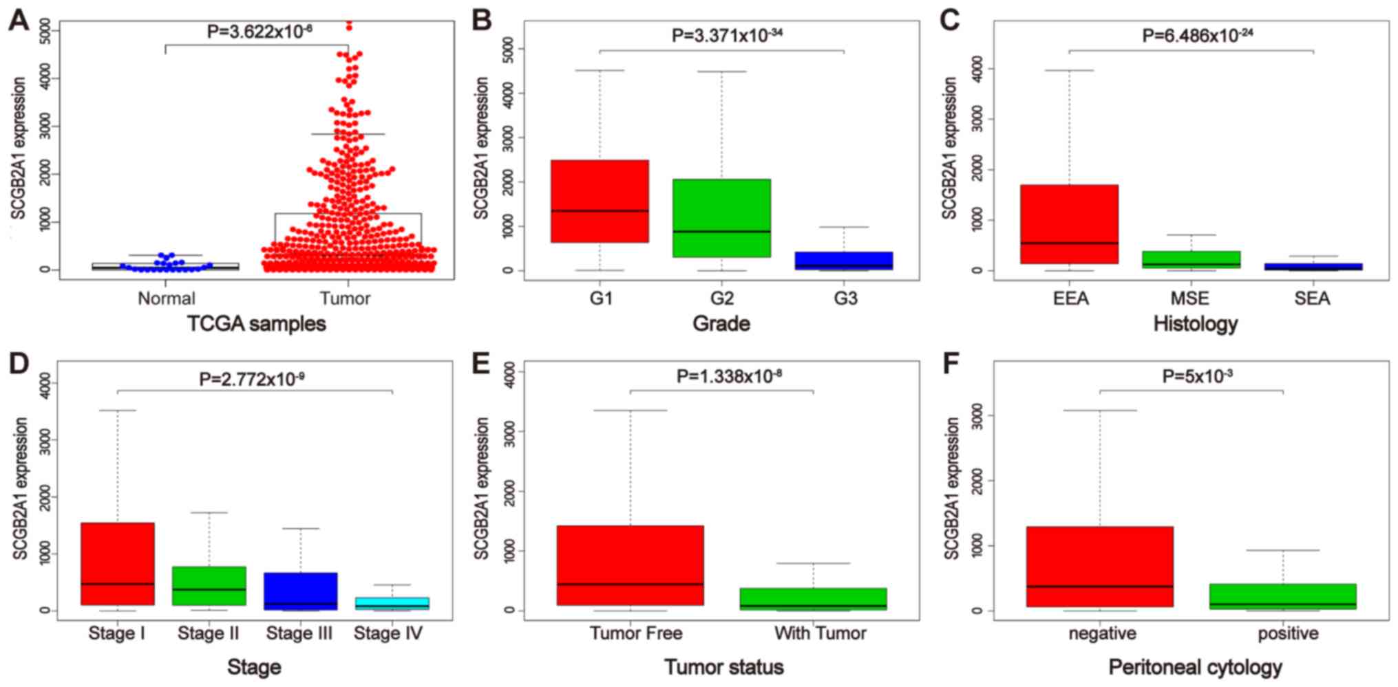 | Figure 2.mRNA expression levels of SCGB2A1
according to The Cancer Genome Atlas database. (A) Differential
mRNA expression of SCGB2A1 between UCEC and normal tissues.
Boxplots of the association between SCGB2A1 mRNA expression and
clinicopathological characteristics, including (B) grade, (C)
histology, (D) stage, (E) tumor status, and (F) peritoneal
cytology. SCGB2A1, secretoglobin family 2A member 1; UCEC, uterine
corpus endometrial carcinoma; EEA, endometrioid endometrial
adenocarcinoma; MSE, mixed serous and endometrioid; SEA, serous
endometrial adenocarcinoma. |
| Table II.Logistic regression on the
association between SCGB2A1 expression and clinical pathological
characteristics. |
Table II.
Logistic regression on the
association between SCGB2A1 expression and clinical pathological
characteristics.
| Clinical
characteristics | Total (N) | Odds ratio in
SCGB2A1 expression | P-value |
|---|
| Age
(continuous) | 538 | 0.96
(0.94–0.98) |
<0.01a |
| BMI
(continuous) | 509 | 1.04
(1.02–1.07) |
<0.01a |
| Grade (3 vs. 1 or
2) | 529 | 0.11
(0.08–0.17) |
<0.01a |
| Stage (III or IV
vs. I or II) | 540 | 0.35
(0.23–0.51) |
<0.01a |
| Peritoneal cytology
(positive vs. negative) | 406 | 0.37
(0.20–0.67) | 0.001a |
| Pelvic lymph nodes
(positive vs. negative) | 440 | 0.26
(0.14–0.45) |
<0.01a |
| Para-aortic lymph
nodes (positive vs. negative) | 365 | 0.49
(0.23–0.97) | 0.045a |
| Histology (serous
vs. endometrioid) | 518 | 0.09
(0.05–0.16) |
<0.01a |
| Myometrial invasion
(>50vs. ≤50%) | 468 | 0.47
(0.32–0.70) |
<0.01a |
| Status (with tumor
vs. tumor free) | 503 | 0.31
(0.18–0.53) |
<0.01a |
| Residual tumor (R1
or R2 vs. R0) | 408 | 0.49
(0.24–0.97) | 0.044a |
| Surgical approach
(open vs. minimally invasive) | 518 | 0.95
(0.67–1.36) | 0.787 |
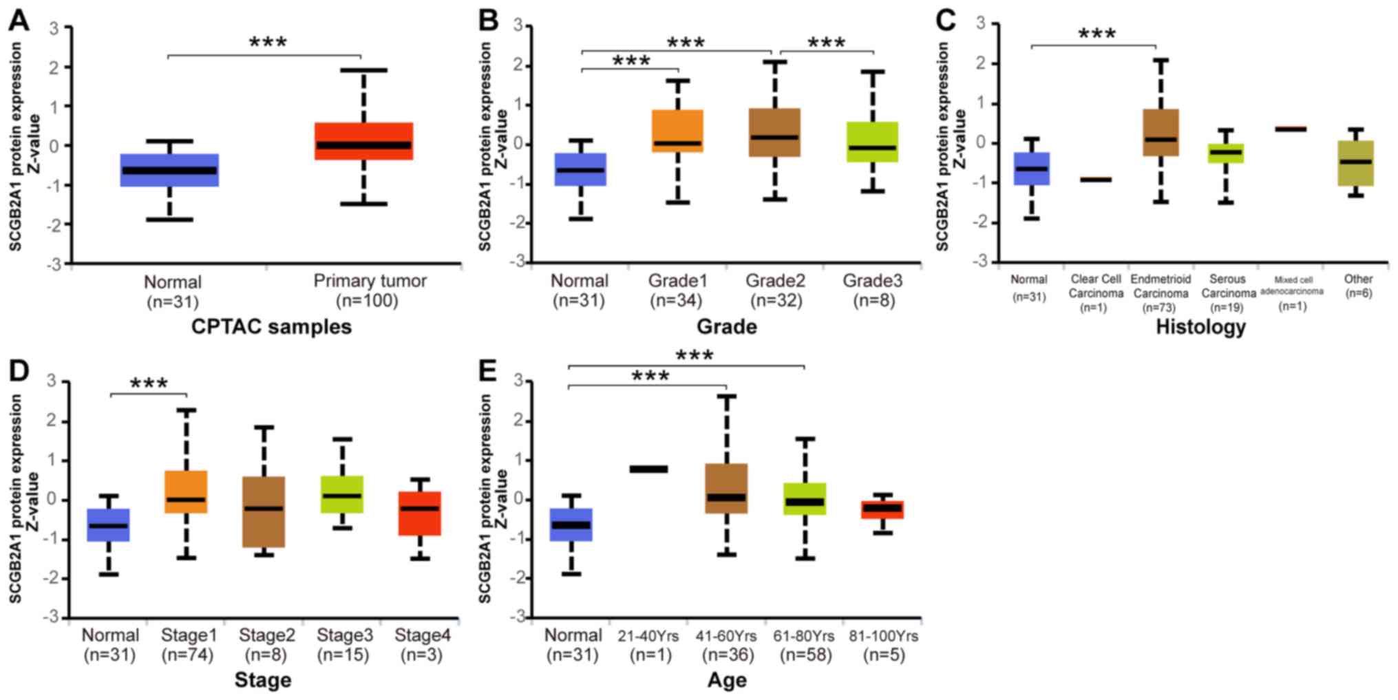
Protein expression levels of SCGB2A1
in UCEC according to CPTAC database
Analysis of the protein expression levels of
SCGB2A1 in UCEC was performed by UALCAN based on the CPTAC
database. As shown in Fig. 3A, the
protein expression levels of SCGB2A1 in UCEC were
significantly increased compared with those in normal tissues
(P<0.05). Furthermore, the association between SCGB2A1
protein expression and clinicopathological variables in UCEC is
shown in Fig. 3B-E. The results
revealed that decreased protein expression levels of SCGB2A1
were associated with high grade (P<0.05). No significant
association was identified between decreased protein expression
levels of SCGB2A1 and serous histological subtype, advanced
stage and advanced age.
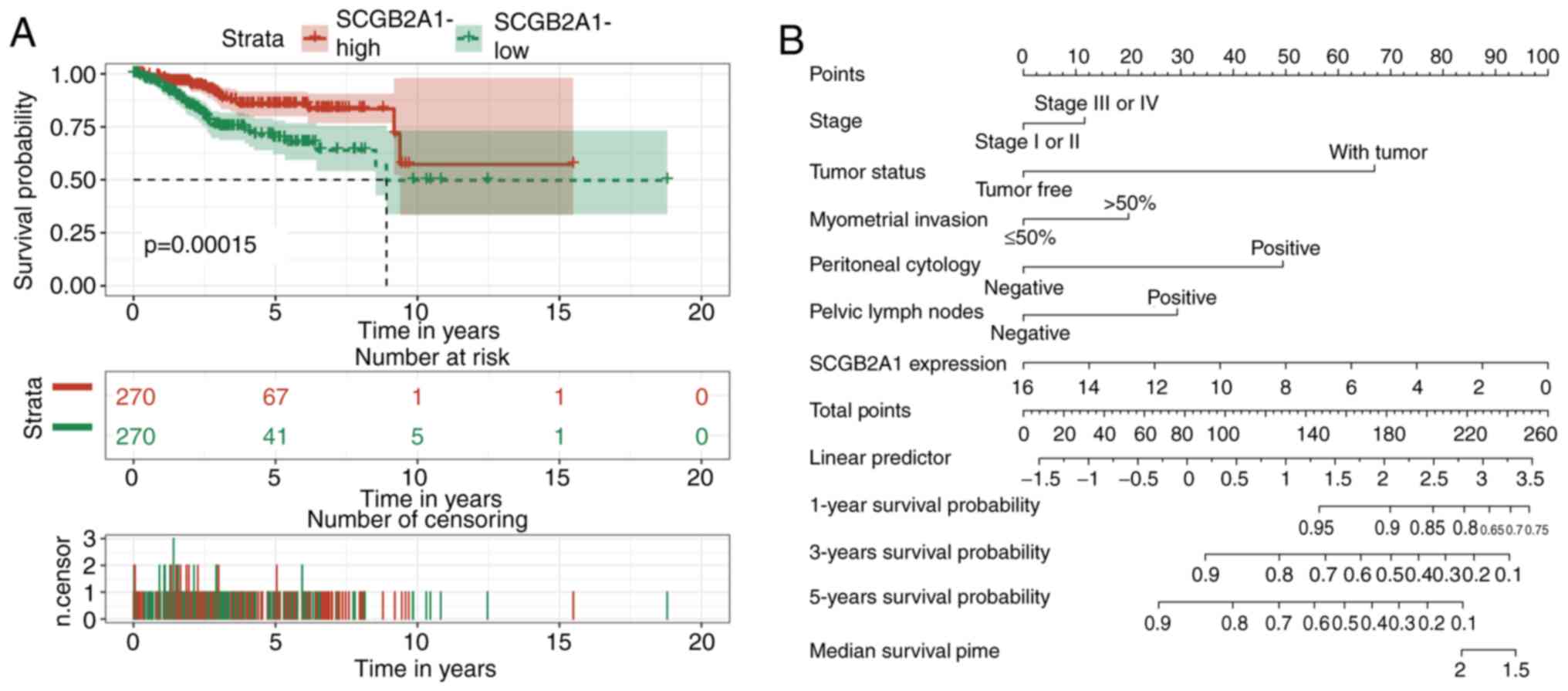
Analysis of the prognostic value of
SCGB2A1 mRNA expression and clinicopathological variables in
UCEC
The survival curve suggested that low expression
levels of SCGB2A1 were associated with poor prognosis in
UCEC (Fig. 4A). Furthermore, the
prognostic value of SCGB2A1 was estimated by univariate Cox
analysis (Table III). It was
revealed that low expression levels of SCGB2A1, advanced
stage, positive peritoneal cytology status and pelvic lymph node
status, deep myometrial invasion, ‘with tumor status’ and residual
tumor were associated with poor prognosis in UCEC (Table III). As defined in TCGA, ‘with
tumor status’ meant that new tumors occurred after operation during
the follow-up, while ‘tumor-free status’ meant that no new tumors
occurred until the follow-up finished. Finally, multivariate Cox
analysis was performed to estimate the independent prognostic value
of SCGB2A1. Considering that residual tumor was uncommon in
clinical practice, this variable was not included in the
multivariate analysis. The results revealed that, in addition to
stage, peritoneal cytology, pelvic lymph node status, myometrial
invasion, and tumor status, SCGB2A1 was independently
associated with poor prognosis in UCEC (hazard ratio, 0.88;
P=0.025; Table III).
| Table III.Univariate and multivariate analyses
of the association between SCGB2A1 expression with overall survival
among patients with uterine corpus endometrial cancer. |
Table III.
Univariate and multivariate analyses
of the association between SCGB2A1 expression with overall survival
among patients with uterine corpus endometrial cancer.
|
| Univariate
analysis | Multivariate
analysis |
|---|
|
|
|
|
|---|
| Parameters | HR (95% CI) | P-value | HR (95% CI) | P-value |
|---|
| Age
(continuous) | 1.03
(0.99–1.08) | 0.156 | ‒ | ‒ |
| BMI
(continuous) | 1.03
(0.97–1.09) | 0.284 | ‒ | ‒ |
| Grade (3 vs. 1 or
2) | 1.91
(0.76–4.81) | 0.167 | ‒ | ‒ |
| Stage (III or IV
vs. I or II) | 5.62
(2.29–13.79) | 0.000a | 1.27
(0.50–3.22) | 0.615 |
| Peritoneal cytology
(positive vs. negative) | 4.21
(1.62–10.97) | 0.003a | 2.75
(1.27–5.97) |
0.010a |
| Pelvic lymph nodes
(positive vs. negative) | 1.58
(1.26–1.98) | 0.000a | 1.82
(0.77–4.33) | 0.174 |
| Para-aortic lymph
nodes (positive vs. negative) | 1.57
(0.36–6.78) | 0.546 | ‒ | ‒ |
| Histology (serous
vs. endometrioid) | 2.35
(0.90–6.14) | 0.081 | ‒ | ‒ |
| Myometrial invasion
(>50 vs. ≤50%) | 2.62
(1.09–6.31) | 0.032a | 1.51
(0.71–3.21) | 0.290 |
| Status (with tumor
vs. tumor-free) | 6.00
(2.49–14.43) | 0.000a | 3.93
(1.97–7.87) |
<0.01a |
| Residual tumor (R1
or R2 vs. R0) | 3.19
(1.16–8.77) | 0.025a | ‒ | ‒ |
| SCGB2A1 expression
(continuous) | 0.82
(0.72–0.93) | 0.003a | 0.88
(0.79–0.98) | 0.025a |
Construction of the nomogram
A nomogram was constructed for the prediction of 1-,
3-, and 5-year survival probabilities of patients with UCEC based
on 6 variables, including stage, tumor status, myometrial invasion,
peritoneal cytology, pelvic lymph node status, and SCGB2A1
expression (Fig. 4B). According to
this nomogram, the variables corresponded to the respective points,
and the sum of the six variable points was defined as the total
points. Additionally, the estimated 1-, 3-, and 5-year survival
probability could be obtained based on the total points.
GSEA
Based on the value of the NES, the most
significantly enriched signaling pathways were selected. As
demonstrated in Fig. 5, the vascular
endothelial growth factor (VEGF) pathway, PTEN pathway,
platelet-derived growth factor (PDGF) pathway, DNA repair,
coactivator associated arginine methyltransferase (CARM) and
estrogen receptor (ER) pathway, KRAS signaling pathway,
PI3K-AKT-mTOR signaling pathway, ataxia-telangiectasia and
Rad3-related (ATR) and BRCA pathway, and G2M checkpoint were
significantly enriched in the SCGB2A1 low-expression
phenotype. The details are shown in Table IV.
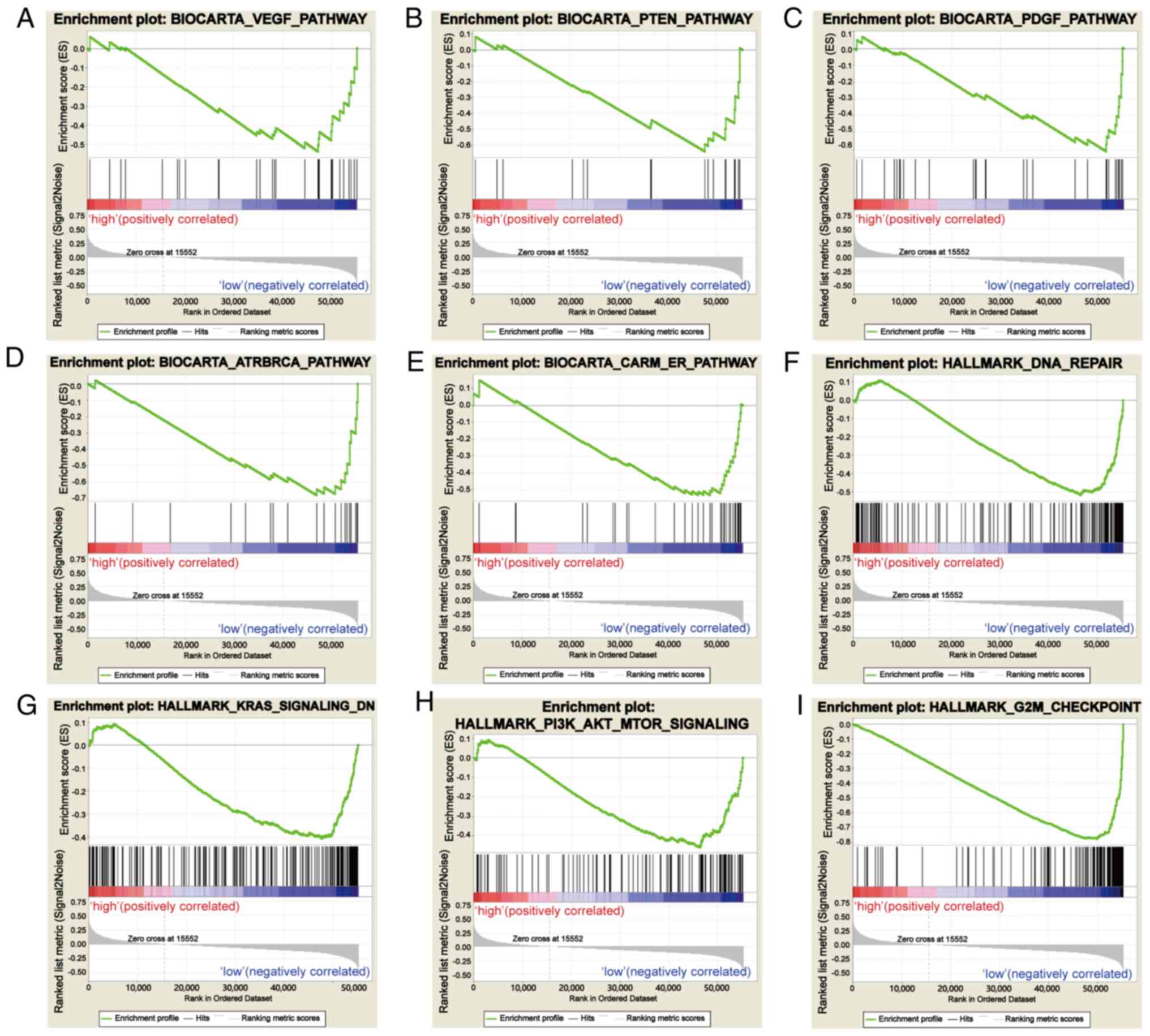 | Figure 5.Enrichment plots from gene set
enrichment analysis. (A) VEGF pathway, (B) PTEN pathway, (C) PDGF
pathway, (D) ATR and BRCA pathway, (E) CARM and ER pathway, (F) DNA
repair, (G) KRAS signaling pathway, (H) PI3K-AKT-mTOR signaling
pathway, and (I) the G2M checkpoint were differentially enriched in
SCGB2A1-associated uterine corpus endometrial carcinoma. VEGF,
vascular endothelial growth factor; PDGF, platelet-derived growth
factor; ATR, ataxia-telangiectasia and Rad3-related; CARM,
coactivator associated arginine methyltransferase; ER, estrogen
receptor; SCGB2A1, secretoglobin family 2A member 1. |
| Table IV.Gene sets enriched in phenotype
low. |
Table IV.
Gene sets enriched in phenotype
low.
| MSigDB
collection | Gene set name | NES | NOM P-value | FDR q-value |
|---|
|
c2.cp.biocarta.v6.2.symbols.gmt |
BIOCARTA_VEGF_PATHWAY | −1.681 | 0.027 | 0.072 |
|
|
BIOCARTA_PTEN_PATHWAY | −1.703 | 0.025 | 0.070 |
|
|
BIOCARTA_PDGF_PATHWAY | −1.896 | 0.000 | 0.042 |
|
|
BIOCARTA_ATRBRCA_PATHWAY | −1.690 | 0.025 | 0.069 |
|
|
BIOCARTA_CARM_ER_PATHWAY | −1.698 | 0.019 | 0.069 |
|
h.all.v6.2.symbols.gmt |
HALLMARK_DNA_REPAIR | −1.722 | 0.044 | 0.069 |
|
|
HALLMARK_KRAS_SIGNALING_DN | −1.675 | 0.009 | 0.073 |
|
|
HALLMARK_PI3K_AKT_MTOR_SIGNALING | −1.777 | 0.008 | 0.057 |
|
|
HALLMARK_G2M_CHECKPOINT | −2.278 | 0.000 | 0.005 |
Systematic analysis of immune
infiltrates associated with SCGB2A1 mRNA expression in UCEC
TIMER was used to further investigate the
association between SCGB2A1 and immune infiltration in UCEC.
SCGB2A1 exhibited a significant positive association with
the infiltration level of CD8+ T cells (P<0.05) and
macrophages (P<0.05), and a negative association with
neutrophils (P<0.05) (Fig. 6A).
Furthermore, high infiltration levels of B cells and
CD8+ T cells were statistically significant in UCEC
according to the cumulative survival analysis (P<0.05; Fig. 6B). Finally, the distribution of tumor
infiltration levels in UCEC with different SCNAs for SCGB2A1
is shown in Fig. 6C. Compared with
those in normal tissues, the infiltration levels of B cells,
CD8+ T cells, CD4+ T cells, macrophages,
neutrophils and dendritic cells for high amplification in UCEC were
significantly different (P<0.05). In addition, the infiltration
levels of CD8+ T cells and dendritic cells for arm-level
gain in UCEC were statistically different from those of the normal
tissues (P<0.05).
Discussion
The present study revealed that decreased expression
levels of SCGB2A1 were associated with poor prognostic
clinicopathological characteristics and short survival time in
UCEC. In addition, the significance of SCGB2A1 in
transcriptomics, proteomics and the immune microenvironment was
explored using Oncomine, CPTAC and TIMER. However, in certain
cancer types, SCGB2A1 expression is controversial. In
breast, kidney, and ovarian cancer, SCGB2A1 was identified
to be highly expressed in some analyses, while in other analyses,
it was identified to be expressed at low levels (Fig. 1A). Based on the detailed information
in Table SI, it was proposed that
different cancer subtypes and the number of samples may affect
SCGB2A1 expression. Additionally, a nomogram based on 6
variables, including SCGB2A1 expression, was developed for
the estimation of the 1-, 3-, and 5-year survival probability in
UCEC. GSEA was utilized to further understand the function of
SCGB2A1, which revealed that the VEGF, PTEN, and PDGF
pathways, DNA repair, CARM and ER, KRAS, and PI3K-AKT-mTOR
signaling pathways, and the ATR and BRCA pathway were
differentially enriched in the low SCGB2A1 expression
phenotype. These results suggested that SCGB2A1 may be
considered as a candidate prognostic marker and a novel therapeutic
target in UCEC.
As a member of the uteroglobin gene family,
SCGB2A1 was first isolated from the human endometrium
(10); however, it has rarely been
investigated in UCEC. Tassi et al (18) reported that SCGB2A1 was
upregulated in endometrioid endometrial cancer tissues compared
with normal tissues; however, the aforementioned study presented
some limitations due to a lack of prognostic analysis and subgroup
analysis in UCEC. The present study revealed the differential
expression of SCGB2A1 in UCEC, and that the mRNA and protein
expression levels of SCGB2A1 in serous carcinoma were
decreased compared with those in endometrioid carcinoma, which
suggested that SCGB2A1 may be involved in the carcinogenesis
of UCEC cells. Although no significant differential expression of
SCGB2A1 was identified between normal tissues and serous
carcinoma and stage IV cancer tissues, the expression levels of
SCGB2A1 in normal tissues were significantly decreased
compared with those in G3 cancer, stage III or IV, with tumor and
peritoneal cytology-positive tissues (P<0.05). The specific
mechanism requires further exploration. In UCEC, genetic
alternations of KRAS and PTEN are common (34,35).
PTEN is an essential tumor suppressor gene in UCEC (36), and changes in PTEN could result in
disorders of the cell cycle, and abnormal proliferation and
differentiation in carcinogenesis (37). As an oncogene, KRAS has a synergistic
effect with PTEN in tumorigenesis and upregulates the expression
levels of ER (38,39). Furthermore, the activation of the
PI3K-AKT-mTOR signaling pathway via the ER signaling pathway
results in cell proliferation (40).
The present results revealed that SCGB2A1 was associated
with the PTEN, KRAS, and PI3K-AKT-mTOR signaling pathways.
Therefore, SCGB2A1 may be involved in the carcinogenesis of
UCEC by mediating cell proliferation via these signaling pathways.
Although these pathways have not been reported to be associated
with SCGB2A1, further exploration is required.
In the past, the prognostic value of SCGB2A1
expression has been analyzed in some specific tumors. Higher
expression levels of SCGB2A1 may decrease the risk of
recurrence of epithelial ovarian cancer (9). However, upregulation of SCGB2A1
in colorectal cancer decreases the sensitivity to 5-fluorouracil
and oxaliplatin, and promotes chemoresistance and radio-resistance,
which results in poor prognosis (16). To the best of our knowledge, the
prognostic value of SCGB2A1 in UCEC remains unclear. The
results of the present study revealed that decreased SCGB2A1
expression was associated with short survival time in UCEC.
Furthermore, a nomogram was constructed to predict the prognosis of
patients with UCEC more accurately. Notably, SCGB2A1
expression levels decreased as age, stage, grade, and level of
myometrial invasion increased, suggesting that SCGB2A1 may
be associated with the progression of UCEC. Furthermore,
SCGB2A1 was downregulated in samples with positive
peritoneal cytology, and positive pelvic lymph node and para-aortic
lymph node statuses, and upregulated in the samples with negative
statuses of these indicators. It has been acknowledged that
angiogenesis is a common process in the development of tumors,
including UCEC (41,42). VEGF acts as a key mediator of tumor
angiogenesis, and it is upregulated by the induction of several
growth factors and hypoxia (43,44). In
addition, overexpression of VEGF in UCEC has been reported to be
associated with deep myometrial invasion and lymph node metastasis
(45). Therefore, SCGB2A1 may
be involved in the progression of UCEC by mediating angiogenesis
via the VEGF signaling pathway. Additionally, serum biomarkers are
critical during the management of patients with cancer in clinical
practice, while advances in UCEC are limited. CA125 and HE4 have
been identified as promising serum biomarkers in guiding the
management of UCEC, but some limitations remain (46). Further analysis of serum levels of
SCGB2A1 may prompt it to become a potential marker for
monitoring the development of UCEC and predicting prognosis
(47).
The present study performed immune infiltration
analysis of SCGB2A1 in UCEC, and the levels of B cells,
CD8+ T cells, macrophages and neutrophils were
identified to be statistically significant. To the best of our
knowledge, no studies have been reported regarding the association
between SCGB2A1 and TIICs in UCEC, but there are some
analyses regarding the effect of TIICs on UCEC (48–50). A
previous study revealed that high levels of CD8+ T
lymphocytes are an independent favorable prognostic predictor in
UCEC (51), which is consistent with
the results of the present study. A high density of macrophages is
associated with type 2 endometrial cancer (52), and tumor-associated macrophages have
been reported to promote the invasion of UCEC cells (53). However, the present results indicated
that the infiltration levels of macrophages were positively
associated with SCGB2A1. As the immune infiltration analysis
by TIMER was limited to the general scope of macrophages, further
specific analysis is required.
One of the limitations of the present study was that
it was primarily based on in silico analysis, while in
vitro and in vivo experiments were lacking. The present
study developed a multi-omics analysis and prognostic module, and
several databases were utilized to validate the results. However,
it remains necessary to conduct further assessments using in
vitro and in vivo analyses. Furthermore, the validation
of the feasibility of serum SCGB2A1 levels is also essential
for clinical practice value.
In conclusion, low expression levels of
SCGB2A1 in UCEC may predict poor prognosis, and these
signaling pathways may be crucial for the regulatory effect of
SCGB2A1 in UCEC. As the present results were primarily based
on bioinformatics analysis, further studies are required to
validate the role of SCGB2A1 in UCEC and to improve the
understanding of the underlying mechanisms.
Supplementary Material
Supporting Data
Acknowledgements
The results shown here are based on the data
generated by The Cancer Genome Atlas Research Network (http://cancergenome.nih.gov/).
Funding
No funding was received.
Availability of data and materials
The datasets used and/or analyzed during the current
study are available from the corresponding author on reasonable
request
Authors' contributions
JL was responsible for the conception and design of
the study, drafting the manuscript, and the acquisition, analysis,
and interpretation of data. WX collected, analyzed and interpreted
the data. YZ made substantial contributions to conception and
design, and he contributed to revising this manuscript critically
for important intellectual content and overall supervision. All
authors read and approved the final manuscript.
Ethics approval and consent to
participate
Not applicable.
Patient consent for publication
Not applicable.
Competing interests
The authors declare that they have no competing
interests.
References
|
1
|
Miller KD, Nogueira L, Mariotto AB,
Rowland JH, Yabroff KR, Alfano CM, Jemal A, Kramer JL and Siegel
RL: Cancer treatment and survivorship statistics, 2019. CA Cancer J
Clin. 69:363–385. 2019. View Article : Google Scholar : PubMed/NCBI
|
|
2
|
Siegel RL, Miller KD and Jemal A: Cancer
statistics, 2019. CA Cancer J Clin. 69:7–34. 2019. View Article : Google Scholar : PubMed/NCBI
|
|
3
|
Brooks RA, Fleming GF, Lastra RR, Lee NK,
Moroney JW, Son CH, Tatebe K and Veneris JL: Current
recommendations and recent progress in endometrial cancer. CA
Cancer J Clin. 69:258–279. 2019.PubMed/NCBI
|
|
4
|
Colombo N, Creutzberg C, Amant F, Bosse T,
González-Martín A, Ledermann J, Marth C, Nout R, Querleu D, Mirza
MR, et al: ESMO-ESGO-ESTRO consensus conference on endometrial
cancer: Diagnosis, treatment and follow-up. Ann Oncol. 27:16–41.
2016. View Article : Google Scholar : PubMed/NCBI
|
|
5
|
Reijnen C, IntHout J, Massuger LFAG,
Strobbe F, Küsters-Vandevelde HVN, Haldorsen IS, Snijders MPLM and
Pijnenborg JMA: Diagnostic accuracy of clinical biomarkers for
preoperative prediction of lymph node metastasis in endometrial
carcinoma: A systematic review and meta-analysis. Oncologist.
24:e880–e890. 2019. View Article : Google Scholar : PubMed/NCBI
|
|
6
|
Lee YC, Lheureux S and Oza AM: Treatment
strategies for endometrial cancer: Current practice and
perspective. Curr Opin Obstet Gynecol. 29:47–58. 2017. View Article : Google Scholar : PubMed/NCBI
|
|
7
|
Tewari KS, Burger RA, Enserro D, Norquist
BM, Swisher EM, Brady MF, Bookman MA, Fleming GF, Huang H, Homesley
HD, et al: Final overall survival of a randomized trial of
bevacizumab for primary treatment of ovarian cancer. J Clin Oncol.
37:2317–2328. 2019. View Article : Google Scholar : PubMed/NCBI
|
|
8
|
Ni J, Kalff-Suske M, Gentz R, Schageman J,
Beato M and Klug J: All human genes of the uteroglobin family are
localized on chromosome 11q12.2 and form a dense cluster. Ann N Y
Acad Sci. 923:25–42. 2000. View Article : Google Scholar : PubMed/NCBI
|
|
9
|
Tassi RA, Calza S, Ravaggi A, Bignotti E,
Odicino FE, Tognon G, Donzelli C, Falchetti M, Rossi E, Todeschini
P, et al: Mammaglobin B is an independent prognostic marker in
epithelial ovarian cancer and its expression is associated with
reduced risk of disease recurrence. BMC Cancer. 9:2532009.
View Article : Google Scholar : PubMed/NCBI
|
|
10
|
Becker RM, Darrow C, Zimonjic DB, Popescu
NC, Watson MA and Fleming TP: Identification of mammaglobin B, a
novel member of the uteroglobin gene family. Genomics. 54:70–78.
1998. View Article : Google Scholar : PubMed/NCBI
|
|
11
|
Wong RL, Wang Q, Treviño LS, Bosland MC,
Chen J, Medvedovic M, Prins GS, Kannan K, Ho SM and Walker CL:
Identification of secretaglobin Scgb2a1 as a target for
developmental reprogramming by BPA in the rat prostate.
Epigenetics. 10:127–134. 2015. View Article : Google Scholar : PubMed/NCBI
|
|
12
|
Ouellette RJ, Richard D and Maicas E:
RT-PCR for mammaglobin genes, MGB1 and MGB2, identifies breast
cancer micrometastases in sentinel lymph nodes. Am J Clin Pathol.
121:637–643. 2004. View Article : Google Scholar : PubMed/NCBI
|
|
13
|
Hassan EM, Willmore WG, McKay BC and
DeRosa MC: In vitro selections of mammaglobin A and mammaglobin B
aptamers for the recognition of circulating breast tumor cells. Sci
Rep. 7:144872017. View Article : Google Scholar : PubMed/NCBI
|
|
14
|
Aihara T, Fujiwara Y, Miyake Y, Okami J,
Okada Y, Iwao K, Sugita Y, Tomita N, Sakon M, Shiozaki H and Monden
M: Mammaglobin B gene as a novel marker for lymph node
micrometastasis in patients with abdominal cancers. Cancer Lett.
150:79–84. 2000. View Article : Google Scholar : PubMed/NCBI
|
|
15
|
Fiegl M, Haun M, Massoner A, Krugmann J,
Müller-Holzner E, Hack R, Hilbe W, Marth C, Duba HC, Gastl G and
Grünewald K: Combination of cytology, fluorescence in situ
hybridization for aneuploidy, and reverse-transcriptase polymerase
chain reaction for human mammaglobin/mammaglobin B expression
improves diagnosis of malignant effusions. J Clin Oncol.
22:474–483. 2004. View Article : Google Scholar : PubMed/NCBI
|
|
16
|
Munakata K, Uemura M, Takemasa I, Ozaki M,
Konno M, Nishimura J, Hata T, Mizushima T, Haraguchi N, Noura S, et
al: SCGB2A1 is a novel prognostic marker for colorectal cancer
associated with chemoresistance and radioresistance. Int J Oncol.
44:1521–1528. 2014. View Article : Google Scholar : PubMed/NCBI
|
|
17
|
Bellone S, Tassi R, Betti M, English D,
Cocco E, Gasparrini S, Bortolomai I, Black JD, Todeschini P, Romani
C, et al: Mammaglobin B (SCGB2A1) is a novel tumour antigen highly
differentially expressed in all major histological types of ovarian
cancer: Implications for ovarian cancer immunotherapy. Br J Cancer.
109:462–471. 2013. View Article : Google Scholar : PubMed/NCBI
|
|
18
|
Tassi RA, Bignotti E, Falchetti M, Calza
S, Ravaggi A, Rossi E, Martinelli F, Bandiera E, Pecorelli S,
Santin AD, et al: Mammaglobin B expression in human endometrial
cancer. Int J Gynecol Cancer. 18:1090–1096. 2008. View Article : Google Scholar : PubMed/NCBI
|
|
19
|
Rhodes DR, Kalyana-Sundaram S, Mahavisno
V, Varambally R, Yu J, Briggs BB, Barrette TR, Anstet MJ,
Kincead-Beal C, Kulkarni P, et al: Oncomine 3.0: Genes, pathways,
and networks in a collection of 18,000 cancer gene expression
profiles. Neoplasia. 9:166–180. 2007. View Article : Google Scholar : PubMed/NCBI
|
|
20
|
Boja E, Tezak Z, Zhang B, Wang P, Johanson
E, Hinton D and Rodriguez H: Right data for right patient-a
precisionFDA NCI-CPTAC Multi-omics mislabeling challenge. Nat Med.
24:1301–1302. 2018. View Article : Google Scholar : PubMed/NCBI
|
|
21
|
Chandrashekar DS, Bashel B, Balasubramanya
SAH, Creighton CJ, Ponce-Rodriguez I, Chakravarthi BVSK and
Varambally S: UALCAN: A portal for facilitating tumor subgroup gene
expression and survival analyses. Neoplasia. 19:649–658. 2017.
View Article : Google Scholar : PubMed/NCBI
|
|
22
|
Chen F, Chandrashekar DS, Varambally S and
Creighton CJ: Pan-cancer molecular subtypes revealed by
mass-spectrometry-based proteomic characterization of more than 500
human cancers. Nat Commun. 10:56792019. View Article : Google Scholar : PubMed/NCBI
|
|
23
|
Huo Q, Li Z, Cheng L, Yang F and Xie N:
SIRT7 is a prognostic biomarker associated with immune infiltration
in luminal breast cancer. Front Oncol. 10:6212020. View Article : Google Scholar : PubMed/NCBI
|
|
24
|
Yang H, Gao S, Chen J and Lou W: UBE2I
promotes metastasis and correlates with poor prognosis in
hepatocellular carcinoma. Cancer Cell Int. 20:2342020. View Article : Google Scholar : PubMed/NCBI
|
|
25
|
Chen W, Dai X, Chen Y, Tian F, Zhang Y,
Zhang Q and Lu J: Significance of STAT3 in immune infiltration and
drug response in cancer. Biomolecules. 10:8342020. View Article : Google Scholar
|
|
26
|
Li T, Fan J, Wang B, Traugh N, Chen Q, Liu
JS, Li B and Liu XS: TIMER: A web server for comprehensive analysis
of tumor-infiltrating immune cells. Cancer Res. 77:e108–e110. 2017.
View Article : Google Scholar : PubMed/NCBI
|
|
27
|
Tomczak K, Czerwinska P and Wiznerowicz M:
The cancer genome atlas (TCGA): An immeasurable source of
knowledge. Contemp Oncol (Pozn). 19A:A68–A77. 2015.
|
|
28
|
Beroukhim R, Mermel CH, Porter D, Wei G,
Raychaudhuri S, Donovan J, Barretina J, Boehm JS, Dobson J,
Urashima M, et al: The landscape of somatic copy-number alteration
across human cancers. Nature. 463:899–905. 2010. View Article : Google Scholar : PubMed/NCBI
|
|
29
|
Mermel CH, Schumacher SE, Hill B, Meyerson
ML, Beroukhim R and Getz G: GISTIC2.0 facilitates sensitive and
confident localization of the targets of focal somatic copy-number
alteration in human cancers. Genome Biol. 12:R412011. View Article : Google Scholar : PubMed/NCBI
|
|
30
|
Sepulveda JL: Using R and Bioconductor in
clinical genomics and transcriptomics. J Mol Diagn. 22:3–20. 2020.
View Article : Google Scholar : PubMed/NCBI
|
|
31
|
Mootha VK, Lindgren CM, Eriksson KF,
Subramanian A, Sihag S, Lehar J, Puigserver P, Carlsson E,
Ridderstråle M, Laurila E, et al: PGC-1alpha-responsive genes
involved in oxidative phosphorylation are coordinately
downregulated in human diabetes. Nat Genet. 34:267–273. 2003.
View Article : Google Scholar : PubMed/NCBI
|
|
32
|
Liberzon A, Subramanian A, Pinchback R,
Thorvaldsdóttir H, Tamayo P and Mesirov JP: Molecular signatures
database (MSigDB) 3.0. Bioinformatics. 27:1739–1740. 2011.
View Article : Google Scholar : PubMed/NCBI
|
|
33
|
Dellinger TH, Smith DD, Ouyang C, Warden
CD, Williams JC and Han ES: L1CAM is an independent predictor of
poor survival in endometrial cancer-An analysis of the cancer
genome atlas (TCGA). Gynecol Oncol. 141:336–340. 2016. View Article : Google Scholar : PubMed/NCBI
|
|
34
|
Minaguchi T, Yoshikawa H, Oda K, Ishino T,
Yasugi T, Onda T, Nakagawa S, Matsumoto K, Kawana K and Taketani Y:
PTEN mutation located only outside exons 5, 6, and 7 is an
independent predictor of favorable survival in endometrial
carcinomas. Clin Cancer Res. 7:2636–2642. 2001.PubMed/NCBI
|
|
35
|
Gibson WJ, Hoivik EA, Halle MK,
Taylor-Weiner A, Cherniack AD, Berg A, Holst F, Zack TI, Werner HM,
Staby KM, et al: The genomic landscape and evolution of endometrial
carcinoma progression and abdominopelvic metastasis. Nat Genet.
48:848–855. 2016. View Article : Google Scholar : PubMed/NCBI
|
|
36
|
Song MS, Salmena L and Pandolfi PP: The
functions and regulation of the PTEN tumour suppressor. Nat Rev Mol
Cell Biol. 13:283–296. 2012. View Article : Google Scholar : PubMed/NCBI
|
|
37
|
Witek L, Janikowski T, Bodzek P, Olejek A
and Mazurek U: Expression of tumor suppressor genes related to the
cell cycle in endometrial cancer patients. Adv Med Sci. 61:317–324.
2016. View Article : Google Scholar : PubMed/NCBI
|
|
38
|
Chen J, Zhao KN, Li R, Shao R and Chen C:
Activation of PI3K/Akt/mTOR pathway and dual inhibitors of PI3K and
mTOR in endometrial cancer. Curr Med Chem. 21:3070–3080. 2014.
View Article : Google Scholar : PubMed/NCBI
|
|
39
|
Tu Z, Gui L, Wang J, Li X, Sun P and Wei
L: Tumorigenesis of K-ras mutation in human endometrial carcinoma
via upregulation of estrogen receptor. Gynecol Oncol. 101:274–279.
2006. View Article : Google Scholar : PubMed/NCBI
|
|
40
|
McDonald ME and Bender DP: Endometrial
cancer: Obesity, genetics, and targeted agents. Obstet Gynecol Clin
North Am. 46:89–105. 2019. View Article : Google Scholar : PubMed/NCBI
|
|
41
|
Lee II, Maniar K, Lydon JP and Kim JJ: Akt
regulates progesterone receptor B-dependent transcription and
angiogenesis in endometrial cancer cells. Oncogene. 35:5191–5201.
2016. View Article : Google Scholar : PubMed/NCBI
|
|
42
|
Vassileva V, Millar A, Briollais L,
Chapman W and Bapat B: Genes involved in DNA repair are mutational
targets in endometrial cancers with microsatellite instability.
Cancer Res. 62:4095–4099. 2002.PubMed/NCBI
|
|
43
|
Shweiki D, Itin A, Soffer D and Keshet E:
Vascular endothelial growth factor induced by hypoxia may mediate
hypoxia-initiated angiogenesis. Nature. 359:843–845. 1992.
View Article : Google Scholar : PubMed/NCBI
|
|
44
|
Carmeliet P: VEGF as a key mediator of
angiogenesis in cancer. Oncology. 69 Suppl 3:S4–S10. 2005.
View Article : Google Scholar
|
|
45
|
Hirai M, Nakagawara A, Oosaki T, Hayashi
Y, Hirono M and Yoshihara T: Expression of vascular endothelial
growth factors (VEGF-A/VEGF-1 and VEGF-C/VEGF-2) in postmenopausal
uterine endometrial carcinoma. Gynecol Oncol. 80:181–188. 2001.
View Article : Google Scholar : PubMed/NCBI
|
|
46
|
Di Cello A, Di Sanzo M, Perrone FM,
Santamaria G, Rania E, Angotti E, Venturella R, Mancuso S, Zullo F,
Cuda G and Costanzo F: DJ-1 is a reliable serum biomarker for
discriminating high-risk endometrial cancer. Tumour Biol.
39:10104283177057462017. View Article : Google Scholar : PubMed/NCBI
|
|
47
|
Bernstein JL, Godbold JH, Raptis G, Watson
MA, Levinson B, Aaronson SA and Fleming TP: Identification of
mammaglobin as a novel serum marker for breast cancer. Clin Cancer
Res. 11:6528–6535. 2005. View Article : Google Scholar : PubMed/NCBI
|
|
48
|
Holub K and Biete A: New pre-treatment
eosinophil-related ratios as prognostic biomarkers for survival
outcomes in endometrial cancer. BMC Cancer. 18:12802018. View Article : Google Scholar : PubMed/NCBI
|
|
49
|
Ning C, Xie B, Zhang L, Li C, Shan W, Yang
B, Luo X, Gu C, He Q, Jin H, et al: Infiltrating macrophages induce
ERα expression through an IL17A-mediated epigenetic mechanism to
sensitize endometrial cancer cells to estrogen. Cancer Res.
76:1354–1366. 2016. View Article : Google Scholar : PubMed/NCBI
|
|
50
|
Chang WC, Li CH, Huang SC, Chang DY, Chou
LY and Sheu BC: Clinical significance of regulatory T cells and
CD8+ effector populations in patients with human
endometrial carcinoma. Cancer. 116:5777–5788. 2010. View Article : Google Scholar : PubMed/NCBI
|
|
51
|
Kondratiev S, Sabo E, Yakirevich E, Lavie
O and Resnick MB: Intratumoral CD8+ T lymphocytes as a
prognostic factor of survival in endometrial carcinoma. Clin Cancer
Res. 10:4450–4456. 2004. View Article : Google Scholar : PubMed/NCBI
|
|
52
|
Kelly MG, Francisco AM, Cimic A, Wofford
A, Fitzgerald NC, Yu J and Taylor RN: Type 2 endometrial cancer is
associated with a high density of tumor-associated macrophages in
the stromal compartment. Reprod Sci. 22:948–953. 2015. View Article : Google Scholar : PubMed/NCBI
|
|
53
|
Jing X, Peng J, Dou Y, Sun J, Ma C, Wang
Q, Zhang L, Luo X, Kong B, Zhang Y, et al: Macrophage ERα promoted
invasion of endometrial cancer cell by mTOR/KIF5B-mediated
epithelial to mesenchymal transition. Immunol Cell Biol.
97:563–576. 2019. View Article : Google Scholar : PubMed/NCBI
|