Introduction
Bladder cancer is the second most common malignancy
involving the urinary system (1)
following prostate cancer. An estimated 549,393 new cases of
bladder cancer were diagnosed in 2018 worldwide, making it the
ninth most common type of cancer (2). Among the different types of bladder
cancer, urothelial cancer (UC) is the predominant histological
subtype in developed countries, accounting for 90% of all cases
(3). Since the 1980s, methotrexate,
vinblastine, doxorubicin, and cisplatin (M-VAC) have been used as
standard chemotherapy treatments for metastatic urothelial cancer
(4). Gemcitabine and cisplatin (GC)
chemotherapy, which was introduced in the late 1990s (5), does not provide an overall survival
advantage over M-VAC (6). These
therapies are not curative and the median survival time of patients
with metastatic disease was only ~13 months in the 1980s (4) and has not improved in the 1990s
(7). Identification of new
therapeutic targets is thus warranted for patients with metastatic
UC. Histone deacetylase (HDAC) inhibition has been proposed as one
alternative strategy for treatment of UC (8).
Histone acetylation and deacetylation are key
processes of the epigenetic machinery that regulates gene
expression. Increased levels of histone acetylation have been
associated with increased transcriptional activity and decreased
levels of acetylation have been associated with repression of gene
expression (9). HDACs remove acetyl
groups from lysine residues on histones, and 18 human HDACs,
belonging to various classes of classic HDAC and SIR2 families,
have been identified (10,11). HDACs are located in different
subcellular compartments, such as the nucleus, cytoplasm, and
mitochondria (12), are included in
the transcription suppression complex, and recruitment of HDACs
suppresses transcription of specific genes in cancer cells, such as
p21, Bax and TGF-βRII (12). Thus,
HDAC inhibition has been proposed as a strategy for the treatment
of cancer (13). Several small
molecule HDAC inhibitors have been developed and are generally
classified as hydroxamates, benzamides,
tetrapeptides/depsipeptides, or sirtuin inhibitors (14–16).
Hematological malignancies, such as lymphoma, multiple myeloma,
myelodysplastic syndrome and acute leukemia, have shown promising
responses to HDAC inhibitors and 4 HDAC inhibitors (vorinostat,
panobinostat, romidepsin and belinostat) have been approved by the
United States Food and Drug Administration for the treatment of
hematological malignancies (17,18). A
high number of clinical trials, using structurally dissimilar
pan-HDAC inhibitors, in solid tumors, as single agents or in
combinations, are underway (13);
however, these inhibitors have been ineffective. Furthermore,
pan-HDAC inhibitors have been found to exhibit several significant
dose-limiting toxicities (DLT) in clinical trials compared with
that in other epigenetic agents, such as inhibitors of DNA
methyltransferases (15,19). In particular, HDAC inhibitors have
been associated with serious cardiotoxicities (19,20), as
well as other common high-grade adverse events, such as
hematological and gastrointestinal toxicities and fatigue/asthenia
(15,19). Application of selective HDAC
inhibitors might reduce the toxicity associated with broad
inhibition of multiple HDAC family members and unwanted off-target
effects of pan-HDAC inhibitors.
Class IIB HDACs, which include HDAC6 and HDAC10, are
predominantly localized in the cytoplasm, controlling non-histone
acetylation, but can also be found in the nucleus (15,21).
HDAC6 is the largest of the HDACs and its structure differs from
that of other members of the family, suggesting its early
separation from other HDACs (21).
It harbors dual deacetylase domains and a C-terminal
ubiquitin-binding motif (21). HDAC6
has multiple nuclear and cytoplasmic targets and is involved in
various physiological and disease processes, such as antigen
presentation, cell migration and cell survival (22). HDAC6 also regulates the stability of
various proteins, including α-tubulin, cortactin, HSP90, β-catenin
and survivin, via the ubiquitin/proteasome system (23). It controls numerous important
biological processes, including cell migration and regulation of
the cytoskeleton through deacetylation of α-tubulin (24), cell-cell interactions and degradation
of misfolded proteins (25). It has
been reported that HDAC6 promotes the migration and invasion of
bladder cancer cells by targeting the cytoskeletal protein,
cortactin (26). In addition, HDAC6
supports progression through the cell cycle by interacting with
Aurora kinases (27) and is involved
in oncogenesis, by controlling endocytosis of oncogenic receptors,
such as the epidermal growth factor receptor (28). It has also been reported that
inhibition or depletion of HDAC6 suppresses PD-L1 expression via
STAT3 (29). Thus, HDAC6 inhibition
might sensitize tumor cells to the host immune response and several
selective HDAC6 inhibitors have been synthesized (10).
In published expression array data (30), HDAC6 expression was either
upregulated or unchanged in bladder cancer compared with that in
benign controls. Furthermore, mRNA and protein HDAC6 expression has
been confirmed in eight cultured UC cell lines using reverse
transcription-quantitative PCR and western blot analysis (30). The expression of HDAC6 mRNA varies in
different types of cancer tissues, and it remains unclear whether
HDAC6 levels are elevated in UC and whether they are associated
with clinicopathological characteristics and survival. In the
present study, the protein expression level of HDAC6 in cultured UC
cell lines was determined using western blot analysis and in
bladder cancer specimens using immunohistochemistry (IHC). UC cells
were treated with a panel of 12 small molecule HDAC6 inhibitors and
a dose-dependent decrease in cancer cell proliferation was found
using MTS assay with IC50 in the low µM range.
Therefore, HDAC6 inhibition, using small molecule inhibitors could
be a novel promising strategy for UC treatment.
Materials and methods
Patients and IHC
The present study was approved by the Ethical
Committee of Niigata University (Institutional review board no.
2169) and consent to participate in the study was obtained using
the opt-out method. A total of 97 UC surgical specimens and paired
normal urothelium tissues (≥1 cm from the tumor) from 80 sequential
patients (43 patients with solitary tumors and 37 patients with
multiple tumors, from which a total of 54 tumors were used) who
underwent transurethral resection of bladder tumor for BT at the
Niigata University Hospital between January 2013 and March 2015
were included in the present study. A total of 59 patients had
primary tumors and 38 patients had recurrent tumors. None of the
patients had lymph node or distant metastasis (negative M status).
The maximum follow-up period was 70 months (median, 46 months).
Follow up data is missing for some patients who dropped out from
follow-up and therefore the number of patients in the survival
analysis is lower than that of total patients. Clinical
characteristics are presented in Table
I. The tumors were fixed in 10% buffered formalin and embedded
in paraffin, and ID numbers were allocated to the samples for
anonymity. Paraffin sections were routinely stained with
hematoxylin and eosin, and pathological diagnoses of UC were made,
according to the Union for International Cancer Control (UICC) TNM
classification of malignant tumors (31), and pathological grades were assigned
according to a system developed in 1997 by the Japanese Urological
Association in 1997 on the basis of UICC criteria (32). The tissue microarrays, BL241a (BTs;
24 cores from 10 cases) and KD483 (48 cores from 48 cases; 13 cases
were upper urinary tract UC) were purchased from US Biomax,
Inc.
 | Table I.Analysis of HDAC6 staining patterns
with various clinicopathological characteristics and PD-L1
status. |
Table I.
Analysis of HDAC6 staining patterns
with various clinicopathological characteristics and PD-L1
status.
|
|
| HDAC6
expression |
|---|
|
|
|
|
|---|
| Clinicopathological
characteristic | Total | Nuclear | Nuc+cyt | Cytoplasmic | Negative |
|---|
| Total (%) | 97 | 5 (5.2) | 36 (37.1) | 54 (55.7) | 2 (2.1) |
| T stage, n (%) |
|
pTa | 52 | 4 (7.7) | 21 (40.4) | 27 (51.9) | 0 (0.0) |
|
pT1 | 25 | 1 (4.0) | 8 (32.0) | 14 (56) | 2 (8.0) |
|
>pT2 | 14 | 0 (0.0) | 4 (28.6) | 10 (71.4) | 0 (0.0) |
|
Tis | 6 | 0 (0.0) | 3 (50.0) | 3 (50.0) | 0 (0.0) |
| Grade, n (%) |
|
Low | 31 | 2 (6.4) | 13 (41.9) | 16 (51.6) | 0 (0.0) |
|
High | 66 | 3 (4.6) | 23 (34.8) | 38 (57.6) | 2 (3.0) |
| PD-L1, n (%) |
|
Negative | 54 | 4 (7.4) | 18 (33.3) | 31 (57.4) | 1 (1.9) |
|
Positive | 43 | 1 (2.3) | 18 (41.9) | 23 (53.5) | 1 (2.3) |
The HDAC6 rabbit monoclonal antibody (D2E5; dilution
1:1,000) and the PD-L1 rabbit monoclonal antibody (E1L3N; dilution
1:200) (both Cell Signaling Technology, Inc.) were used for IHC
analyses. IHC staining was performed as described previously
(33). For each stain, two, 5-µm
thick paraffin sections from different parts of each tumor,
representative of the entire tumor, were mounted on silanized glass
slides (Dako; Agilent Technologies, Inc.). After deparaffination
and re-hydration, epitopes were reactivated by autoclaving sections
in 10 mM citric buffer (pH 6.0) for 10 min. The slides were
incubated with the primary antibodies for 1 h, at ambient
temperature in a moist chamber. After washing with PBS, bound
antibody was detected using the peroxidase method and the
Histofine® simple stain MAX-PO (MULTI; Nichirei
Corporation). The staining reaction was developed using
3,3′-diaminobenzidine in the presence of hydrogen peroxide for 10
min at room temperature. Nuclear counterstaining was performed
using hematoxylin for 10 min at room temperature. Positive and
negative controls were included in each staining series. Normal
kidney and normal human placenta tissues served as positive
controls for HDAC6 and PD-L1, respectively. The results were
observed using an Olympus BX50 microscope equipped with an Olympus
DP20 digital microscope camera. All slides were evaluated for
immunostaining in a blinded manner. There were no inter- or
intra-sample fluctuations in staining intensity. IHC evaluation was
performed by two medical doctors trained in pathology blinded to
the clinical data, with complete observer agreement. HDAC-6
immunoreactivity was scored according to the percentage of positive
tumor cells (0, negative; 1, 0–4; 2, 5–24; 2, 25–49; 3, 50–100% of
cells positive), and intensity (0, negative; 1, mild; 2,
intermediate; 3, intense) (34). The
HDAC IHC scores were calculated by multiplying intensity and
percentage scores. The expression level of HDAC-6 was classified as
low if the total score was <2 and high if the total score was
≥3. PD-L1 immunostaining was reported as positive if >1% of all
tumor cells presented with any membrane PD-L1 staining (35). Cytoplasmic staining and inflammatory
cell staining were not scored.
Cell culture and reagents
The established UC cell lines T24, HT1376, and RT4
were obtained from the American Type Culture Collection and were
cultured as previously described (28). A panel of 12 HDAC6 inhibitors
(Table II) was obtained from the
Department of Medicinal Chemistry and Pharmacognosy, University of
Illinois at Chicago. The cells were treated with the inhibitors at
concentrations of 1, 5, 10 and 20 µM for 24, 48, 72 or 96 h at
37°C.
| Table II.Names, structure and IC50
values of the HDAC6 inhibitors in the 3 urothelial cancer cell
lines. |
Measurement of cell viability
Cell viability was measured with a colorimetric
CellTiter 96® AQueous one solution cell proliferation
assay (Promega Corporation) according to the manufacturer's
instructions. Experiments were performed in three replicates using
a flat-bottom 96-well plate (Corning, Inc.). Absorbance was
measured at 490 nm. The graphs are presented as the mean ± SD.
IC50, i.e. the drug concentration that inhibits the
growth of cancer cells by 50%, was calculated using GraphPad Prism
v7 (GraphPad Software Inc.).
Western blot analysis
Western blot analysis was performed as previously
described (36). A HDAC6 (D2E5; cat.
no. 7558; dilution 1:1,000) rabbit monoclonal antibody and a goat
anti-rabbit HRP-labeled secondary antibody (cat. no. 7074; dilution
1:10,000) (both from Cell Signaling Technology, Inc.) were detected
using a SuperSignal west pico substrate (Pierce; Thermo Fisher
Scientific, Inc.) according to the manufacturer's instructions. A
β-actin (13E5) rabbit monoclonal antibody (dilution 1:1,000; cat.
no. 4970; Cell Signaling Technology, Inc.) was used as the loading
control. Images were analyzed using AN UN-SCAN-IT gel automated
digitizing system software (v5.1; Silk Scientific Inc.).
Flow cytometry
Cells were cultured in medium and then harvested by
trypsinization. T24 cells were treated with HDACi F (5 and 10 µM)
for 72 h. Protein expression of PD-L1 was detected using PD-L1
(E1L3N; cat. no. 14772) rabbit monoclonal antibody conjugated to
Alexa Fluor 488 (Cell Signaling Technology, Inc.). The analysis was
performed using a BD Accuri C6 flow cytometer system, equipped with
a 488 nm argon laser light source and a 515 nm bandpass filter
(FL1; Becton, Dickinson and Company) and analyzed using the C6
software (v1.0.264.21; Becton, Dickinson and Company).
Statistical analysis
The absolute and percentage values are presented as
categorical variables. Fisher's exact test (including Fisher's
exact test for larger tables) was used to determine the association
between IHC staining and pathological or clinical characteristics,
and between HDAC6 expression and PD-L1 staining. Survival curves
were constructed using the Kaplan-Meier method and the differences
between the curves were compared using the log-rank test. All tests
were two-sided and P<0.05 were considered to indicate a
statistically significant difference. Statistical analyses were
performed using the Prism software package for Windows (GraphPad
Software, Inc.).
Results
Expression and subcellular
localization of HDAC6 in bladder cancer
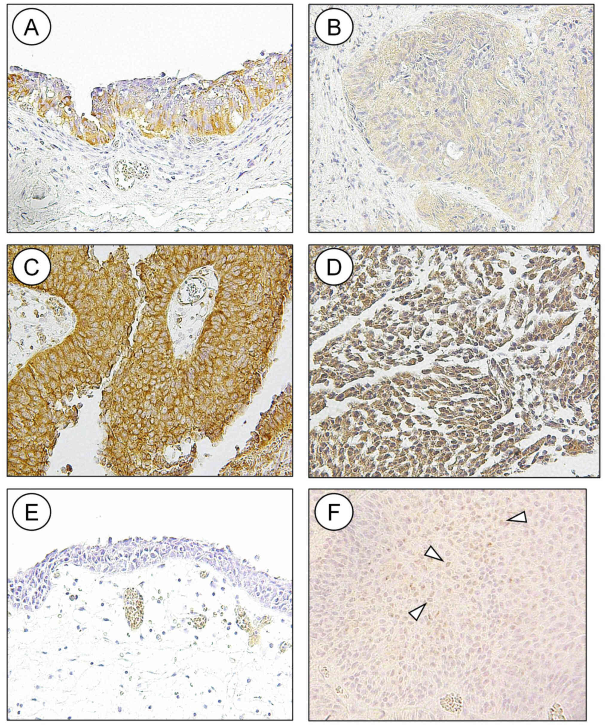
In the tissue microarrays, HDAC6 was detected in the
cytoplasm of high-grade bladder cancer, while low-grade bladder
cancer and upper urinary tract UC did not stain positive for HDAC6
(data not shown). A subsequent IHC staining experiment, using a
larger number of surgically resected UC tissues detected HDAC6 in
the cytoplasm of normal urothelium (Fig.
1A) and in the nuclei and cytoplasm of UC (Fig. 1C and D). A total of 54 tumors had
pure cytoplasmic staining without nuclear staining, 5 had only
nuclear staining, and the remaining 36 had various combinations of
nuclear and cytoplasmic staining (Fig.
1B-D and Table I). Nuclear
staining was only observed in stages Ta and Ta; however, invasive
tumors and Tis presented with either a combination of nuclear and
cytoplasmic staining or cytoplasmic staining (Table I); however, this was not
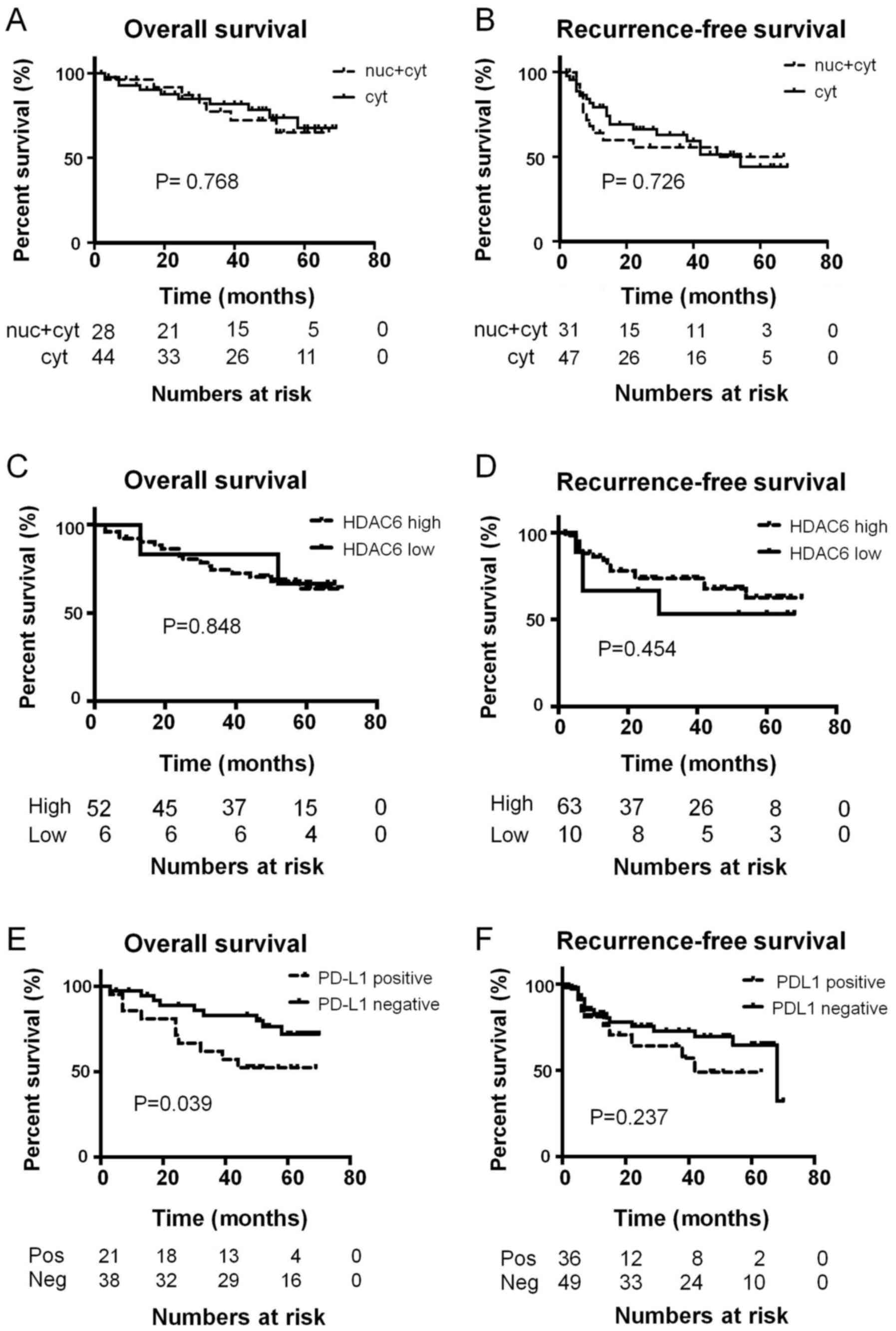
statistically significant. HDAC6 subcellular localization was not a
predictor of overall or recurrence-free survival times in patients
with UC (Fig. 2A and B). The
expression of HDAC6 was classified as low or high based on the
product of the intensity and percentage of positive tumor scores.
Staining for HDAC6 was either low (Fig.
1B) or high (Fig. 1C and D).
High HDAC6 expression was detected in 84 (86.6%) specimens
(Fig. 1B and Table III) and was observed more
frequently in multiple tumors compared with that in solitary ones
(94.3 vs. 79.0%), as well as in all Tis samples; however, the
difference was not statistically significant. Overall, HDAC6
expression was not associated with any pathological or clinical
characteristics. There was no association between HDAC6 expression
level and overall or recurrence-free survival (Fig. 2C and D).
| Table III.Immunohistochemical staining of HDAC6
and PD-L1, and the clinicopathological characteristics of the
patients with urothelial cancer. |
Table III.
Immunohistochemical staining of HDAC6
and PD-L1, and the clinicopathological characteristics of the
patients with urothelial cancer.
|
|
| HDAC6 | PD-L1 |
|---|
|
|
|
|
|
|---|
| Clinicopathological
characteristic | Value | Low | High | Negative | Positive |
|---|
| Total number | 97 tumors | 13 (13.4) | 84 (86.6) | 55 (56.7) | 42 (43.3) |
| Mean age ± SD,
years | 75.48±8.79 | 75.46±8.79 | 75.49±8.84 | 76.65±8.75 | 75.26±8.94 |
| Sex, n (%) |
|
Male | 61 | 3 (15.8) | 54 (88.5) | 39 (64) | 22 (36.0) |
|
Female | 19 | 7 (13.5) | 16 (84.2) | 10 (52.6) | 9 (47.4) |
| Number of tumors, n
(%) |
|
Solitary | 43 | 9 (21.0) | 34 (79.0) | 24 (55.8) | 19 (44.1) |
|
Multiple | 54 | 4 (7.5) | 50 (94.3) | 31 (58.4) | 23 (43.4) |
| Primary or
recurrent, n (%) |
|
Primary | 59 | 7 (11.9) | 52 (88.1) | 34 (57.6) | 25 (42.4) |
|
Recurrent | 38 | 6 (15.8) | 32 (84.2) | 18 (47.4) | 20 (52.6) |
| Tumor size, cm
(%) |
|
<3 | 79 | 11 (14) | 68 (86) | 34 (43.0) | 45 (56.9) |
|
>3 | 18 | 2 (11.2) | 16 (88.8) | 10 (55.6) | 8 (44.4) |
| Grade, n (%) |
|
Low | 31 | 4 (13) | 27 (87) | 18 (58.0) | 13 (41.9) |
|
High | 66 | 9 (13.6) | 57 (86.4) | 37 (56.0) | 29 (43.9) |
| T stage, n (%) |
|
pTa | 52 | 7 (13.5) | 45 (86.5) | 32 (61.5) | 20 (38.5) |
|
pT1 | 25 | 4 (19.3) | 21 (80.7) | 13 (53.8) | 12 (46.2) |
|
>pT2 | 14 | 2 (14.3) | 12 (85.7) | 5 (35.7) | 9 (64.3) |
|
Tis | 6 | 0 | 6 (100) | 5 (83.3) | 1 (16.7) |
PD-L1 IHC staining and its prognostic
significance
Normal urothelium was not stained by the anti-PD-L1
antibody (Fig. 1E). Membranous
staining of PD-L1 was detected in 42 (43.3%) UC samples (Fig. 1F and Table III). The expression of PD-L1 was
not associated with any pathological or clinical characteristics
(Table III). There was also no
association between HDAC6 expression and PD-L1 (Table I); however, PD-L1 positivity was a
predictor of shorter overall survival times (Fig. 2E; P=0.039). In addition, patients
with tumors positive for PD-L1 tended to have shorter
recurrence-free survival times (Fig.
2F), but the difference was not statistically significant.
Expression of HDAC6 and PD-L1 in
cultured cell lines and HDAC6 inhibition of PD-L1
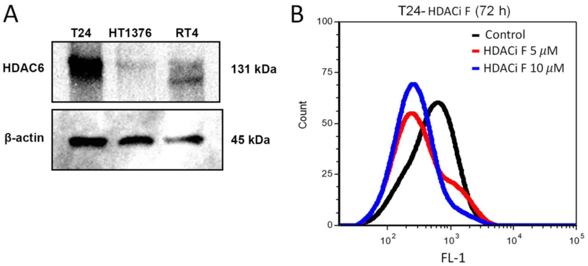
Western blot analysis of HDAC6 protein expression
was performed in the T24, HT1376, and RT4 bladder cancer cell lines
(Fig. 3A). PD-L1 expression was also
detected in the T24 cell line using flow cytometry (Fig. 3B). It has been reported that
targeting HDAC6 downregulated the expression of PD-L1 via the STAT3
pathway in cell lines (29).
Pharmacological inhibition of HDAC6 by HDACi F in T24 cells
downregulated the expression of PD-L1 as detected by flow cytometry
(Fig. 3B).
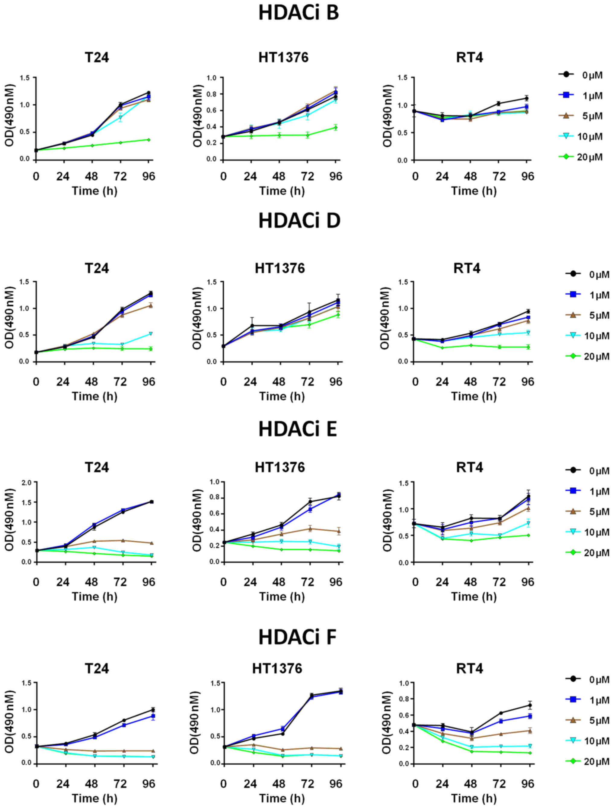
HDAC6 inhibitors induce growth
inhibition of bladder cancer cell lines
A total of 12 selective HDAC6 inhibitors (Table II) were investigated in the T24,
HT1376 and RT4 bladder cancer cell lines. Treatment with the
inhibitors (HDACi B, D, E and F) decreased the viability (detected
by MTS assay) of bladder cancer cells with the IC50
being as low as 2.20 µM (Figs. 4 and
S1; Table II). The median IC50
(13.06) for T24, a high-grade invasive UC, was lower (range,
2.20–50.71) compared with that in HT1376, an intermediate grade
cell line (median 17.96; range, 3.10–68.79) and in particular for
RT4, a low-grade model cell line (median 36.47; range, 4.90–371.20)
(Figs. 4 and S1; Table
II).
Discussion
Histone acetylation and deacetylation are key
processes of the epigenetic machinery regulating gene expression
(9). Increased levels of histone
acetylation have been associated with increased transcriptional
activity, whereas decreased levels of acetylation have been
associated with repression of gene expression (9). HDACs are included in the transcription
suppression complex and recruitment of HDACs suppresses the
transcription of specific genes in cancer cells. HDACs also
regulate multiple nuclear and cytoplasmic proteins and, thus, play
an essential role in protein degradation, cell survival and cell
migration (21,37). A total of 18 members of the HDAC
family have been discovered so far (11,16),
among which HDAC6 belongs to the Class II HDACs, which can move in
and out of the nucleus. HDAC6 is widely expressed in normal and
tumor tissues, and is predominantly localized in the cytoplasm,
where it controls non-histone acetylation (21,38) and
regulates numerous important biological processes, including cell
migration (16,30), while HDAC inhibitors are a new class
of anti-cancer drugs, that can induce apoptosis and cell cycle
arrest in cancer cells (16,39). The mechanism of HDAC inhibitors
remains unknown; however, the effect could be either epigenetic or
non-epigenetic (10).
HDACs are potential drug targets for chemotherapy;
however, a previous study has shown only modest antineoplastic
activity with pan-HDAC inhibitors in UC (30). The reported IC50 of
vorinostat, a pan-HDAC inhibitor, in cultured UC cells is up to 6.0
µM, and sensitivity to vorinostat in UC is not associated with the
expression levels of HDACs (30).
Therefore, inhibition of specific HDAC isoenzymes might be more
efficacious and tumor-specific. HDAC6 causes epigenetic alterations
by deacetylation of histones but also controls cell metabolism by
direct deacetylation of non-histone targets (10,22). Two
bands of HDAC6 were detected in RT4 cells, which presumably
corresponded to phosphorylated and dephosphorylated HDAC6 (40). To the best of our knowledge, the
present study is the first report on the subcellular localization
of HDAC6 in clinical UC samples, demonstrating that HDAC6 was
predominantly a cytoplasmic protein, which is in agreement that it
has non-histone targets (10,22).
HDAC6 was previously found to be overexpressed in UC tissues, using
reverse transcription-quantitative PCR (41); however, HDAC6 specific inhibitors
were found to be active at high µM concentrations in UC cell lines
(10–100 µM) (41). A different
previous study concluded that targeting HDAC6 might not be
effective in UC (8). In the present
study, HDAC6 inhibitors reduced proliferation of cultured human UC
cell lines at low µM concentrations, as low as 2.20 µM, and that
high-grade invasive UC cells were more susceptible to
pharmacological HDAC6 inhibition (Figs.
4 and S1; Table II). Thus, we hypothesized that
HDAC6-specific inhibitors might be effective in bladder cancer;
however, currently there are no biomarkers for HDAC inhibitors, and
it has been reported that HDAC6 protein expression levels did not
determine sensitivity to its inhibitors (41).
It has been reported that HDAC6 also induces
upregulation of several tumor-associated antigens (gp100, MART1,
TYRP1 and TYRP2) and major histocompatibility complex class I,
suggesting a potential improvement in the immunogenicity of tumor
cells (42). Immune checkpoint
blockade with anti-CTLA-4, anti-PD-1 and anti-PD-L1 antibodies have
changed the paradigm of cancer treatment, and it has been
demonstrated that selective HDAC6 inhibition improves the antitumor
activity of anti-PD-1 immune checkpoint blockade (42). PD-L1 is an important molecule
expressed in cancer cells that activates the inhibitory PD-1
pathway in T-cells (43). HDAC6 is
important for cytokine-mediated upregulation of PD-L1 protein
expression, which is primarily mediated by the recruitment and
activation of STAT3 (29). In the
active state, HDAC6 forms a complex with STAT3 and pharmacological
inhibition of HDAC6 results in disruption of this complex; this
disruption leads to decreased STAT3 phosphorylation but no changes
in STAT3 acetylation, as well as, presumably, diminished
recruitment of STAT3 to the PD-L1 gene promoter region (29,44). In
the present study, the pharmacological HDAC6 inhibition decreased
PD-L1 protein expression in cultured T24 UC cells (Fig. 3B).
Experiments using HDAC6 knockout mouse embryonic
fibroblasts (MEFs) have demonstrated that some effects of the
HDAC6-specific inhibitor, tubacin were independent of HDAC6,
whereas the effects of tubastatin A on MEF and in a cultured
bladder cancer cell line were HDAC6-dependent (45). Therefore, further studies are
required to clarify the effects of specific HDAC6 inhibitors in UC,
which could be involved in the modulation of multiple intracellular
pathways that are involved in apoptosis, tumor growth, antitumor
immune responses and immune surveillance (10,12,16,22,44). The
exact mechanism and factors influencing the modulation of PD-L1 by
HDAC6 requires further investigation.
In conclusion, the present study showed cytoplasmic
expression of HDAC6 in resected UC tumor specimens using IHC
staining and investigated the effects of 12 selective HDAC6 small
molecule inhibitors in vitro. It was found that pharmacological
inhibition of HDAC6 reduced the proliferation of UC cells with some
inhibitors (HDACi D, HDACi E and HDACi F) demonstrating low
IC50 values. PD-L1 protein expression was also
investigated using IHC staining of surgical specimens, and with
flow cytometry and western blot analysis in cultured cell lines.
The present study is a pilot study and demonstrated the efficacy of
selective HDAC6 inhibitors in UC. Specific pharmacological HDAC6
inhibition could be a promising new strategy for the treatment of
metastatic UC.
Supplementary Material
Supporting Data
Acknowledgements
The authors would like to thank the Department of
Medicinal Chemistry and Pharmacognosy (University of Illinois at
Chicago), for providing the inhibitors. The authors would also like
to thank Dr Irina Gaisina (Department of Medicinal Chemistry and
Pharmacognosy, University of Illinois at Chicago) for the fruitful
discussion and comments, which improved the manuscript.
Funding
The present study was supported by grants-in-aid
from Niigata University (grant nos. CH28015, June 2018 and CH29018,
December 2018).
Availability of data and materials
The datasets used and/or analyzed during the current
study are available from the corresponding author on reasonable
request.
Authors' contributions
HK, TA, AK and YS acquired, analyzed and interpreted
the patient data. HK drafted the manuscript and revised it
critically for important intellectual content. TA, AK and YS
participated in fruitful discussion on the manuscript. VB and YT
made substantial contributions to conception and design. VB
analyzed and interpreted the patient data, and drafted and revised
the manuscript for important intellectual content. YT revised the
manuscript for important intellectual content and gave final
approval of the version to be published. All authors read and
approved the final manuscript.
Ethics approval and consent to
participate
This study was approved by the Human Research Ethics
Committee of School of Medicine, Niigata University (Niigata,
Japan). All the patients provided written informed consent
according to the Declaration of Helsinki, and all experimental
methods were conducted according to relevant guidelines.
Patient consent for publication
Not applicable.
Competing interests
The authors declare that they have no competing
interests.
Glossary
Abbreviations
Abbreviations:
|
HDACs
|
histone deacetylases
|
|
PD-L1
|
programmed death-ligand 1
|
|
IHC
|
Immunohistochemistry
|
|
UC
|
urothelial cancer
|
|
BT
|
bladder tumor
|
|
GC
|
gemcitabine and cisplatin
|
|
M-VAC
|
methotrexate, vinblastine,
doxorubicin, and cisplatin
|
References
|
1
|
Ebrahimi H, Amini E, Pishgar F, Moghaddam
SS, Nabavizadeh B, Rostamabadi Y, Aminorroaya A, Fitzmaurice C,
Farzadfar F, Nowroozi MR, et al: Global, regional and national
burden of bladder cancer, 1990 to 2016: Results from the GBD Study
2016. J Urol. 201:893–901. 2019. View Article : Google Scholar : PubMed/NCBI
|
|
2
|
Bray F, Ferlay J, Soerjomataram I, Siegel
RL, Torre LA and Jemal A: Global cancer statistics 2018: GLOBOCAN
estimates of incidence and mortality worldwide for 36 cancers in
185 countries. CA Cancer J Clin. 68:394–424. 2018. View Article : Google Scholar : PubMed/NCBI
|
|
3
|
Droller MJ: Bladder cancer: Current
diagnosis and treatment (Current Clinical Urology). Humana.
2010.
|
|
4
|
Sternberg CN, Yagoda A, Scher HI, Watson
RC, Herr HW, Morse MJ, Sogani PC, Vaughan ED Jr, Bander N,
Weiselberg LR, et al: M-Vac (methotrexate, vinblastine, doxorubicin
and cisplatin) for advanced transitional cell carcinoma of the
urothelium. J Urol. 139:461–469. 1988. View Article : Google Scholar : PubMed/NCBI
|
|
5
|
Moore MJ, Winquist EW, Murray N, Tannock
IF, Huan S, Bennett K, Walsh W and Seymour L: Gemcitabine plus
cisplatin, an active regimen in advanced urothelial cancer: A phase
II trial of the National cancer institute of Canada clinical trials
group. J Clin Oncol. 17:2876–2881. 1999. View Article : Google Scholar : PubMed/NCBI
|
|
6
|
Seront E and Machiels JP: Molecular
biology and targeted therapies for urothelial carcinoma. Cancer
Treat Rev. 41:341–353. 2015. View Article : Google Scholar : PubMed/NCBI
|
|
7
|
Scher HI, Geller NL, Curley T and Tao Y:
Effect of relative cumulative dose-intensity on survival of
patients with urothelial cancer treated with M-VAC. J Clin Oncol.
11:400–407. 1993. View Article : Google Scholar : PubMed/NCBI
|
|
8
|
Pinkerneil M, Hoffmann MJ, Schulz WA and
Niegisch G: HDACs and HDAC inhibitors in urothelial
carcinoma-perspectives for an antineoplastic treatment. Curr Med
Chem. 24:4151–4165. 2017. View Article : Google Scholar : PubMed/NCBI
|
|
9
|
Jaenisch R and Bird A: Epigenetic
regulation of gene expression: How the genome integrates intrinsic
and environmental signals. Nat Genet. 33 (Suppl):S245–S254. 2003.
View Article : Google Scholar
|
|
10
|
Gaisina IN, Tueckmantel W, Ugolkov A, Shen
S, Hoffen J, Dubrovskyi O, Mazar A, Schoon RA, Billadeau D and
Kozikowski AP: Identification of HDAC6-selective inhibitors of low
cancer cell cytotoxicity. Chem Med Chem. 11:81–92. 2016. View Article : Google Scholar : PubMed/NCBI
|
|
11
|
Renaud JP: Structural biology in drug
discovery: Methods, Techniques, and Practices. Wiley; 2020,
View Article : Google Scholar
|
|
12
|
Glozak MA and Seto E: Histone deacetylases
and cancer. Oncogene. 26:5420–5432. 2007. View Article : Google Scholar : PubMed/NCBI
|
|
13
|
Halsall JA and Turner BM: Histone
deacetylase inhibitors for cancer therapy: An evolutionarily
ancient resistance response may explain their limited success.
Bioessays. 38:1102–1110. 2016. View Article : Google Scholar : PubMed/NCBI
|
|
14
|
Benedetti R, Conte M and Altucci L:
Targeting histone deacetylases in diseases: Where are we? Antioxid
Redox Signal. 23:99–126. 2015. View Article : Google Scholar : PubMed/NCBI
|
|
15
|
Subramanian S, Bates SE, Wright JJ,
Espinoza-Delgado I and Piekarz RL: Clinical toxicities of histone
deacetylase inhibitors. Pharmaceuticals (Basel). 3:2751–2767. 2010.
View Article : Google Scholar : PubMed/NCBI
|
|
16
|
Zhang J and Zhong Q: Histone deacetylase
inhibitors and cell death. Cell Mol Life Sci. 71:3885–3901. 2014.
View Article : Google Scholar : PubMed/NCBI
|
|
17
|
simplehttps://chemoth.com/
|
|
18
|
Rana Z, Diermeier S, Hanif M and Rosengren
RJ: Understanding failure and improving treatment using HDAC
inhibitors for prostate cancer. Biomedicines. 8:222020. View Article : Google Scholar
|
|
19
|
Cavenagh JD and Popat R: Optimal
management of histone deacetylase inhibitor-related adverse events
in patients with multiple myeloma: A focus on panobinostat. Clin
Lymphoma Myeloma Leuk. 18:501–507. 2018. View Article : Google Scholar : PubMed/NCBI
|
|
20
|
Shah MH, Binkley P, Chan K, Xiao J,
Arbogast D, Collamore M, Farra Y, Young D and Grever M:
Cardiotoxicity of histone deacetylase inhibitor depsipeptide in
patients with metastatic neuroendocrine tumors. Clin Cancer Res.
12:3997–4003. 2006. View Article : Google Scholar : PubMed/NCBI
|
|
21
|
de Ruijter AJ, van Gennip AH, Caron HN,
Kemp S and van Kuilenburg AB: Histone deacetylases (HDACs):
Characterization of the classical HDAC family. Biochem J.
370:737–749. 2003. View Article : Google Scholar : PubMed/NCBI
|
|
22
|
Li Y, Shin D and Kwon SH: Histone
deacetylase 6 plays a role as a distinct regulator of diverse
cellular processes. FEBS J. 280:775–793. 2013.PubMed/NCBI
|
|
23
|
Pernet L, Faure V, Gilquin B,
Dufour-Guérin S, Khochbin S and Vourc'h C: HDAC6-ubiquitin
interaction controls the duration of HSF1 activation after heat
shock. Mol Biol Cell. 25:4187–4194. 2014. View Article : Google Scholar : PubMed/NCBI
|
|
24
|
Boyault C, Sadoul K, Pabion M and Khochbin
S: HDAC6, at the crossroads between cytoskeleton and cell signaling
by acetylation and ubiquitination. Oncogene. 26:5468–5476. 2007.
View Article : Google Scholar : PubMed/NCBI
|
|
25
|
Valenzuela-Fernández A, Cabrero JR,
Serrador JM and Sánchez-Madrid F: HDAC6: A key regulator of
cytoskeleton, cell migration and cell-cell interactions. Trends
Cell Biol. 18:291–297. 2008. View Article : Google Scholar : PubMed/NCBI
|
|
26
|
Zuo Q, Wu W, Li X, Zhao L and Chen W:
HDAC6 and SIRT2 promote bladder cancer cell migration and invasion
by targeting cortactin. Oncol Rep. 27:819–824. 2012.PubMed/NCBI
|
|
27
|
Cha TL, Chuang MJ, Wu ST, Sun GH, Chang
SY, Yu DS, Huang SM, Huan SK, Cheng TC, Chen TT, et al: Dual
degradation of Aurora A and B kinases by the histone deacetylase
inhibitor LBH589 Induces G2-M arrest and apoptosis of renal cancer
cells. Clin Cancer Res. 15:840–850. 2009. View Article : Google Scholar : PubMed/NCBI
|
|
28
|
Gao YS, Hubbert CC and Yao TP: The
microtubule-associated histone deacetylase 6 (HDAC6) regulates
epidermal growth factor receptor (EGFR) endocytic trafficking and
degradation. J Biol Chem. 285:11219–11226. 2010. View Article : Google Scholar : PubMed/NCBI
|
|
29
|
Lienlaf M, Perez-Villarroel P, Knox T,
Pabon M, Sahakian E, Powers J, Woan KV, Lee C, Cheng F, Deng S, et
al: Essential role of HDAC6 in the regulation of PD-L1 in melanoma.
Mol Oncol. 10:735–750. 2016. View Article : Google Scholar : PubMed/NCBI
|
|
30
|
Niegisch G, Knievel J, Koch A, Hader C,
Fischer U, Albers P and Schulz WA: Changes in histone deacetylase
(HDAC) expression patterns and activity of HDAC inhibitors in
urothelial cancers. Urol Oncol. 31:1770–1779. 2013. View Article : Google Scholar : PubMed/NCBI
|
|
31
|
Brierley JD, Gospodarowicz MK and
Wittekind C: TNM classification of malignant Tumours. 8th edition.
John Wiley & Sons, Ltd.; 2017
|
|
32
|
The Japanese Society of Pathology,
Japanese Society of Urology (ed.), . General Rule for Clinical and
Pathological Studies on Bladder Cancer. Third edition (in
Japanese). Kanahara Publishing; Tokyo, Japan: pp. 1022001
|
|
33
|
Bilim V, Yuuki K, Itoi T, Muto A, Kato T,
Nagaoka A, Motoyama T and Tomita Y: Double inhibition of XIAP and
Bcl-2 axis is beneficial for retrieving sensitivity of renal cell
cancer to apoptosis. Br J Cancer. 98:941–949. 2008. View Article : Google Scholar : PubMed/NCBI
|
|
34
|
Seo J, Min SK, Park HR, Kim DH, Kwon MJ,
Kim LS and Ju YS: Expression of Histone Deacetylases HDAC1, HDAC2,
HDAC3, and HDAC6 in invasive ductal carcinomas of the breast. J
Breast Cancer. 17:323–331. 2014. View Article : Google Scholar : PubMed/NCBI
|
|
35
|
Brahmer J, Reckamp KL, Baas P, Crinò L,
Eberhardt WE, Poddubskaya E, Antonia S, Pluzanski A, Vokes EE,
Holgado E, et al: Nivolumab versus docetaxel in advanced
squamous-cell non-small-cell lung cancer. N Engl J Med.
373:123–135. 2015. View Article : Google Scholar : PubMed/NCBI
|
|
36
|
Bilim V, Kawasaki T, Katagiri A, Wakatsuki
S, Takahashi K and Tomita Y: Altered expression of beta-catenin in
renal cell cancer and transitional cell cancer with the absence of
beta-catenin gene mutations. Clin Cancer Res. 6:460–466.
2000.PubMed/NCBI
|
|
37
|
Choudhary C, Kumar C, Gnad F, Nielsen ML,
Rehman M, Walther TC, Olsen JV and Mann M: Lysine acetylation
targets protein complexes and co-regulates major cellular
functions. Science. 325:834–840. 2009. View Article : Google Scholar : PubMed/NCBI
|
|
38
|
Liu Y, Peng L, Seto E, Huang S and Qiu Y:
Modulation of histone deacetylase 6 (HDAC6) nuclear import and
tubulin deacetylase activity through acetylation. J Biol Chem.
287:29168–29174. 2012. View Article : Google Scholar : PubMed/NCBI
|
|
39
|
Finzer P, Kuntzen C, Soto U, zur Hausen H
and Rösl F: Inhibitors of histone deacetylase arrest cell cycle and
induce apoptosis in cervical carcinoma cells circumventing human
papillomavirus oncogene expression. Oncogene. 20:4768–4776. 2001.
View Article : Google Scholar : PubMed/NCBI
|
|
40
|
Du Y, Seibenhener ML, Yan J, Jiang J and
Wooten MC: aPKC Phosphorylation of HDAC6 results in increased
deacetylation activity. PLoS One. 10:e01231912015. View Article : Google Scholar : PubMed/NCBI
|
|
41
|
Rosik L, Niegisch G, Fischer U, Jung M,
Schulz WA and Hoffmann MJ: Limited efficacy of specific HDAC6
inhibition in urothelial cancer cells. Cancer Biol Ther.
15:742–757. 2014. View Article : Google Scholar : PubMed/NCBI
|
|
42
|
Knox T, Sahakian E, Banik D, Hadley M,
Palmer E, Noonepalle S, Kim J, Powers J, Gracia-Hernandez M,
Oliveira V, et al: Selective HDAC6 inhibitors improve anti-PD-1
immune checkpoint blockade therapy by decreasing the
anti-inflammatory phenotype of macrophages and down-regulation of
immunosuppressive proteins in tumor cells. Sci Rep. 9:61362019.
View Article : Google Scholar : PubMed/NCBI
|
|
43
|
Patsoukis N, Wang Q, Strauss L and
Boussiotis VA: Revisiting the PD-1 pathway. Sci Adv.
6:eabd27122020. View Article : Google Scholar : PubMed/NCBI
|
|
44
|
Keremu A, Aimaiti A, Liang Z and Zou X:
Role of the HDAC6/STAT3 pathway in regulating PD-L1 expression in
osteosarcoma cell lines. Cancer Chemother Pharmacol. 83:255–264.
2019. View Article : Google Scholar : PubMed/NCBI
|
|
45
|
Ota S, Zhou ZQ and Hurlin PJ: Suppression
of FGFR3- and MYC-dependent oncogenesis by tubacin: Association
with HDAC6-dependent and independent activities. Oncotarget.
9:3172–3187. 2017. View Article : Google Scholar : PubMed/NCBI
|
|
46
|
Tavares MT, Shen S, Knox T, Hadley M,
Kutil Z, Bařinka C, Villagra A and Kozikowski AP: Synthesis and
pharmacological evaluation of selective histone deacetylase 6
inhibitors in melanoma models. ACS Med Chem Lett. 8:1031–1036.
2017. View Article : Google Scholar : PubMed/NCBI
|
|
47
|
Shen S, Benoy V, Bergman JA, Kalin JH,
Frojuello M, Vistoli G, Haeck W, Van Den Bosch L and Kozikowski AP:
Bicyclic-capped histone deacetylase 6 inhibitors with improved
activity in a model of axonal charcot-marie-tooth disease. ACS Chem
Neurosci. 7:240–258. 2016. View Article : Google Scholar : PubMed/NCBI
|
|
48
|
Kozikowski AS and Bergman J: Preparation
of tetrahydroquinoline substituted hydroxamic acids as selective
histone deacetylase 6 inhibitors (ed.) AP (ed.). 2017.
|
|
49
|
Gaisina IN, Lee SH, Kaidery NA, Ben Aissa
M, Ahuja M, Smirnova NN, Wakade S, Gaisin A, Bourassa MW, Ratan RR,
et al: Activation of Nrf2 and hypoxic adaptive response contribute
to neuroprotection elicited by phenylhydroxamic acid selective
HDAC6 inhibitors. ACS Chem Neurosci. 9:894–900. 2018. View Article : Google Scholar : PubMed/NCBI
|
|
50
|
Osipyants AI, Poloznikov AA, Smirnova NA,
Hushpulian DM, Khristichenko AY, Chubar TA, Zakhariants AA, Ahuja
M, Gaisina IN, Thomas B, et al: L-ascorbic acid: A true substrate
for HIF prolyl hydroxylase? Biochimie. 147:46–54. 2018. View Article : Google Scholar : PubMed/NCBI
|
|
51
|
Shen S, Hadley M, Ustinova K, Pavlicek J,
Knox T, Noonepalle S, Tavares MT, Zimprich CA, Zhang G, Robers MB,
et al: Discovery of a new Isoxazole-3-hydroxamate-Based histone
deacetylase 6 inhibitor SS-208 with antitumor activity in syngeneic
melanoma mouse models. J Med Chem. 62:8557–8577. 2019. View Article : Google Scholar : PubMed/NCBI
|
|
52
|
Segretti MC, Vallerini GP, Brochier C,
Langley B, Wang L, Hancock WW and Kozikowski AP: Thiol-based potent
and selective HDAC6 inhibitors promote tubulin acetylation and
T-regulatory cell suppressive function. ACS Med Chem Lett.
6:1156–1161. 2015. View Article : Google Scholar : PubMed/NCBI
|
|
53
|
Lv W, Zhang G, Barinka C, Eubanks JH and
Kozikowski AP: Design and synthesis of mercaptoacetamides as
potent, selective, and brain permeable histone deacetylase 6
inhibitors. ACS Med Chem Lett. 8:510–515. 2017. View Article : Google Scholar : PubMed/NCBI
|