Introduction
Endometrial cancer is the most common type of
neoplasm affecting one of the female reproductive organs, and
primarily occurs in postmenopausal women. It has been revealed that
there is an increase in the incidence rate of endometrial cancer
with age, particularly in developing countries. In 2006 to 2007,
rates varied 10-fold across countries, with the highest rates in
North America (United States), Eastern and Northern Europe
(Slovakia), and the lowest rates were in middle-income countries
(South Africa and India) (1). In
2012, endometrial cancer was the 6th most frequently diagnosed
carcinoma, and the 14th most common cause of cancer-associated
deaths in women worldwide (2).
Furthermore, risk factors, such as early menarche, late menopause,
infertility, taking menopausal hormones and obesity play a major
role in endometrial cancer etiology (3).
Endometrial cancer has been associated with
invasiveness and metastasis formation, which are multistage
processes in which malignant cells detach from the primary tumor
mass, travel via the lymph and blood to target tissues, where tumor
cells can adhere to and penetrate the vascular endothelium; thus,
forming new blood vessels in the developing neoplastic foci. Each
of these stages requires specific interactions between cancer cells
and the surrounding intercellular substance, and also with
endothelial cells of blood and lymphatic vessels (4). These interactions are mediated by
specific adhesion molecules, such as cadherins, which constitute a
group of 100 classical and non-classical cadherins. E- and
N-cadherin are well-known classical cadherins and can mediate
homotypic intercellular interactions by interacting in a
Ca2+-dependent manner with the same cadherin types on
adjacent cells (5). E-cadherin is
typically located on epithelial cell membranes, and promotes cell
adhesion and integrity; thus, mediates differentiation of healthy
epithelial tissue architecture (6).
Furthermore, N-cadherin is characteristic of mesenchymal cells,
particularly in cells with greater motility and reduced
polarization. It has been shown that N-cadherins are representative
markers of epithelial-mesenchymal transformation (EMT) (6,7).
P-cadherin is present at adherens junctions, and is found, similar
to E-cadherin, in several mature tissue types, including the
epidermis, breast, prostate and mesothelium (8–10).
Cadherin switching, which is important for the formation of the
cell phenotype, occurs during normal processes of cell development
(11). Furthermore, switching of
E-cadherin to N-cadherin has been observed in carcinomas of the
prostate (12), breast (13) and melanoma (14), whereas changes to P-cadherin were
found in pancreatic (15) or gastric
cancer types (16).
Therefore, the aim of the present study was to
examine the protein expression levels of E-, N- and P-cadherin in
cancer tissue from patients with endometrial cancer. In addition,
the present study investigated the association of these proteins
with clinicopathological parameters.
Materials and methods
Patients and tissue samples
The present study enrolled 38 patients (mean age
68.6 years; range 44–84 years old) (Table I) with endometrial cancer, who
underwent hysterectomy at the Medical Center ‘Żelazna’ in Warsaw
(Poland) between January 2008 and December 2015. The inclusion
criteria for the study group was endometrial cancer in any of The
International Federation of Gynecology and Obstetrics (FIGO) stages
(17) with full histopathological
documentation. The exclusion criteria were any cases with
unspecified status of lymph node or distant organ involvement and
refusal to participate. The postoperative tissue was fixed in 10%
neutral, buffered formalin for 24 h at room temperature and
embedded in paraffin. Sections (4-µm) were cut from the paraffin
blocks and stained with hematoxylin for 5 min and eosin for 1 min
at room temperature. Slides were assessed under a light microscope
Olympus BX40 under magnifications ×200 and ×400. Routine
histopathological analysis was performed to determine tumor
histological type, malignancy grade (G), staging according to FIGO,
the presence of tumor budding, local lymph node involvement and the
presence of distant metastases (ovary, vagina and colon) (17) (Table
I). Five sections with atypical endometrial proliferation from
resected material of the study group were used as controls. A total
of 12 patients (out of 38) had metastases to the lymph nodes and 8
to distant organs (2 of them had metastases to two or more organs
at the same time). Tumor budding was counted in 5 high-power fields
(magnification, ×40), and were classified as low-grade (<5 buds)
or high-grade (≥5 buds; Table I).
The study was approved by the local Bioethics Committee (Medical
University of Bialystok; approval no. R-I-002/68/2016) and written
informed consent, regarding the use of the tissue, was provided by
each patient in the study.
 | Table I.Clinicopathological characteristics of
the patients with endometrial cancer. |
Table I.
Clinicopathological characteristics of
the patients with endometrial cancer.
| Clinicopathological
parameters | Number (%) |
|---|
| Age, years |
|
|
<65 | 19 (50) |
| ≥65 | 19 (50) |
| Tumor type |
|
|
Endometrioid | 31 (81.6) |
|
Serous | 7 (18.4) |
| Tumor grade,
differentiated |
|
| Well | 4 (10.5) |
|
Medium | 25 (65.8) |
|
Poorly | 9 (23.7) |
| Tumor stage |
|
| I | 15 (39.5) |
| II | 3 (7.9) |
| III | 15 (39.5) |
| IV | 5 (13.1) |
| Tumor budding,
grade |
|
| Low | 27 (71) |
| High | 11 (29) |
| pN |
|
|
Absent | 26 (68.4) |
|
Present | 12 (31.6) |
| pM |
|
|
Absent | 30 (78.9) |
|
Present | 8 (21.1) |
Immunohistochemistry
Immunohistochemical analyses were performed using 38
primary endometrial tumors tissues and 20 metastatic tissues: 9
metastases to the lymph nodes (3 were removed due to
micrometastases) and 11 distant metastases (7 ovaries, 2 vaginas
and 2 colons). For comparison, five cases of atypical endometrial
proliferation were also stained. Tissue blocks were cut on a
microtome into 4-µm thick sections on silanized glass slides. The
sections were deparaffinized in xylene and hydrated in a series of
alcohols of decreasing concentration (two treatments with 99.9,
then 96 and 70% ethanol) at room temperature. Then, sections were
heated in a microwave oven for 15 min in citrate buffer (pH, 6.0)
for antigen retrieval. For blocking endogenous peroxidase activity
3% hydrogen peroxide solution was used for 10 min at room
temperature. For blocking non-specific antibody binding horse serum
was used (anti-mouse/rabbit serum produced in horse; Vector
Laboratories, Inc.) for 10 min at room temperature. Next, the
sections were incubated with murine monoclonal anti-E-cadherin
(cat. no. 36B5; 1:100; Leica Microsystems GmbH), anti-N-cadherin
(cat. no. IAR06; 1:100; Leica Microsystems GmbH) and
anti-P-cadherin (cat. no. HPA001767; 1:100; Sigma-Aldrich; Merck
KGaA) antibodies for 60 min at room temperature. Next, the one-step
system ImmPRESS™ Universal Antibody Polymer Reagent (30 min at room
temperature; catalog no. : MP-7500; Vector Laboratories, Inc.) and
chromogen ImmPACT DAB (5 min at room temperature; catalog no. :
SK-4105, Vector Laboratories, Inc.) were used. Cellular nuclei were
stained with hematoxylin for 5 min at room temperature. Positive
and negative controls were performed according to the
manufacturer's protocol (Leica Microsystems, Inc.; Sigma-Aldrich;
Merck KGaA).
The results were determined by two independent
pathologists (Department of General Pathomorphology, Medical
University of Bialystok) under a light microscope. Protein
expression was observed at random using 10 fields of view (FOV) and
a high-power lens (magnification, 10×40), with each FOV counting
≥100 cells. The expression level was observed as cytoplasmic,
membranous or mixed, and was classified as positive in the
cytoplasm (only cytoplasmic reaction was visible) or positive in
the membrane (only membranous or mixed reaction). The cytoplasm or
membrane staining was calculated based on the percentage of
immunoreactive cells and a range 1–100% indicated positive
expression. Data obtained from the immunohistochemical analysis are
presented as the mean percentage of the expression level.
Statistical analysis
Statistical analyses were conducted using the 38
primary tumors and 20 metastatic tumors of endometrial cancer. The
Statistica 11 (v4.0; StatSoft; TIBCO Software, Inc.) and GraphPad
Prism (v5.04; GraphPad Software, Inc.) programs were used for
statistical analysis. The protein expression levels were compared
between two groups using the Mann-Whitney U test. E-, N-,
P-cadherin protein expression levels did not follow a normal
distribution and therefore non-parametric statistical analyses were
performed. Spearman's rank correlation analysis was used to assess
the correlation between membrane and cytoplasmic E-, N-, P-cadherin
expression level in primary and metastatic tumor of endometrial
cancer. The association between membrane and cytoplasmic E-, N-,
P-cadherin protein expression level between the primary tumor and
the clinicopathological features of patients with endometrial
cancer was analyzed using the Mann-Whitney test for two groups,
while the Kruskal-Wallis test was used for three or more groups
plus Dunn's post hoc test. P<0.05 was considered to indicate a
statistically significant difference.
Results
E-, N- and P-cadherin protein
expression levels in endometrial hyperplasia with atypia
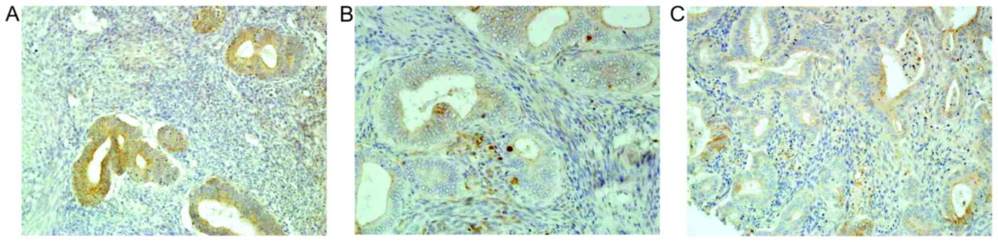
In hyperplastic endometrial lesions, the protein
expression levels of E-, N- and P-cadherin were similar, and weak
cytoplasmic staining was observed. Furthermore, the expression
level was equally distributed throughout the hyperplastic
epithelium. The membrane staining of E-cadherin was present in 3/5
cases (Fig. 1A), whereas that of
P-cadherin was only observed in a few cells and was <10%
(Fig. 1C). In addition, the protein
expression level of N-cadherin was found to be negative (Fig. 1B).
E-, N- and P-cadherin protein
expression levels in the primary tumor and metastatic tissues in
endometrial cancer
The present results indicated that the three
cadherin proteins were expressed either in the membrane and/or in
the cytoplasm and were unevenly distributed in endometrial cancer
tissue (Fig. 2A, B, D, E, G and H).
Furthermore, the staining of the three cadherin proteins was
stronger at the tumor front compared with that in the main tumor
mass (N-cadherin staining; Fig. 2D and
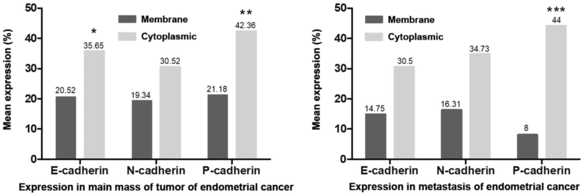
E). The cytoplasmic expression level was significantly higher
compared with that in the membrane, with respect to E-cadherin
(P<0.05) and P-cadherin (P<0.01) in the primary tumor
tissues, while P-cadherin expression level was also significantly
higher in the metastatic tissue (P<0.001) (Fig. 3). Furthermore, it was found that the
membrane expression levels of the 3 cadherin proteins were lower in
metastatic cancer cells compared with that in the primary tumor
cells (Fig. 3). Moreover, a decrease
in the membranous protein expression level of P-cadherin was found
in the metastatic tumor (mean expression, 8%) compared with that in
the primary endometrial cancer tissue (mean expression, 21.18%),
although these findings were not statistically significant
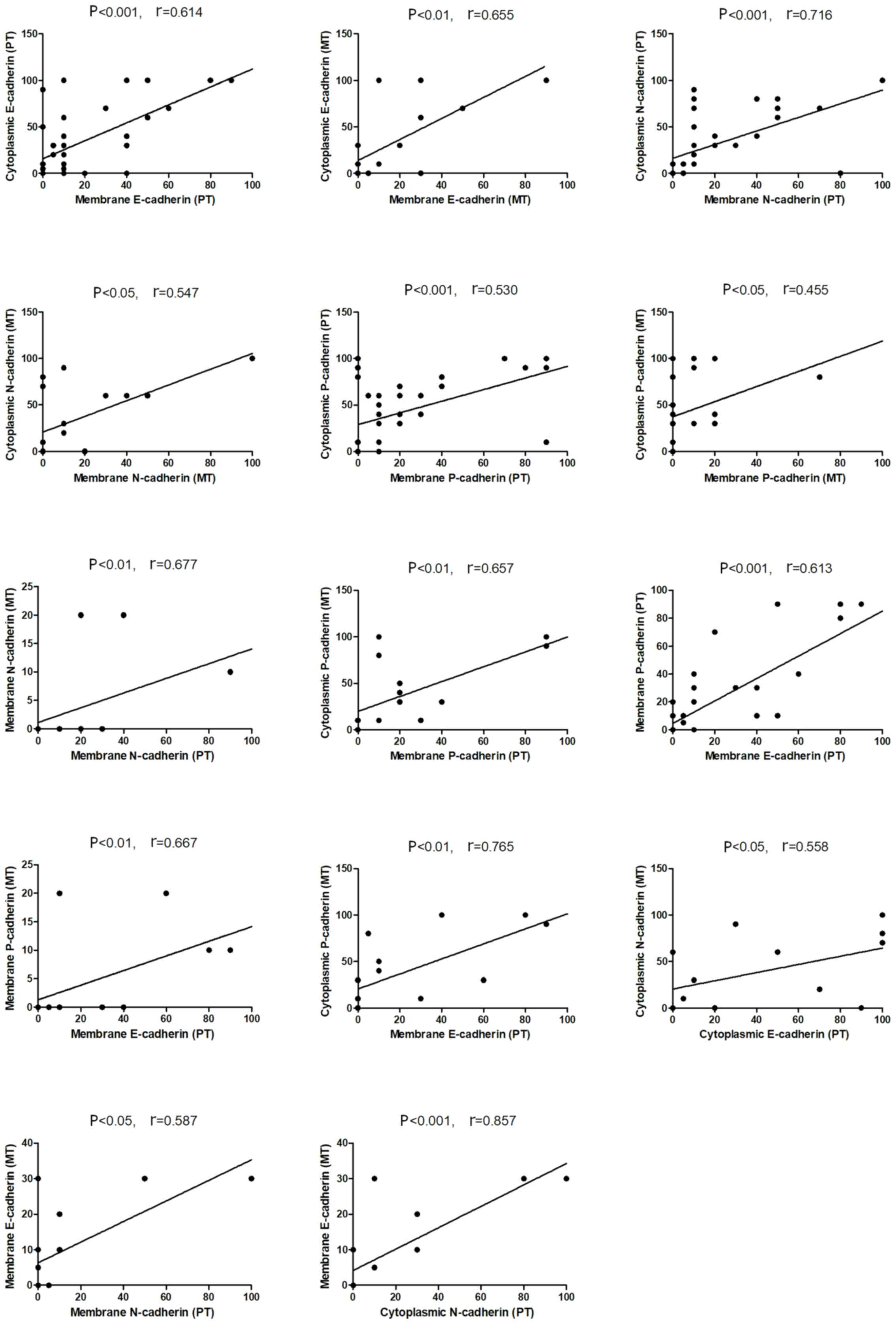
(Fig. 3). As shown in Fig. 4, the protein expression levels of
cytoplasmic E-, N- and P-cadherin were also correlated with
membrane expression level the in primary and metastatic tumors.
Furthermore, positive correlations were found between membrane
N-cadherin expression level in the primary tumor and with the
membrane expression level in the metastatic tumor, also between
membrane P-cadherin expression level in the primary tumor and with
the membrane expression level in the metastatic tumor, and between
membrane P-cadherin expression level in the primary tumor and with
cytoplasmic expression level in the metastatic tumor. In addition,
correlations were also identified between membrane E-cadherin and
membrane P-cadherin expression level in the primary tumor; membrane
P-cadherin in metastatic tumor and membrane E-cadherin in primary
tumor; cytoplasmic P-cadherin in metastatic tumor and membrane
E-cadherin in primary tumor, cytoplasmic N-cadherin in metastatic
tumor and cytoplasmic E-cadherin in primary tumor; membrane
E-cadherin in metastatic tumor and membrane N-cadherin in primary
tumor; and also membrane E-cadherin in metastatic cancer and
cytoplasmic N-cadherin in primary tumor (Fig. 4).
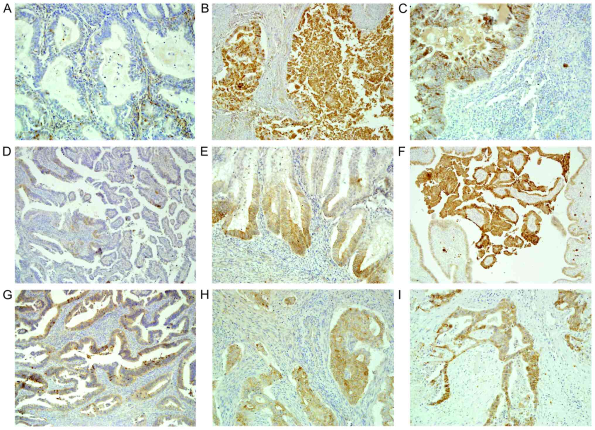 | Figure 2.E-, N-, P-cadherin expression levels
in the primary and metastasis tissues from endometrial cancer. The
3 aforementioned cadherin proteins were expressed in the membranes
and/or in the cytoplasm and were unevenly distributed in the
endometrial cancer tissue. The staining of the three cadherin
proteins were stronger at the tumor front compared with that in the
main tumor mass. Cytoplasmic expression level was higher compared
with that in the membrane. In the main mass of the primary tumor
there was (A) negative and (B) strong cytoplasmic E-cadherin
staining in cancer cells (magnification, ×200 and ×100,
respectively). (C) Positive E-cadherin expression level in the
metastatic cancer cells in the lymph node. Magnification, ×200. (D)
Cytoplasmic N-cadherin expression level was found in a few cells in
the main mass of the tumor. Magnification, ×100. (E) Stronger
membranous and cytoplasmic N-cadherin expression level at the tumor
front. Magnification, ×200. (F) N-cadherin expression level in the
metastasis tissue of the ovary. Magnification, ×100. (G) Weak
(magnification, ×200) and (H) medium cytoplasmic P-cadherin
expression level in main mass of the primary tumor (magnification,
×200). (I) P-cadherin expression level in the metastasis to the
vagina. Magnification, ×100. |
Association between E-, P- and
N-cadherin protein expression levels in primary endometrial cancer
and clinicopathological parameters
The present results suggested that there were no
significant associations between the age of the patients,
histological type of the tumor, FIGO grade and the presence of
distant metastases and the E-, N- and P-cadherin protein expression
levels (Table II). Furthermore, a
significant association was found between the membrane expression
level of P-cadherin and endometrial carcinoma grade; a higher
percentage of P-cadherin membrane expression level was associated
with histologically poorly differentiated cancer types (P=0.023).
In addition, a significant association was found between high-grade
tumor budding and higher cytoplasmic expression level of E-cadherin
(P=0.042), and higher membrane and cytoplasmic expression levels of
P-cadherin (P=0.012 and P=0.002, respectively). The present results
indicated that a higher membrane expression of E-cadherin was also
associated with high-grade tumor budding; however, the result was
not statistically significant. Furthermore, increases in the
membrane expression level of E-cadherin was associated with the
presence of local lymph node involvement (P=0.044; Table II).
| Table II.Association between E-, P- and
N-cadherin protein expression level in the primary tumor tissue and
clinicopathological parameters. |
Table II.
Association between E-, P- and
N-cadherin protein expression level in the primary tumor tissue and
clinicopathological parameters.
|
|
| E-cadherin | N-cadherin | P-cadherin |
|---|
|
|
|
|
|
|
|---|
|
|
| Membrane | Cytoplasmic | Membrane | Cytoplasmic | Membrane | Cytoplasmic |
|---|
|
|
|
|
|
|
|
|
|
|---|
| Clinicopathological
parameters | Number | Mean
percentage | P-value | Mean
percentage | P-value | Mean
percentage | P-value | Mean
percentage | P-value | Mean
percentage | P-value | Mean
percentage | P-value |
|---|
| Age, years |
|
| 0.752 |
| 0.315 |
| 0.904 |
| 0.247 |
| 0.940 |
| 0.410 |
|
<65 | 19 | 19.5 |
| 40.3 |
| 19.7 |
| 36.3 |
| 17.1 |
| 47.4 |
|
|
≥65 | 19 | 21.6 |
| 31.3 |
| 18.9 |
| 24.7 |
| 25.3 |
| 37.4 |
|
| Tumor type |
|
| 0.522 |
| 0.746 |
| 0.449 |
| 0.329 |
| 0.061 |
| 0.436 |
|
Endometrioid | 31 | 21.9 |
| 35.9 |
| 19.2 |
| 28.7 |
| 24.7 |
| 44.2 |
|
|
Serous | 7 | 14.3 |
| 34.3 |
| 20 |
| 38.6 |
| 5.7 |
| 34.3 |
|
| Tumor grade,
differentiated |
|
| 0.521 |
| 0.383 |
| 0.277 |
| 0.331 |
| 0.023a |
| 0.595 |
|
Well | 4 | 15 |
| 26.7 |
| 40 |
| 40 |
| 8.3 |
| 36.7 |
|
|
Medium | 25 | 18.9 |
| 32.5 |
| 15.7 |
| 29.1 |
| 15 |
| 35.9 |
|
|
Poorly | 9 | 33.7 |
| 61.2 |
| 30 |
| 32.5 |
| 41.2 |
| 50 |
|
| Tumor stage |
|
| 0.924 |
| 0.938 |
| 0.063 |
| 0.373 |
| 0.620 |
| 0.236 |
| I | 15 | 17 |
| 30.3 |
| 20.7 |
| 31.3 |
| 13.7 |
| 33.3 |
|
| II | 3 | 6.7 |
| 18.3 |
| 23.3 |
| 40 |
| 13.3 |
| 23.3 |
|
|
III | 15 | 25 |
| 41 |
| 23 |
| 34.7 |
| 28 |
| 48 |
|
| IV | 5 | 26 |
| 46 |
| 2 |
| 10 |
| 28 |
| 64 |
|
| Tumor budding,
grade |
|
| 0.153 |
| 0.042a |
| 0.288 |
| 0.432 |
| 0.012a |
| 0.002a |
|
Low | 27 | 15.5 |
| 26.8 |
| 15 |
| 27 |
| 14.2 |
| 31.5 |
|
|
High | 11 | 32.7 |
| 57.3 |
| 30 |
| 39.1 |
| 38.2 |
| 69.1 |
|
| pN |
|
| 0.044a |
| 0.556 |
| 0.323 |
| 0.786 |
| 0.122 |
| 0.303 |
|
Absent | 26 | 14.8 |
| 34.4 |
| 22.1 |
| 28.5 |
| 15.6 |
| 38.5 |
|
|
Present | 12 | 32.9 |
| 38.3 |
| 13.3 |
| 35 |
| 33.3 |
| 50.8 |
|
| pM |
|
| 0.506 |
| 0.293 |
| 0.171 |
| 0.135 |
| 0.239 |
| 0.104 |
|
Absent | 30 | 18 |
| 31.7 |
| 19.8 |
| 34 |
| 17.8 |
| 37.3 |
|
|
Present | 8 | 30 |
| 50.6 |
| 17.5 |
| 17.5 |
| 33.7 |
| 61.2 |
|
Discussion
E-cadherin has been widely investigated and is
therefore the best described cadherin. Previous studies have shown
that in the healthy endometrium, the expression level of E-cadherin
was moderate to strong in 60–90% of cases and did not differ
between the proliferative and secretion phases (18–20).
Furthermore, E-cadherin was stained in the membrane, and was
located on the borders between polarized cells (18–20).
Nguyen et al (21) revealed
that N- and P-cadherin were found in the endometrium. In addition,
N-cadherin was primarily visualized in the apical surface and at
the lateral junctions of the plasma membrane of the epithelial
cells in the basalis, while it was strongest in the basalis glands
adjacent to the myometrium and found to a lesser extent in the
functional glands. However, P-cadherin was shown to be located in
the basal surface of epithelial glands in both the functionalis and
basalis (21).
The present results suggested that E-, N- and
P-cadherins in hyperplastic endometrial lesions with atypia were
equally weakly expressed in the cytoplasm. Furthermore, the
membrane staining was observed in a few cells, with <10%. The
protein expression levels of E-, N- and P-cadherins in endometrial
cancer were located in the membrane and/or in the cytoplasm, and
were unevenly distributed in the neoplastic tissue. Furthermore,
stronger staining of the three cadherins was identified at the
tumor front compared with that in the main mass of endometrial
cancer tissue. However, no significant differences were found in
E-cadherin protein expression level between primary and metastasis
tumors. The present results were consistent with those from
previous studies, which have shown similar cadherin expression
levels. Ahmed and Muhammad (18)
revealed membranous protein expression level of E-cadherin in
non-neoplastic endometrial lesions, along with proliferative,
secretory and hyperplastic endometrial changes, while neoplastic
endometrial lesions showed mixed membranous-cytoplasmic staining.
In addition, Carico et al (19) showed that E-cadherin protein
expression level in normal endometrial growth was reduced, but was
not homogenous. However, in atypical endometrium and in endometrial
cancer cells, the membranous and cytoplasmic protein expression
level of E-cadherin was weaker compared with normal endometrial
cells. Furthermore, various regions of neoplastic tissue (for
example tumor front, main mass of tumor and free-floating tumor
cells in the ascitic fluid) showed differentiated expression level
of E-cadherin, which reflected the heterogeneity of the neoplastic
epithelium (21–24). Therefore, the loss of E-cadherin
interaction with the cadherin-catenin complex could be attenuated
at an early stage of the hyperplastic process and could be involved
in endometrial cancer progression.
Only one study compared expression of N-cadherin in
normal and neoplastic cells of uterus. Xie et al (20) observed moderate and strong N-cadherin
protein expression level in endometrial cancer, compared with low
or moderate expression level in normal endometrial epithelium.
Comparison of N-cadherin expression with E-cadherin expression in
endometrial cancer did not reveal any statistical significance
(20). The present study found
differences in the protein expression level of cytoplasmic
N-cadherin in metastatic and cytoplasmic E-cadherin in primary
tumors; membrane E-cadherin in metastatic and membrane N-cadherin
in primary tumors and also membrane E-cadherin in metastatic cancer
and cytoplasmic N-cadherin in primary tumors.
It has been demonstrated that P-cadherin protein
expression level is increasing in endometrial cancer cells in
comparison with normal cells. Moreno-Bueno et al (24) identified positive P-cadherin
expression in <10% cases of atypical endometrial hyperplasia.
Furthermore, P-cadherin staining was higher in endometrioid cancer
types and in non-endometrioid neoplasms, accounting for ~46% of
cases. However, positive expression level of P-cadherin was
considered when ≥10% of cells had immunohistochemical staining, due
to low expression of this protein. The present results suggested
that the membranous protein expression level of P-cadherin was
similar to that of E- and N-cadherin, whereas its cytoplasmic
expression was significantly higher. Furthermore, a decrease in the
membranous protein expression level of P-cadherin was observed in
the metastatic tumor compared with that in the primary endometrial
cancer tissue, although these findings were not statistically
significant.
However, changes in the expression levels of 2 of
the cadherin proteins were associated with clinicopathological
factors in endometrial carcinoma. The present results indicated
that higher cytoplasmic protein expression level of E-cadherin and
increased membranous and cytoplasmic expression level of
P-cadherin, was associated with high-grade tumor budding.
Furthermore, Koyuncuoglu et al (25) conducted a similar analysis of the
association between E-cadherin and tumor budding, revealing that
its positive expression was higher in low-grade tumor budding, even
though the results were not significant. While loss of membranous
E-cadherin expression level was frequently observed, histological
analysis of the immunohistochemical reaction identified higher
expression at the tumor front in the present study research. In
other carcinomas, such as colorectal cancer, a decrease in
membranous E-cadherin has been shown at the tumor front, which
allows for the loss of stability of intercellular junctions and
enables cells to detach from the main tumor mass. Thus, in
colorectal cancer epithelial-mesenchymal transformation occurs
during tumor budding (26,27). The present results indicated the
opposite E-cadherin protein expression level in endometrial cancer,
which suggested that EMT does not occur in tumor budding of
endometrial cancer. However, this requires verification in further
studies, also detailed analysis of E-cadherin expression at the
front and in the main mass of endometrial cancer.
In addition, it was found that the increase in the
membranous protein expression level of E-cadherin in endometrial
cancer was associated with local lymph node involvement. However,
there was no statistically significant association between
E-cadherin expression level and local lymph node metastases.
Previous studies have shown associations between E-cadherin and
other prognostic factors of endometrial cancer; however, the
results have been inconsistent. Ahmed and Muhammad (18) revealed an association between lower
E-cadherin expression level and infiltration of lymphatic and blood
vessels in endometrial cancer cells. In addition, Koyuncuoglu et
al (25) identified an
association between low E-cadherin protein expression level and
FIGO stage III+IV, compared with that in stage I+II. However, a
meta-analysis investigating the reduced expression level of
E-cadherin in endometrial cancer revealed a statistically
significant association with total postoperative survival time
(28). Thus, female patients with
endometrial cancer and a reduced E-cadherin expression level may
have poorer prognosis, compared with that in patients with
endometrial cancer and normal or higher E-cadherin expression
levels.
Furthermore, the present results suggested that the
higher percentage of P-cadherin membrane protein expression level
was associated with histologically poorly differentiated cancer
types, which was in line with Piura et al (29). However, Stefansson et al
(30) revealed an association
between higher P-cadherin expression level and high FIGO grade,
increasing FIGO stage, vascular invasion and depth of myometrial
invasion.
The present study found no association between
N-cadherin protein expression level and clinicopathological
parameters. However, Singh et al (31) showed that higher protein expression
level of N-cadherin was more frequent in non-endometrial cancer
types. Furthermore, Xie et al (20) showed that positive N-cadherin protein
expression level was associated with infiltration depth, higher
FIGO stage and lower histological differentiation of the tumor.
The present study has a limitation. For the control
group, endometrial hyperplasia with atypia was chosen from the
vicinity of the tumor obtained during standard surgical procedures.
A total of 5 sections of atypical endometrial proliferation were
analyzed qualitatively to visualize differences with the cancer
cells. For significant differences a larger sample size should be
considered to ensure a representative distribution.
In conclusion, the present results indicated the
involvement of the cadherin family adhesion proteins in the
development of endometrial cancer. Furthermore, loss of E-cadherin
membrane protein expression level and the appearance of
membrane-cytoplasmic expression levels were identified. Contrary to
E-cadherin, an increase in membrane and cytoplasmic staining of N-
and P-cadherin proteins in endometrial cancer was found. Therefore,
differences in the protein expression levels of the cell adhesion
molecules may be involved in differentiation of the histological
type of the tumor and the formation of tumor budding. Thus, the
present results may provide potential prognosis targets,
particularly with respect to changes in P-cadherin expression level
in cancer cells, which may be associated with tumor budding and
aggressiveness.
Acknowledgements
Not applicable.
Funding
The study was supported by the Medical University of
Bialystok, Poland (grant no. N/ST/ZB/16/007/3314).
Availability of data and materials
All data generated or analyzed during this study are
included in this published article.
Author's contributions
ŁL performed the experiment and analyzed data, and
was a major contributor in writing the manuscript. AP performed the
experiment, analyzed data and prepared figures. KGU designed the
experiment, analyzed and interpreted the data. AP and KGU confirm
the authenticity of all the raw data. All authors read and approved
the final manuscript.
Ethics approval and consent to
participate
The study was approved by the local Bioethics
Committee (Medical University of Bialystok; approval no.
R-I-002/68/2016) and written informed consent, regarding the use of
tissue, was provided by each patient in the study.
Patient consent for publication
Not applicable.
Competing interests
The authors declare that they have no competing
interests.
References
|
1
|
Lortet-Tieulent J, Ferlay J, Bray F and
Jemal A: International patterns and trends in endometrial cancer
incidence, 1978–2013. J Natl Cancer Inst. 110:354–361. 2018.
View Article : Google Scholar : PubMed/NCBI
|
|
2
|
Ferlay J, Soerjomataram I, Ervik M,
Dikshit R, Eser S, Mathers C, Rebelo M, Parkin DM, Forman D and
Bray F: GLOBOCAN 2012: Estimated Cancer Incidence, Mortality and
Prevalence Worldwide in 2012 v1.0. IARC CancerBase. No. 11. Lyon:
2013
|
|
3
|
Prat J, Franceschi S, Denny L, Lazcano
Ponce E, Stewart BW and Wild CP: Endometrial cancer. World Cancer
Report 2014. Steward BW and Wild CP: International Agency for
Research on Cancer; Lyon: pp. 465–481. 2014
|
|
4
|
Wallez Y and Huber P: Endothelial adherens
and tight junctions in vascular homeostasis, inflammation and
angiogenesis. Biochim Biophys Acta. 1778:794–809. 2008. View Article : Google Scholar : PubMed/NCBI
|
|
5
|
Halbleib JM and Nelson WJ: Cadherins in
development: Cell adhesion, sorting, and tissue morphogenesis.
Genes Dev. 20:3199–3214. 2006. View Article : Google Scholar : PubMed/NCBI
|
|
6
|
van Roy F: Beyond E-cadherin: Roles of
other cadherin superfamily members in cancer. Nat Rev Cancer.
14:121–134. 2014. View
Article : Google Scholar : PubMed/NCBI
|
|
7
|
Pal M, Bhattacharya S, Kalyan G and Hazra
S: Cadherin profiling for therapeutic interventions in epithelial
mesenchymal transition (EMT) and tumorigenesis. Exp Cell Res.
368:137–146. 2018. View Article : Google Scholar : PubMed/NCBI
|
|
8
|
Son H and Moon A: Epithelial-mesenchymal
transition and cell invasion. Toxicol Res. 26:245–252. 2010.
View Article : Google Scholar : PubMed/NCBI
|
|
9
|
Oda H, Tsukita S and Takeichi M: Dynamic
behavior of the cadherin-based cell-cell adhesion system during
Drosophila gastrulation. Dev Biol. 203:435–450. 1998. View Article : Google Scholar : PubMed/NCBI
|
|
10
|
Rosivatz E, Becker I, Specht K, Fricke E,
Luber B, Busch R, Höfler H and Becker KF: Differential expression
of the epithelial-mesenchymal transition regulators snail, SIP1,
and twist in gastric cancer. Am J Pathol. 161:1881–1891. 2002.
View Article : Google Scholar : PubMed/NCBI
|
|
11
|
Wheelock MJ, Shintani Y, Maeda M, Fukumoto
Y and Johnson KR: Cadherin switching. J Cell Sci. 121:727–735.
2008. View Article : Google Scholar : PubMed/NCBI
|
|
12
|
Gravdal K, Halvorsen OJ, Haukaas SA and
Akslen LA: A switch from E-cadherin to N-cadherin expression
indicates epithelial to mesenchymal transition and is of strong and
independent importance for the progress of prostate cancer. Clin
Cancer Res. 13:7003–7011. 2007. View Article : Google Scholar : PubMed/NCBI
|
|
13
|
Nieman MT, Prudoff RS, Johnson KR and
Wheelock MJ: N-cadherin promotes motility in human breast cancer
cells regardless of their E-cadherin expression. J Cell Biol.
147:631–644. 1999. View Article : Google Scholar : PubMed/NCBI
|
|
14
|
Li G, Satyamoorthy K and Herlyn M:
N-cadherin-mediated intercellular interactions promote survival and
migration of melanoma cells. Cancer Res. 61:3819–3825.
2001.PubMed/NCBI
|
|
15
|
Taniuchi K, Nakagawa H, Hosokawa M,
Nakamura T, Eguchi H, Ohigashi H, Ishikawa O, Katagiri T and
Nakamura Y: Overexpressed P-cadherin/CDH3 promotes motility of
pancreatic cancer cells by interacting with p120ctn and activating
rho-family GTPases. Cancer Res. 65:3092–3099. 2005. View Article : Google Scholar : PubMed/NCBI
|
|
16
|
Shimoyama Y and Hirohashi S: Expression of
E- and P-cadherin in gastric carcinomas. Cancer Res. 51:2185–2192.
1991.PubMed/NCBI
|
|
17
|
Pecorelli S: Revised FIGO staging for
carcinoma of the vulva, cervix, and endometrium. Int J Gynaecol
Obstet. 105:103–104. 2009. View Article : Google Scholar : PubMed/NCBI
|
|
18
|
Ahmed AR and Muhammad EM: E-cadherin and
CD10 expression in atypical hyperplastic and malignant endometrial
lesions. J Egypt Natl Canc Inst. 26:211–217. 2014. View Article : Google Scholar : PubMed/NCBI
|
|
19
|
Carico E, Atlante M, Giarnieri E, Raffa S,
Bucci B, Giovagnoli MR and Vecchione A: E-cadherin and
alpha-catenin expression in normal, hyperplastic and neoplastic
endometrium. Anticancer Res. 30:4993–4997. 2010.PubMed/NCBI
|
|
20
|
Xie X, Zheng X, Wang J and Chen L:
Clinical significance of Twist, E-cadherin, and N-cadherin protein
expression in endometrioid adenocarcinoma. J Cancer Res Ther.
13:817–822. 2017. View Article : Google Scholar : PubMed/NCBI
|
|
21
|
Nguyen HP, Xiao L, Deane JA, Tan KS,
Cousins FL, Masuda H, Sprung CN, Rosamilia A and Gargett CE:
N-cadherin identifies human endometrial epithelial progenitor cells
by in vitro stem cell assays. Hum Reprod. 32:2254–2268. 2017.
View Article : Google Scholar : PubMed/NCBI
|
|
22
|
Veatch AL, Carson LF and Ramakrishnan S:
Differential expression of the cell-cell adhesion molecule
E-cadherin in ascites and solid human ovarian tumor cells. Int J
Cancer. 58:393–399. 1994. View Article : Google Scholar : PubMed/NCBI
|
|
23
|
Risinger JI, Berchuck A, Kohler MF and
Boyd J: Mutations of the E-cadherin gene in human gynecologic
cancers. Nat Genet. 7:98–102. 1994. View Article : Google Scholar : PubMed/NCBI
|
|
24
|
Moreno-Bueno G, Hardisson D, Sarrió D,
Sánchez C, Cassia R, Prat J, Herman JG, Esteller M, Matías-Guiu X
and Palacios J: Abnormalities of E- and P-cadherin and catenin
(beta-, gamma-catenin, and p120ctn) expression in endometrial
cancer and endometrial atypical hyperplasia. J Pathol. 199:471–478.
2003. View Article : Google Scholar : PubMed/NCBI
|
|
25
|
Koyuncuoglu M, Okyay E, Saatli B, Olgan S,
Akin M and Saygili U: Tumor budding and E-cadherin expression in
endometrial carcinoma: Are they prognostic factors in endometrial
cancer? Gynecol Oncol. 125:208–213. 2012. View Article : Google Scholar : PubMed/NCBI
|
|
26
|
Lee SJ, Choi SY, Kim WJ, Ji M, Lee TG, Son
BR, Yoon SM, Sung R, Lee EJ, Youn SJ and Park SM: Combined aberrant
expression of E-cadherin and S100A4, but not β-catenin is
associated with disease-free survival and overall survival in
colorectal cancer patients. Diagn Pathol. 8:992013. View Article : Google Scholar : PubMed/NCBI
|
|
27
|
Karamitopoulou E, Zlobec I, Panayiotides
I, Patsouris ES, Peros G, Rallis G, Lapas C, Karakitsos P,
Terracciano LM and Lugli A: Systematic analysis of proteins from
different signaling pathways in the tumor center and the invasive
front of colorectal cancer. Hum Pathol. 42:1888–1896. 2011.
View Article : Google Scholar : PubMed/NCBI
|
|
28
|
Zheng X, Du XL and Jiang T: Prognostic
significance of reduced immunohistochemical expression of
E-cadherin in endometrial cancer-results of a meta-analysis. Int J
Clin Exp Med. 8:18689–18696. 2015.PubMed/NCBI
|
|
29
|
Piura B, Rabinovich A, Aizenberg N and
Wolfson M: Cadherins in malignancies of the female genital tract.
Harefuah. 144:261–265, 303, 302. 2005.(In Hebrew). PubMed/NCBI
|
|
30
|
Stefansson IM, Salvesen HB and Akslen LA:
Prognostic impact of alterations in P-cadherin expression and
related cell adhesion markers in endometrial cancer. J Clin Oncol.
22:1242–1252. 2004. View Article : Google Scholar : PubMed/NCBI
|
|
31
|
Singh M, Darcy KM, Brady WE, Clubwala R,
Weber Z, Rittenbach JV, Akalin A, Whitney CW, Zaino R, Ramirez NC
and Leslie KK: Cadherins, catenins and cell cycle regulators:
Impact on survival in a gynecologic oncology group phase II
endometrial cancer trial. Gynecol Oncol. 123:320–328. 2011.
View Article : Google Scholar : PubMed/NCBI
|