Introduction
Non-small cell lung cancer (NSCLC) is one of the
most common types of lung cancer, which accounts for the majority
of cancer-associated mortalities with the incidence rat of 1.3
million cases per year according to the statistical data until 2018
(1,2). Advancements in molecular biology and
therapeutic strategies for NSCLC have allowed identification of
novel drugs and treatments; however, the 5-year survival rate
(<15%) of patients with NSCLC remains poor (3,4). Current
therapies in the clinic, such as surgical resection, chemotherapy
and radiotherapy, have been established with limited positive
effects on the long-term survival of patients (5). Poor patients prognosis is predominantly
due to advanced local invasion and distant metastasis at diagnosis
(6). Recently, a number of studies
have focused on investigating novel therapeutic strategies for
patients with NSCLC, and identifying effective therapeutic targets
(7–9).
MicroRNAs (miRNAs/miRs) are a class of short, highly
conserved non-coding RNAs that regulate gene expression by binding
to the 3′-untranslated region (UTR) of target mRNAs (10,11).
miRNAs have the ability to predict clinical outcomes, detect cancer
and monitor disease conditions (10). Previous studies have reported that
miRNAs participate in the progression and development of different
types of cancer, including NSCLC (12,13). A
previous study demonstrated that miR-1301-3p inhibits cell
proliferation, and induces cell cycle arrest and apoptosis of
breast cancer (14). In prostate
cancer, miR-1301-3p promotes the expansion of prostate cancer stem
cells by targeting GSK3β and SFRP1, and activating the Wnt pathway
(15). In addition, miR-1301-3p
expression is associated with the pathological stages of colorectal
cancer (16). A recent miRNA
expression profile revealed that miR-1301-3p expression is
significantly upregulated in NSCLC tissues compared with normal
clinical samples, suggesting that miR-1301-3p participates in the
development of NSCLC (17), which
has not yet been fully investigated. Thy-1 is a
glycosylphosphatidylinositol-linked outer membrane leaflet
glycoprotein that is closely associated with idiopathic pulmonary
fibrosis, which can increase the risk of lung cancer (18). Thus, it is speculated that Thy-1 may
play a vital role in the development of NSCLC.
The present study aimed to investigate the role of
miR-1301-3p in NSCLC to determine its association with the
prognosis and progression of NSCLC. In addition, the effects of the
underlying molecular mechanisms of miR-1301-3p were also
investigated.
Materials and methods
Patients
The present study recruited 124 patients with NSCLC
at Binzhou Medical University Hospital, between January 2013 and
December 2015. The inclusion criteria was as following: i) 18 years
old or above; ii) histologically diagnosed with NSLC and were
amenable to surgery; iii) had never undergone any kinds of
anti-cancer therapy, such as chemotherapy and radiotherapy prior to
surgery; iv) clinical data were completed. Patients diagnosed with
other cancers were excluded. NSCLC tissues and adjacent normal
tissues (about 2 cm from the lesion) were collected via surgical
resection. Collected tissues were immediately frozen in liquid
nitrogen and stored at −80°C until subsequent experimentation. The
clinicopathological characteristics of the patients are presented
in Table I. Survival analysis was
performed via a 5-year follow-up survey at 6, 9, 12, 15, 18, 21,
24, 30, 36, 42, 48, and 60 months after surgery by telephone. The
present study was approved by the Ethics Committee of Binzhou
Medical University Hospital (Binzhou, China; approval no. 201212),
and written informed consent was provided by all patients prior to
tissue collection.
 | Table I.Association between miR-1301-3p
expression and the clinicopathological characteristics of patients
with non-small cell lung cancer (n=124). |
Table I.
Association between miR-1301-3p
expression and the clinicopathological characteristics of patients
with non-small cell lung cancer (n=124).
| Characteristic | Patient, n | Low miR-1301-3p
expression (n=51) | High miR-1301-3p
expression (n=73) | P-value |
|---|
| Age, years |
|
|
| 0.704 |
|
<60 | 59 | 30 | 29 |
|
|
≥60 | 65 | 21 | 44 |
|
| Sex |
|
|
| 0.453 |
|
Male | 70 | 31 | 39 |
|
|
Female | 54 | 20 | 34 |
|
| TNM stage |
|
|
| 0.025a |
|
I–II | 86 | 39 | 47 |
|
|
III–IV | 38 | 12 | 26 |
|
| Lymph node
metastasis |
|
|
| 0.027a |
|
Negative | 84 | 34 | 50 |
|
|
Positive | 40 | 17 | 23 |
|
|
Differentiation |
|
|
| 0.136 |
|
Well-moderate | 79 | 39 | 40 |
|
|
Poor | 45 | 12 | 33 |
|
| Smoking |
|
|
| 0.270 |
| No | 61 | 23 | 38 |
|
|
Yes | 63 | 28 | 35 |
|
| Tumor size, cm |
|
|
| 0.262 |
|
<4 | 67 | 35 | 32 |
|
| ≥4 | 57 | 16 | 41 |
|
Cell culture and transfection
The NSCLC cell lines, A549, H1299, MRC5 and SK-LU-1,
and the lung epithelial cell line, BEAS-2B, were purchased from the
American Typical Culture Collection. Cells were maintained in DMEM
medium (Thermo Fisher Scientific, Inc.) supplemented with 10% fetal
bovine serum (FBS, Gibco; Thermo Fisher Scientific, Inc.), at 37°C
with 5% CO2.
miR-1301-3p mimic, miR-1301-3p inhibitor or the
corresponding negative controls (25 nM; Guangzhou RiboBio Co.,
Ltd.) were transfected into A549 and H1299 cells using
Lipofectamine® 3000 transfection reagent (cat. no.
L3000-015, Invitrogen; Thermo Fisher Scientific, Inc.) at room
temperature for the overexpression or knockdown of miR-1301-3p.
Transfected cells were available for following experiments after 24
h of the transfection. The following sequences were used:
miR-1301-3p mimic, 5′-UUGCAGCUGCCUGGGAGUGACUUC-3′; and miR-1301-3p
inhibitor, 5′-GAAGUCACUCCCGGCAAGCUGCAA-3′.
Reverse transcription-quantitative
(RT-q)PCR
Total RNA was extracted from tissues and cultured
cells using TRIzol® reagent (Thermo Fisher Scientific,
Inc.), according to the manufacturer's protocol. Total RNA was
reverse transcribed into cDNA using the TaqMan microRNA reverse
transcription kit (cat. no. 4366596, Applied Biosystems; Thermo
Fisher Scientific, Inc.), according to the manufacturer's protocol.
qPCR was subsequently performed using the SYBR Green I Master Mix
kit (cat. no. 12223012, Invitrogen; Thermo Fisher Scientific, Inc.)
and the 7300 Real-Time PCR System (Applied Biosystems; Thermo
Fisher Scientific, Inc.). The following primer sequences were used
for qPCR: miR-1301-3p forward, 5′-TTACAGCTGCCTGAGAGTGACTTA-3′ and
reverse, 5′-CTCTACAGCTATATTGCCAGCCA-3′; and U6 forward
5′-CGCTTCGGCAGGCATTATATAC-3′ and reverse 5′-AAGGGGCCATGCTAATCTT-3′.
The following thermocycling conditions were used for qPCR: 10 sec
at 95°C, followed by 40 cycles of 5 sec at 95°C and 20 sec at 60°C.
Relative expression levels were calculated using the
2−ΔΔCq method (19) and
normalized to the internal reference gene U6. All experiments were
performed in triplicate.
Cell proliferation assay
The proliferative ability of transfected cells was
assessed via the Cell Counting Kit-8 (CCK-8) assay. Briefly, A549
and H1299 cells were seeded into 96-well plates at a density of
5×10³ and incubated at 37°C with 5% CO2 for
0, 24, 48 and 72 h. Subsequently, CCK-8 reagent (Dojindo Molecular
Technologies, Inc.) was added to each well and incubated for 1 h at
37°C. Cell proliferation was measured at a wavelength of 450 nm
using a microplate reader (Synergy 4, BioTek Instruments,
Inc.).
Migration and invasion assays
A total of 1×10⁵ A549 and H1299 cells
were plated in the upper chambers of 24-well Transwell plates in
DMEM culture medium without FBS, whereas culture medium
supplemented with 10% FBS was plated into the lower chambers as the
chemoattractant. For the invasion assay, Transwell membranes were
precoated with Matrigel (BD Biosciences) at 37°C for 1 h. Following
incubation at 37°C for 48 h, the migratory/invasive cells were
stained with 0.1% crystal violet at 37°C for 5 min and counted
under a light microscope (magnification, ×400).
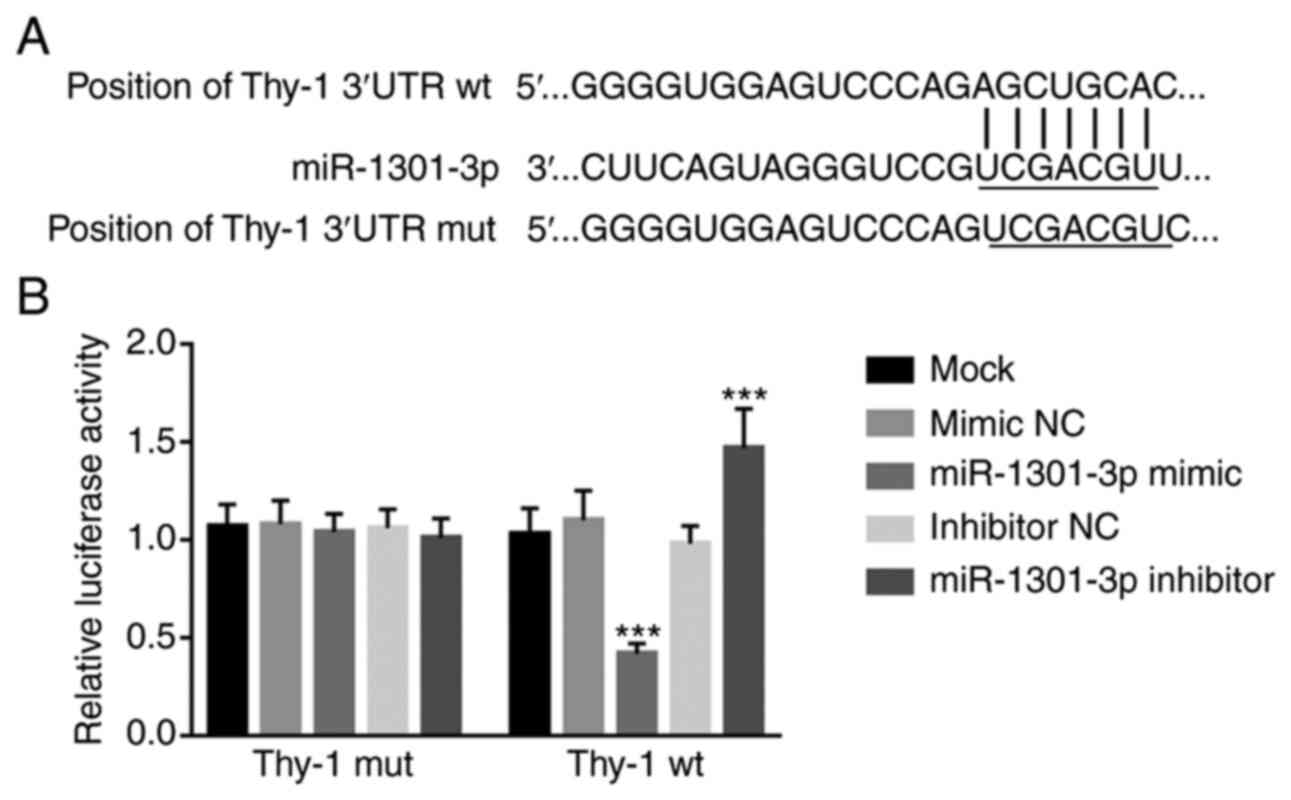
Target prediction and dual-luciferase
reporter assay
The TargetScan database (http://www.targetscan.org/vert_71) was used to predict
the target of miR-1301-3p and verify the 3′-UTR binding region of
miR-1301-3p.
The dual-luciferase reporter assay was performed
using a dual-luciferase reporter assay system (Promega
Corporation), according to the manufacturer's protocol. The
recombinant vectors, pGL3-thy-1-wt and pGL3-thy-1-mut were
generated using the pmirGLO vector (Promega Corporation). A549
cells were seeded into 96-well plates and co-transfected with
miR-1301-3p mimic or miR-1301-3p inhibitor, and pGL-thy-1-3′UTR-wt
or pGL-thy-1-3′UTR-mut using Lipofectamine® 2000
transfection reagent (Invitrogen; Thermo Fisher Scientific, Inc.)
and incubated at 37°C. Luciferase activities were detected 24 h
post-transfection with reference to Renilla luciferase
activity.
Statistical analysis
Statistical analysis was performed using SPSS 20.0
software (IBM Corp.) and GraphPad Prism 7.0 software (GraphPad
Software, Inc.). Data are presented as the mean ± standard
deviation obtained from at least triplicate experiments or
determination. Paired Student's t-test was used to compare
differences between two groups, while one-way ANOVA followed by
Tukey's post hoc test were used to compare differences between
multiple groups. The average expression level of miR-1301-3p (3.63)
in NSCLC tissues was used as the cut-off value to divide 124
patients with NSCLC into high (n=73) and low (n=51) miR-1301-3p
expression groups. The association between miR-1301-3p expression
and the clinicopathological characteristics of patients with NSCLC
was estimated by the Pearson's χ2 test. The Kaplan-Meier
method followed by log-rank test, and Cox regression analyses were
performed to assess survival and determine the prognostic value of
miR-1301-3p, respectively.
Results
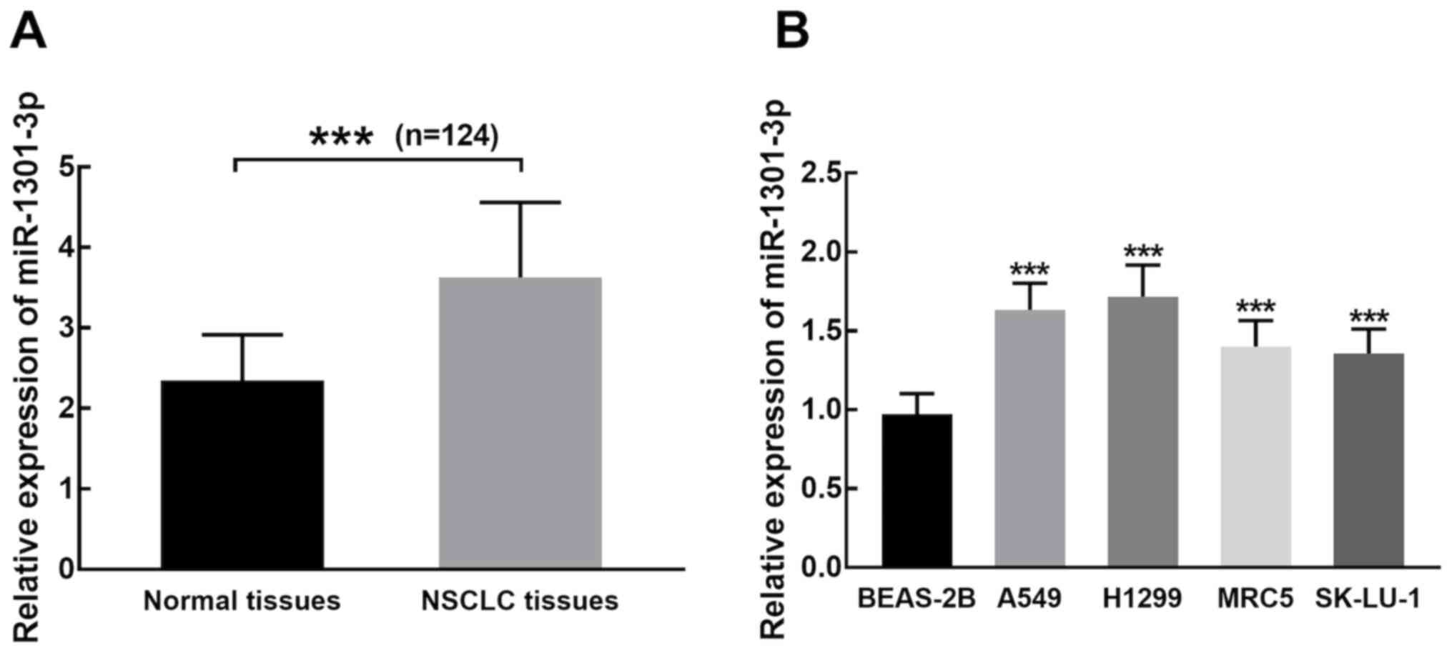
miR-1301-3p expression is
significantly upregulated in NSCLC tissues and cells
miR-1301-3p expression was significantly higher in
NSCLC tissues compared with adjacent normal tissues (P<0.001;
Fig. 1A). Similarly, miR-1301-3p
expression was significantly upregulated in NSCLC cells compared
with BEAS-2B cells (P<0.001; Fig.
1B).
miR-1301-3p expression is
significantly associated with TNM stage and lymph node
metastasis
The average expression level of miR-1301-3p (3.63)
in NSCLC tissues was used as the cut-off value to divide 124
patients with NSCLC into high (n=73) and low (n=51) miR-1301-3p
expression groups. As presented in Table
I, patients with high miR-1301-3p expression were significantly
associated with positive lymph node metastasis (P=0.027) and an
advanced clinical stage (P=0.025).
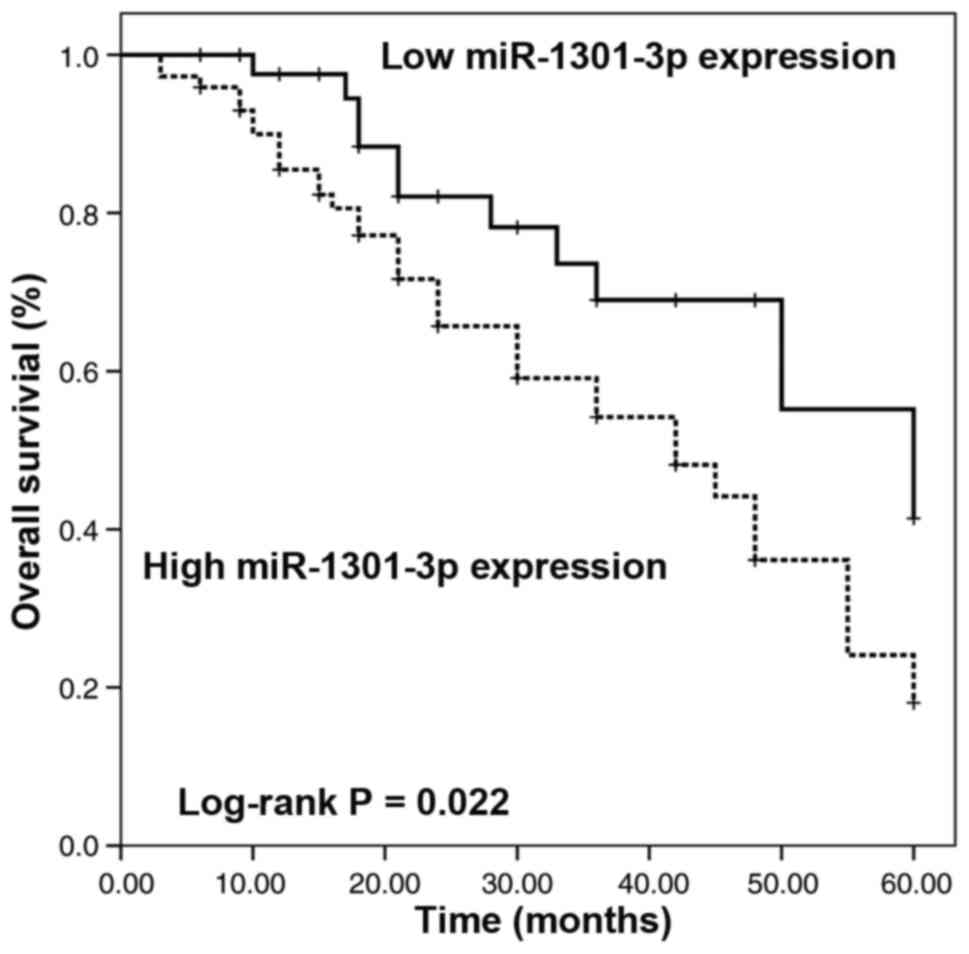
Overexpression of miR-1301-3p is
associated with a poor prognosis of patients with NSCLC
Survival information was obtained via a 5-year
follow-up survey and plotted using the Kaplan-Meier method. As
presented in Fig. 2, patients with
high miR-1301-3p expression had a significantly shorter overall
survival time than those with low miR-1301-3p expression (Log-rank
P=0.022).
Cox regression analysis was performed to determine
the prognostic value of miR-1301-3p. The results demonstrated that
miR-1301-3p expression [hazard ration (HR), 2.450; 95% confidence
interval (CI), 1.166–5.150; P=0.018], TNM stage (HR, 2.162; 95% CI,
1.093–4.276; P=0.027) and lymph node metastasis (HR, 2.181; 95% CI,
1.087–4.374; P=0.028) were independent factors for the clinical
prognosis of patients with NSCLC (Table
II).
| Table II.Cox regression analysis of
miR-1301-3p and survival of patients with NSCLC. |
Table II.
Cox regression analysis of
miR-1301-3p and survival of patients with NSCLC.
| Characteristic | HR factor | 95% CI | P-value |
|---|
| miR-1301-3p | 2.450 | 1.166–5.150 | 0.018 |
| Age | 1.223 | 0.657–2.276 | 0.525 |
| Sex | 1.290 | 0.671–2.481 | 0.445 |
| TNM stage | 2.162 | 1.093–4.276 | 0.027 |
| Lymph node
metastasis | 2.181 | 1.087–4.374 | 0.028 |
|
Differentiation | 1.548 | 0.824–2.907 | 0.175 |
| Smoking | 1.502 | 1.785–2.873 | 0.219 |
| Tumor size | 1.445 | 0.758–2.756 | 0.263 |
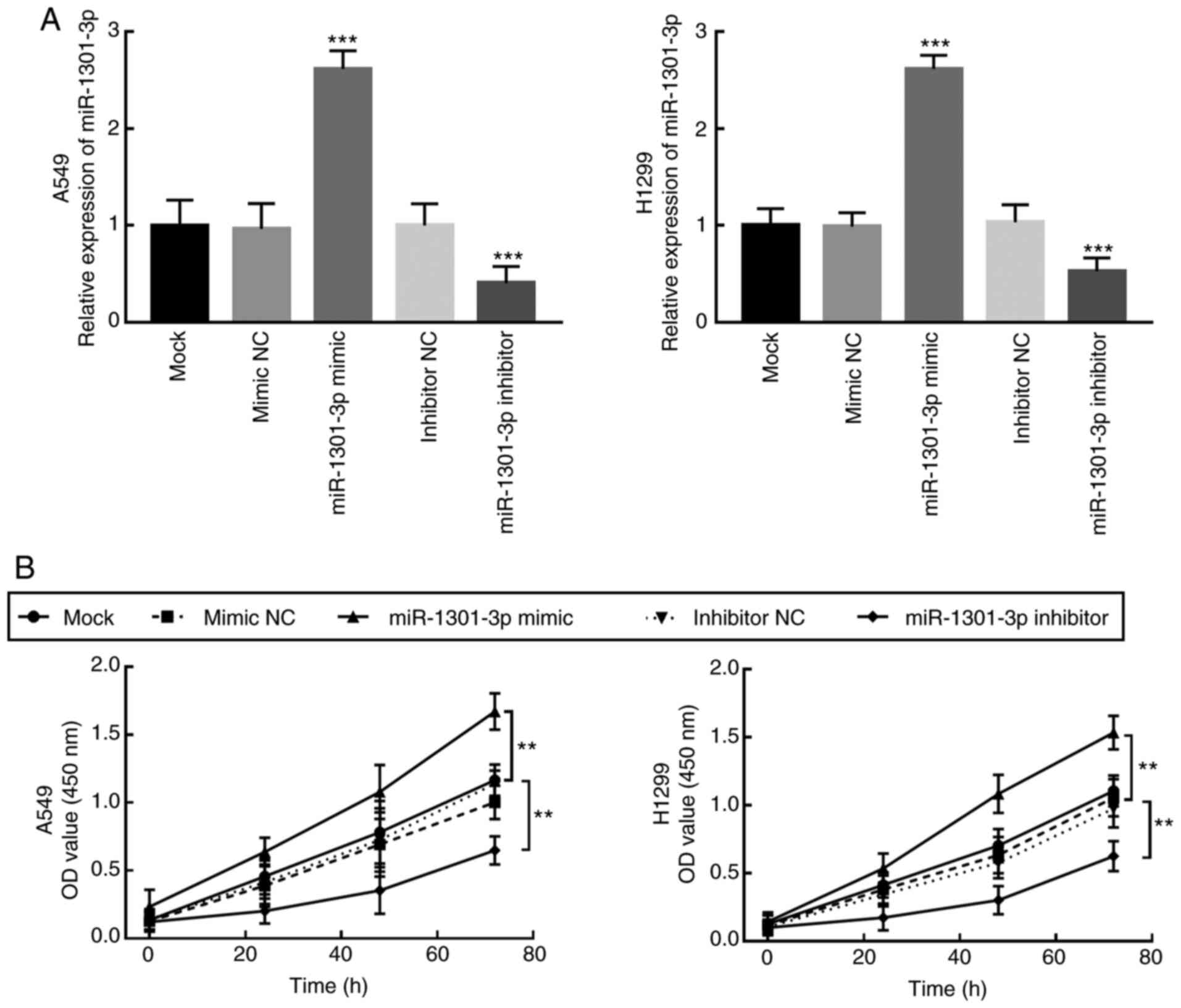
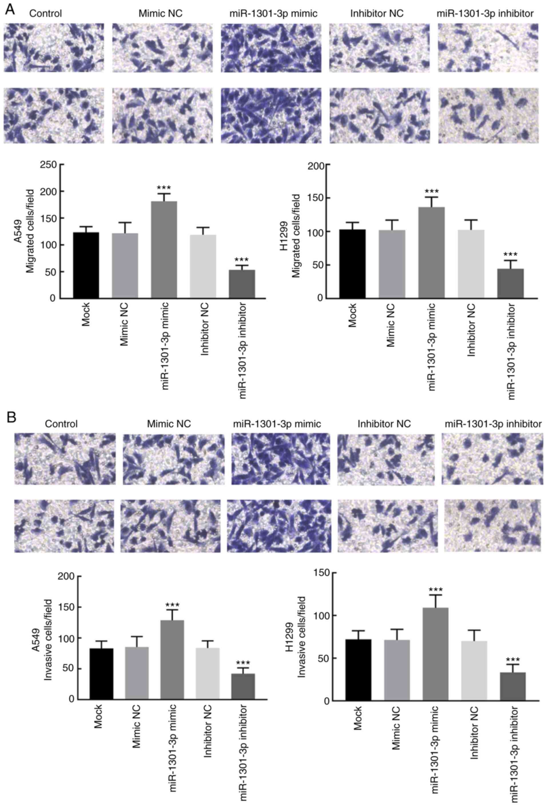
Overexpression of miR-1301-3p promotes
proliferation, migration and invasion of NSCLC cells
To determine the biological function of miR-1301-3p
in NSCLC, miR-1301-3p expression was regulated by transfection with
miR-1301-3p mimic or miR-1301-3p inhibitor. RT-qPCR analysis
demonstrated that transfection with miR-1301-3p mimic significantly
increased miR-1301-3p expression in A549 and H1299 cells, while
miR-1301-3p inhibitor significantly decreased miR-1301-3p
expression compared with the mock and negative controls
(P<0.001; Fig. 3A).
Proliferation of the transfected cells was detected
via the CCK-8 assay. Overexpression of miR-1301-3p promoted A549
and H1299 cell proliferation, whereas miR-1301-3p knockdown
significantly suppressed the proliferation of A549 and H1299 cells
(P<0.01; Fig. 3B). Similarly, the
migratory and invasive abilities of NSCLC cells significantly
enhanced following overexpression of miR-1301-3-3p, the effects of
which were reversed following miR-1301-3-3p knockdown (P<0.001;
Fig. 4A and B).
Thy-1 is a direct target of
miR-1301-3p
To determine the molecular mechanism underlying the
biological function of miR-1301-3p in NSCLC, the target of
miR-1301-3p was investigated. The target of miR-1301-3p was first
predicted using the TargetScan database, and the potential binding
sites are presented in Fig. 5A,
which was further confirmed via the dual-luciferase reporter
assay.
The luciferase activity of Thy-1 was inhibited
following overexpression of miR-1301-3p and increased following
miR-1301-3p knockdown in the Thy-1 wild-type group (P<0.001;
Fig. 5B). Conversely, neither
miR-1301-3p overexpression or knockdown affected the luciferase
activity of Thy-1 in the Thy-1 mutant group (Fig. 5B), suggesting that Thy-1 is a direct
target of miR-1301-3p.
Discussion
NSCLC is a common malignant tumor that accounts for
the majority of lung cancer cases, including adenocarcinoma,
squamous cell carcinoma, large cell carcinoma and several other
subtypes (20). The clinical outcome
of NSCLC remains unsatisfactory due to the high metastasis and
recurrence rates (4). It is
well-known that miRNAs play important roles in the progression and
development of several diseases, including cancers (21–23). For
example, miR4766-5p, miR-1915-3p and miR-615-3p are associated with
tumor development in gastric cancer (24–26).
Several miRNAs have been identified as effective biomarkers for the
progression of NSCLC. For example, miR-330-3p has been identified
as an upregulated miRNA in NSCLC, in which upregulation remarkably
promotes the proliferation, invasion and migration of NSCLC cells
by activating the MAPK/ERK signaling pathway (27). The prognostic value of several miRNAs
has also been reported in NSCLC and different types of cancer
(28,29). For example, downregulated miR-5702
expression is associated with the clinical progression and poor
prognosis of patients with NSCLC (30). Furthermore, miR-552 has been
identified as a prognostic predictor for patients with colorectal
cancer (31).
In the present study, miR-1301-3p expression was
significantly upregulated in NSCLC tissues and cell lines, which is
consistent with the miRNA expression profile of NSCLC (17). Dysregulation of miR-1301-3p
expression was significantly associated with the TNM stage and
lymph node metastasis of patients, suggesting that miR-1301-3p may
be involved in the development of NSCLC. In addition,
overexpression of miR-1301-3p was associated with the poor
prognosis of patients and was identified as an independent
indicator for the clinical prognosis of NSCLC, along with advanced
TNM stage and lymph node metastasis, which is consistent with a
previous study on colorectal cancer (32,33).
The levels of miR-1301 and its biological functions
vary in different types of cancer. For example, downregulated
miR-1301-3p expression in breast cancer acts as a tumor suppressor
that inhibits cell proliferation and induces cell apoptosis
(14). Similarly, in colorectal
cancer, overexpression of miR-1301-3p suppresses cell proliferation
and invasion, induces cell apoptosis, and decreases the volume and
weight of colorectal tumors (33).
Conversely, miR-1301-3p promotes cell expansion of prostate cancer
stem cells by inhibiting GSK3β and SFRP1 and activating the Wnt
pathway (15).
In the present study, overexpression of miR-1301-3p
promoted the proliferation, migration and invasion of NSCLC cells,
suggesting that miR-1301-3p may exert a tumor promoting role in the
progression of NSCLC. In addition, thy-1 was identified as a direct
target of miR-1301-3p. Thy-1 is a
glycosylphosphatidylinositol-linked outer membrane leaflet
glycoprotein (34). In a previous
study, thy-1 has been reported to suppress myofibroblastic
differentiation of lung fibroblasts, which is closely associated
with the occurrence of lung cancer (35). In addition, thy-1 has also been
reported to exert adverse impacts on the prognosis of lung cancer
(36). In melanoma, thy-1
contributes to cell metastasis by mediating the adhesion of
melanoma cells (37). Thus, it was
concluded that the promoting effects of miR-1301-3p were mediated
by regulating thy-1.
Among the clinicopathological characteristics of
patients with NSCLC, tumor size, differentiation and smoking status
are important factors to assess the clinical outcome of patients
(38–40). However, the small sample size limited
the results of the present study, which failed to exhibit a
significant association between these factors and the overall
survival of patients with NSCLC. Furthermore, in vivo
experiments are required in prospective studies to confirm the
results presented here.
In conclusion, the results of the present study
demonstrated that miR-1301-3p expression was significantly
upregulated in NSCLC tissues and cells, which was closely
associated with the TNM stage and lymph node metastasis of
patients. In addition, miR-1301-3p expression, advanced TNM stage
and lymph node metastasis served as independent prognostic factors
for NSCLC. Overexpression of miR-1301-3p significantly promoted the
proliferation, migration and invasion of NSCLC cells by targeting
thy-1. MiR-1301-3p was identified as a tumor promoter in NSCLC by
regulating thy-1, which requires further in vivo
validation.
Acknowledgements
Not applicable.
Funding
The present study was funded by Binzhou Medical
University Research Program and Research Startup Fund (grant no.
BY2014KJ48).
Availability of data and materials
The datasets used and/or analyzed during the present
study are available from the corresponding author upon reasonable
request.
Authors' contributions
All authors contributed to the conception and design
of the present study. LX, NN and PH performed the experiments. LX
contributed to analysis and interpretation of the data. HG revised
the manuscript for important intellectual content. All authors
drafted the initial manuscript, and have read and approved the
final manuscript.
Ethics approval and consent to
participate
The present study was approved by the Ethics
Committee of Binzhou Medical University Hospital (Binzhou, China;
approval no. 201212), and written informed consent was provided by
all patients prior to tissue collection.
Patient consent for publication
Not applicable.
Competing interests
The authors declare that they have no competing
interests.
References
|
1
|
Kumar M, Ernani V and Owonikoko TK:
Biomarkers and targeted systemic therapies in advanced non-small
cell lung cancer. Mol Aspects Med. 45:55–66. 2015. View Article : Google Scholar : PubMed/NCBI
|
|
2
|
Bray F, Ferlay J, Soerjomataram I, Siegel
RL, Torre LA and Jemal A: Global cancer statistics 2018: GLOBOCAN
estimates of incidence and mortality worldwide for 36 cancers in
185 countries. CA Cancer J Clin. 68:394–424. 2018. View Article : Google Scholar : PubMed/NCBI
|
|
3
|
Bilfinger T, Keresztes R, Albano D and
Nemesure B: Five-year survival among Stage IIIA lung cancer
patients receiving two different treatment modalities. Med Sci
Monit. 22:2589–2594. 2016. View Article : Google Scholar : PubMed/NCBI
|
|
4
|
Goldstraw P, Chansky K, Crowley J,
Rami-Porta R, Asamura H, Eberhardt WE, Nicholson AG, Groome P,
Mitchell A, Bolejack V, et al: The IASLC lung cancer staging
project: Proposals for revision of the TNM stage groupings in the
forthcoming (Eighth) edition of the TNM classification for lung
cancer. J Thorac Oncol. 11:39–51. 2016. View Article : Google Scholar : PubMed/NCBI
|
|
5
|
Marra A, Richardsen G, Wagner W,
Muller-Tidow C, Koch OM and Hillejan L: Prognostic factors of
resected node-positive lung cancer: Location, extent of nodal
metastases, and multimodal treatment. Thorac Surg Sci.
8:Doc012011.PubMed/NCBI
|
|
6
|
Ettinger DS, Wood DE, Aisner DL, Akerley
W, Bauman J, Chirieac LR, D'Amico TA, DeCamp MM, Dilling TJ,
Dobelbower M, et al: Non-small cell lung cancer, version 5.2017,
NCCN clinical practice guidelines in oncology. J Natl Compr Canc
Netw. 15:504–535. 2017. View Article : Google Scholar : PubMed/NCBI
|
|
7
|
Yu SL, Koo H, Lee SI, Kang J, Han YH, Yeom
YI and Lee DC: A synthetic CPP33-conjugated HOXA9 active domain
peptide inhibits invasion ability of non-small lung cancer cells.
Biomolecules. 10:15892020. View Article : Google Scholar
|
|
8
|
Mielgo-Rubio X, Calvo V, Luna J, Remon J,
Martín M, Berraondo P, Jarabo JR, Higuera O, Conde E, De Castro J,
et al: Immunotherapy moves to the early-stage setting in non-small
cell lung cancer: Emerging evidence and the role of biomarkers.
Cancers (Basel). 12:34592020. View Article : Google Scholar
|
|
9
|
Mustachio LM and Roszik J: Current
targeted therapies for the fight against non-small cell lung
cancer. Pharmaceuticals (Basel). 13:3742020. View Article : Google Scholar
|
|
10
|
Mohr AM and Mott JL: Overview of microRNA
biology. Semin Liver Dis. 35:3–11. 2015. View Article : Google Scholar : PubMed/NCBI
|
|
11
|
Lu TX and Rothenberg ME: MicroRNA. J
Allergy Clin Immunol. 141:1202–1207. 2018. View Article : Google Scholar : PubMed/NCBI
|
|
12
|
Rupaimoole R and Slack FJ: MicroRNA
therapeutics: Towards a new era for the management of cancer and
other diseases. Nat Rev Drug Discov. 16:203–222. 2017. View Article : Google Scholar : PubMed/NCBI
|
|
13
|
Romano G, Veneziano D, Acunzo M and Croce
CM: Small non-coding RNA and cancer. Carcinogenesis. 38:485–491.
2017. View Article : Google Scholar : PubMed/NCBI
|
|
14
|
Peng X, Yan B and Shen Y: MiR-1301-3p
inhibits human breast cancer cell proliferation by regulating cell
cycle progression and apoptosis through directly targeting ICT1.
Breast Cancer. 25:742–752. 2018. View Article : Google Scholar : PubMed/NCBI
|
|
15
|
Song XL, Huang B, Zhou BW, Wang C, Liao
ZW, Yu Y and Zhao SC: miR-1301-3p promotes prostate cancer stem
cell expansion by targeting SFRP1 and GSK3β. Biomed Pharmacother.
99:369–374. 2018. View Article : Google Scholar : PubMed/NCBI
|
|
16
|
Wang L, Zhao Y, Xu M, Zhou F and Yan J:
Serum miR-1301-3p, miR-335-5p, miR-28-5p, and their target B7-H3
may serve as novel biomarkers for colorectal cancer. J BUON.
24:1120–1127. 2019.PubMed/NCBI
|
|
17
|
Zhang YQ, Wang WY, Xue JX, Xu Y, Fan P,
Caughey BA, Tan WW, Cao GQ, Jiang LL, Lu Y, et al: MicroRNA
expression profile on solid subtype of invasive lung adenocarcinoma
reveals a panel of four miRNAs to Be associated with poor prognosis
in Chinese patients. J Cancer. 7:1610–1620. 2016. View Article : Google Scholar : PubMed/NCBI
|
|
18
|
Huston SF, Abdelmalik AG, Nguyen NC,
Farghaly HR and Osman MM: Whole-body 18F-FDG PET/CT: The need for a
standardized field of view-a referring-physician aid. J Nucl Med
Technol. 38:123–127. 2010. View Article : Google Scholar : PubMed/NCBI
|
|
19
|
Livak KJ and Schmittgen TD: Analysis of
relative gene expression data using real-time quantitative PCR and
the 2(-Delta Delta C(T)) method. Methods. 25:402–408. 2001.
View Article : Google Scholar : PubMed/NCBI
|
|
20
|
Pao W and Chmielecki J: Rational,
biologically based treatment of EGFR-mutant non-small-cell lung
cancer. Nat Rev Cancer. 10:760–774. 2010. View Article : Google Scholar : PubMed/NCBI
|
|
21
|
Ahn YH and Ko YH: Diagnostic and
therapeutic implications of microRNAs in non-small cell lung
cancer. Int J Mol Sci. 21:87822020. View Article : Google Scholar
|
|
22
|
El Aamri M, Yammouri G, Mohammadi H, Amine
A and Korri-Youssoufi H: Electrochemical biosensors for detection
of MicroRNA as a cancer biomarker: Pros and Cons. Biosensors.
10:1862020. View Article : Google Scholar
|
|
23
|
Ortiz-Quintero B: Extracellular MicroRNAs
as intercellular mediators and noninvasive biomarkers of cancer.
Cancers (Basel). 12:34552020. View Article : Google Scholar
|
|
24
|
Wang J, Liu L, Sun Y, Xue Y, Qu J, Pan S,
Li H, Qu H, Wang J and Zhang J: miR-615-3p promotes proliferation
and migration and inhibits apoptosis through its potential target
CELF2 in gastric cancer. Biomed Pharmacother. 101:406–413. 2018.
View Article : Google Scholar : PubMed/NCBI
|
|
25
|
Cui HW, Han WY, Hou LN, Yang L, Li X and
Su XL: miR-1915-3p inhibits Bcl-2 expression in the development of
gastric cancer. Biosci Rep. 39:BSR201823212019. View Article : Google Scholar : PubMed/NCBI
|
|
26
|
Wei Y, Wang Y, Zang A, Wang Z, Fang G and
Hong D: miR-4766-5p inhibits the development and progression of
gastric cancer by targeting NKAP. Onco Targets Ther. 12:8525–8536.
2019. View Article : Google Scholar : PubMed/NCBI
|
|
27
|
Wei CH, Wu G, Cai Q, Gao XC, Tong F, Zhou
R, Zhang RG, Dong JH, Hu Y and Dong XR: MicroRNA-330-3p promotes
cell invasion and metastasis in non-small cell lung cancer through
GRIA3 by activating MAPK/ERK signaling pathway. J Hematol Oncol.
10:1252017. View Article : Google Scholar : PubMed/NCBI
|
|
28
|
Jia Y, Tan W and Zhou Y: Transfer
RNA-derived small RNAs: Potential applications as novel biomarkers
for disease diagnosis and prognosis. Ann Transl Med. 8:10922020.
View Article : Google Scholar : PubMed/NCBI
|
|
29
|
Parizi PK, Yarahmadi F, Tabar HM, Hosseini
Z, Sarli A, Kia N, Tafazoli A and Esmaeili SA: MicroRNAs and target
molecules in bladder cancer. Med Oncol. 37:1182020. View Article : Google Scholar : PubMed/NCBI
|
|
30
|
Li K, Xu Y and Yuan LN: Down-regulation of
miR-5702 is associated with clinical progression and poor prognosis
in patients with non-small-cell lung cancer. Eur Rev Med Pharmacol
Sci. 23:2047–2052. 2019.PubMed/NCBI
|
|
31
|
Wang N and Liu W: Increased expression of
miR-552 acts as a potential predictor biomarker for poor prognosis
of colorectal cancer. Eur Rev Med Pharmacol Sci. 22:412–416.
2018.PubMed/NCBI
|
|
32
|
Wen J, Wang H, Dong T, Gan P, Fang H, Wu
S, Li J, Zhang Y, Du R and Zhu Q: STAT3-induced upregulation of
lncRNA ABHD11-AS1 promotes tumour progression in papillary thyroid
carcinoma by regulating miR-1301-3p/STAT3 axis and PI3K/AKT
signalling pathway. Cell Prolif. 52:e125692019. View Article : Google Scholar : PubMed/NCBI
|
|
33
|
Xu G, Wang H, Yuan D, Yao J, Meng L, Li K,
Zhang Y, Dang C and Zhu K: RUNX1-activated upregulation of lncRNA
RNCR3 promotes cell proliferation, invasion, and suppresses
apoptosis in colorectal cancer via miR-1301-3p/AKT1 axis in vitro
and in vivo. Clin Transl Oncol. 22:1762–1777. 2020. View Article : Google Scholar : PubMed/NCBI
|
|
34
|
Rege TA and Hagood JS: Thy-1 as a
regulator of cell-cell and cell-matrix interactions in axon
regeneration, apoptosis, adhesion, migration, cancer, and fibrosis.
FASEB J. 20:1045–1054. 2006. View Article : Google Scholar : PubMed/NCBI
|
|
35
|
Shentu TP, Huang TS, Cernelc-Kohan M, Chan
J, Wong SS, Espinoza CR, Tan C, Gramaglia I, van der Heyde H, Chien
S and Hagood JS: Thy-1 dependent uptake of mesenchymal stem
cell-derived extracellular vesicles blocks myofibroblastic
differentiation. Sci Rep. 7:180522017. View Article : Google Scholar : PubMed/NCBI
|
|
36
|
Schliekelman MJ, Creighton CJ, Baird BN,
Chen Y, Banerjee P, Bota-Rabassedas N, Ahn YH, Roybal JD, Chen F,
Zhang Y, et al: Thy-1+ Cancer-associated fibroblasts
adversely impact lung cancer prognosis. Sci Rep. 7:64782017.
View Article : Google Scholar : PubMed/NCBI
|
|
37
|
Schubert K, Gutknecht D, Koberle M,
Anderegg U and Saalbach A: Melanoma cells use Thy-1 (CD90) on
endothelial cells for metastasis formation. Am J Pathol.
182:266–276. 2013. View Article : Google Scholar : PubMed/NCBI
|
|
38
|
Hsu HH, Ko KH, Chou YC, Lin LF, Tsai WC,
Lee SC, Chang H and Huang TW: SUVmax and tumor size predict
surgical outcome of synchronous multiple primary lung cancers.
Medicine (Baltimore). 95:e23512016. View Article : Google Scholar : PubMed/NCBI
|
|
39
|
Petrovic M, Baskic D, Bankovic D and Ilic
N: Neuroendocrine differentiation as an indicator of
chemosensitivity and prognosis in nonsmall cell lung cancer.
Biomarkers. 16:311–320. 2011. View Article : Google Scholar : PubMed/NCBI
|
|
40
|
Fukui M, Suzuki K, Matsunaga T, Oh S and
Takamochi K: Importance of smoking cessation on surgical outcome in
primary lung cancer. Ann Thorac Surg. 107:1005–1009. 2019.
View Article : Google Scholar : PubMed/NCBI
|