Introduction
Gastric cancer (GC) is the fifth most commonly
diagnosed type of cancer worldwide, and the majority of patients
with early GC are curable by appropriate treatments (1). Patients with advanced GC, however, have
a poor prognosis, despite the progress achieved in various
treatment strategies, including extended surgical resections and
intensive chemotherapy, with or without the use of molecular
targeted treatments. Preoperative chemotherapy has been
demonstrated to be beneficial in certain subgroups of patients with
GC (2,3). Therefore, early detection of this
lethal disease is critical, and the preoperative identification of
tumor characteristics can guide decision-making regarding the
selection of the most appropriate treatment strategies. Toward
these ends, various serum tumor markers have been established and
utilized in clinical practice (4).
Although they are widely used as supplementary information for
diagnosis, their diagnostic accuracy, as well as their specificity
and sensitivity for GC, have yet to be optimized (5).
Comprehensive molecular analyses have recently
elucidated various genetic and epigenetic alterations in several
types of cancer, and numerous studies in which circulating
cell-free nucleic acids have been analyzed have reported the
potential utility of blood molecular biomarkers (6). Blood biomarkers identified in the
serum, plasma or other biological fluids derived from patients have
several advantages, such as overcoming the undependability due to
the tumor heterogeneities, and the feasibility of repeated sample
collection. It has also been demonstrated that numerous metabolites
are involved in carcinogenesis and cancer progression. In order to
comprehensively identify and analyze the genetic and metabolic
variations that are involved in carcinogenesis and cancer
development, mass spectrometry (MS) techniques and devices are
useful in the medical field (7). In
addition, results of integrative metabolomic analyses using machine
learning methods have been reported (8,9). It is
expected that these technologies will become generally available,
particularly for establishing a cancer diagnosis. Endoscopic
examination, the gold standard for a definitive GC diagnosis, has
revealed that a white opaque substance is a novel endoscopic
finding in gastric neoplasms, indicating that there is
intracellular accumulation of lipid droplets in GC (10,11).
Therefore, a lipidomic approach to GC has attracted great research
attention.
In recent clinical settings, not only surgical or
endoscopic curative resection, but also perioperative combined
treatments, such as neoadjuvant and/or adjuvant chemotherapies, are
often practiced and have greatly affected survival outcomes
(2,3). However, it is challenging to select the
best treatment at the best time, as clinical situations vary
constantly. Thus, in addition to achieving early cancer detection,
it is necessary to identify and develop novel biomarkers for
precision medicine to provide a highly effective and low-risk
treatment to each individual patient. Various studies have been
published on the expression of cancer-specific biomolecules in
non-cancerous and cancerous tissues, including studies using MS
(12–16). It is important to compare the
differences between cancerous and non-cancerous tissues, or between
patients with cancer and healthy volunteers (HVs) without any
history of cancer, in order to identify cancer-specific molecules
that can be useful in the early detection of cancer. In the future,
personalized medicine will be more important in order to select the
best treatment strategies for individual cases. Therefore, the
present study conducted a comprehensive lipidomic approach using MS
and a machine learning method to provide the basis of precision
medicine for GC. Lipid metabolism is attracting increasing
attention in tumor development and numerous other diseases. In
particular, phospholipids have been reported to play important
roles in various cancers (17–19). In
addition, peripheral blood samples have a broad diagnostic utility
due to their ease of use and accessibility, in contrast to tissue
samples, which can be obtained only by surgical resection or biopsy
with relatively highly invasiveness (20,21).
Thus, methods such as liquid biopsy, which can be used to analyze
primary tumors using body fluids, including blood, urine, digestive
juice and cerebral spinal fluid, are an attractive research focus
(22,23).
The present study analyzed the levels of
phospholipids in plasma derived from patients with GC by using
liquid chromatography/electrospray ionization-MS (LC/ESI-MS), a
method which accurately identifies and quantifies lipid molecules.
Furthermore, the present study aimed to establish a machine
learning-based diagnostic algorithm for GC.
Materials and methods
Biobank establishment
A human biobank named ‘SHINGEN (Yamanashi Biobank of
Gastroenterological Cancers)’, was established, which included
frozen tissue, cell and fluid samples obtained from January 2018
until the present date. The tissue samples were derived from
primary and metastatic malignant tumors, and from adjacent
non-tumor tissues. Cellular and liquid components from the
intraperitoneal lavage of patients collected during surgical
procedures were obtained following centrifugation at 2,000 × g for
10 min at 4°C. Peripheral blood samples, including plasma and
serum, were obtained from patients at various time points during
clinical interventions or observations without treatment. For the
preparation of the plasma samples, 5 ml peripheral whole blood was
collected from each patient using vacuum blood sampling tubes
containing the anticoagulant reagent EDTA (NP-EN0507; NIPRO Corp.).
The tubes were immediately centrifuged at 2,000 × g for 10 min at
4°C. Serum samples were collected using vacuum blood sampling tubes
containing separating agents and coagulation accelerators
(VP-AS074K; Terumo Corp.). After incubation at 4°C for 30 min, the
tubes were centrifuged following the aforementioned method. The
plasma and serum samples obtained upon centrifugation were stored
at −80°C until further analysis. Samples from patients with various
digestive malignancies, such as GC, esophageal cancer, colorectal
cancer, pancreatic cancer and hepatocellular carcinoma were
included in the SHINGEN biobank. Furthermore, samples from patients
who underwent subsequent surgical resection according to clinical
guidelines after radical resection for their early cancer, were
also included in this biobank. Of note, the majority of such
patients had no residual malignancies at the time of surgery and/or
blood sample collection.
Patient clinical information and
ethical concerns
Patient clinical information, including pathological
findings, was obtained from the electronic medical recording system
at University of Yamanashi Hospital (Yamanashi, Japan). The
pathological findings of GC were defined according to the Union for
International Cancer Control classification of malignant tumors
(8th edition) (24). This
information was stored in the database of the biobank. The
biological sample collection in the SHINGEN (#1665) biobank and the
study design (#2192) were approved by the Ethics Committee of the
University of Yamanashi, and the study was performed in accordance
with the ethical standards of the Declaration of Helsinki and its
later amendments (25). Written
informed consent for the use of biological samples and clinical
data was obtained from all the patients.
Inclusion and exclusion criteria
Between January 2018 and April 2020, a total of
1,592 blood plasma samples from patients who were treated at the
First Department of Surgery at the University of Yamanashi
Hospital, were collected prospectively in the SHINGEN biobank. A
total of 910 plasma samples were obtained during the intra- or
post-intervention period, and 682 were collected during the
preoperative phase. Of these, 16 patient samples were set as the
control group. These samples were obtained from patients who
received additional resection after endoscopic resection for early
tumors (12 patients with GC, 2 with esophageal cancer and 2 with
colorectal cancer) and who were confirmed to have no residual
malignancies based on their postoperative pathological findings. Of
the remaining 666 plasma samples, 152 were from patients with GC
and 514 from patients with other malignancies. After excluding
patients with GC who had no lymph node metastasis based on their
preoperative clinical findings and those who underwent non-radical
resection with residual tumors, 20 patients with advanced GC were
analyzed in the present study. The patient flow diagram is shown in
Fig. S1.
LC/ESI-MS
A total of 10 µl human plasma was added into 990 µl
0.1% formic acid in methanol, and the sample solution was mixed
using ThermoMixer C (Eppendorf) for 5 min at 4°C. After incubation
on ice for 10 min, the sample solution was centrifuged at 15,000 ×
g for 10 min. The resultant supernatant was 2-fold diluted using
0.1% formic acid in methanol, and 300 µl diluted supernatant was
applied into a LabTotal Vial (Shimadzu Corporation). The vial was
inserted to the sample rack of the autosampler (Nexera X2 SIL-30AC;
Shimadzu Corporation), and 3 µl sample was injected into the column
for LC separation.
LC/ESI-MS was performed using the high-pressure LC
installed LCMS-8060 (Shimadzu Corporation) system. To analyze the
lipid components in human plasma, the LC/MS/MS Method Package for
Phospholipid Profiling (Shimadzu Corporation) was used following
the manufacturer's instructions. Kinetex C8 column (Kinetex C8,
150×2.1 mm i.d., 3.6-µm particle size; Phenomenex), mobile phase A
(20 mM ammonium formate in water) and mobile phase B (acetonitrile:
Isopropanol 1:1 v/v) were used for LC separation. The concentration
of mobile phase B was programmed as 20% (0 min)-20% (1 min)-40% (2
min)-92.5% (25 min). The oven temperature was 45°C. Data processing
and molecular identification/quantification were performed
automatically by using the LabSolutions software (version 5.82 SP1;
Shimadzu Corporation).
Statistical analysis
Age and tumor size were represented as the mean ±
standard error of the mean. Statistical evaluation of the gender
ratio between the two groups was performed using the χ2
test. The relative ion intensities of each molecule between the
control and GC groups were calculated as the ratio of the mean
values in each group. The variance of continuous values was
confirmed by unpaired t-test. All statistical analyzes, receiver
operating characteristic (ROC) graphs and box plots were performed
using EZR (Saitama Medical Center, Jichi Medical University,
Saitama, Japan), which is a graphical user interface for R (The R
Foundation for Statistical Computing, Vienna, Austria) (26). Comparisons among groups were
performed using partial least squares (PLS) regression according to
a previously reported protocol (27). PLS regression is usually performed to
compare the characteristics of the samples and to classify certain
groups. In the present study, this method was used to determine the
discriminability between the GC group and the control group before
applying the machine learning approach.
Diagnostic algorithm
To construct the diagnostic algorithm of GC,
logistic regression (LR), a type of machine learning method, was
used for discriminant analysis. The expression levels (peak area in
the chromatogram) of 536 lipid molecules obtained from each plasma
sample were individually normalized by the median value. The
normalized datasets of control and cancer were learned by LR, and
blinded samples were classified as cancer or not. The cancer
possibility was indicated as the probability value (0.0–1.0). The
procedure and mathematical formulae used were those described in
our previous study (27). The
predictive accuracy of the LR classifier was evaluated by using a
leave-one-out cross validation (LOOCV) procedure (28). These procedures are shown in Fig. S2.
Results
Patient characteristics
The clinicopathological characteristics of the
patients are summarized in Table I.
There were no differences in the age or sex of the patients between
the two groups. The GC group included numerous patients with
advanced GC who exhibited deep invasion, extensive lymph node
metastasis and severe lymphovascular invasion. By contrast, tumor
markers such as carcinoembryonic antigen (CEA) and carbohydrate
antigen 19-9 (CA19-9) were not higher than the standard values in
the majority of patients, despite being generally used as
biomarkers of GC. Although three patients in the GC group had
postoperative cancer recurrence during the follow-up period, none
of the patients in the control group had residual malignancies or
recurrences.
 | Table I.Characteristics of controls and
patients with GC. |
Table I.
Characteristics of controls and
patients with GC.
| Variables | Control (n=16) | GC (n=20) | P-value |
|---|
| Comparison between
control and GC |
|
|
|
| Age
(mean ± SD) | 71.2±10.3 | 67.1±12.1 |
0.285 |
| Sex, n
(male/female) | 9/7 | 11/9 | 0.999 |
| Cancer specific
variables |
|
|
|
| Tumor
size, mm (mean ± SD) |
| 66.4±49.9 |
|
| CEA, n
(<5/≥5 ng/ml) |
| 17/3 |
|
| CA19-9,
n (<37/≥37 U/ml) |
| 14/6 |
|
|
T-factor, n (T1/2/3/4) |
| 2/3/8/7 |
|
|
N-factor (N0/1/2/3) |
| 2/5/6/7 |
|
|
M-factor, n (M0/1) |
| 19/1 |
|
| Stage,
n (I/II/III/IV) |
| 0/10/9/1 |
|
|
Lymphatic invasion, n
(negative/positive) |
| 1/19 |
|
| Venous
invasion, n (negative/positive) |
| 2/18 |
|
|
Pathological subtypes, n
(differentiated/undifferentiated) |
| 10/10 |
|
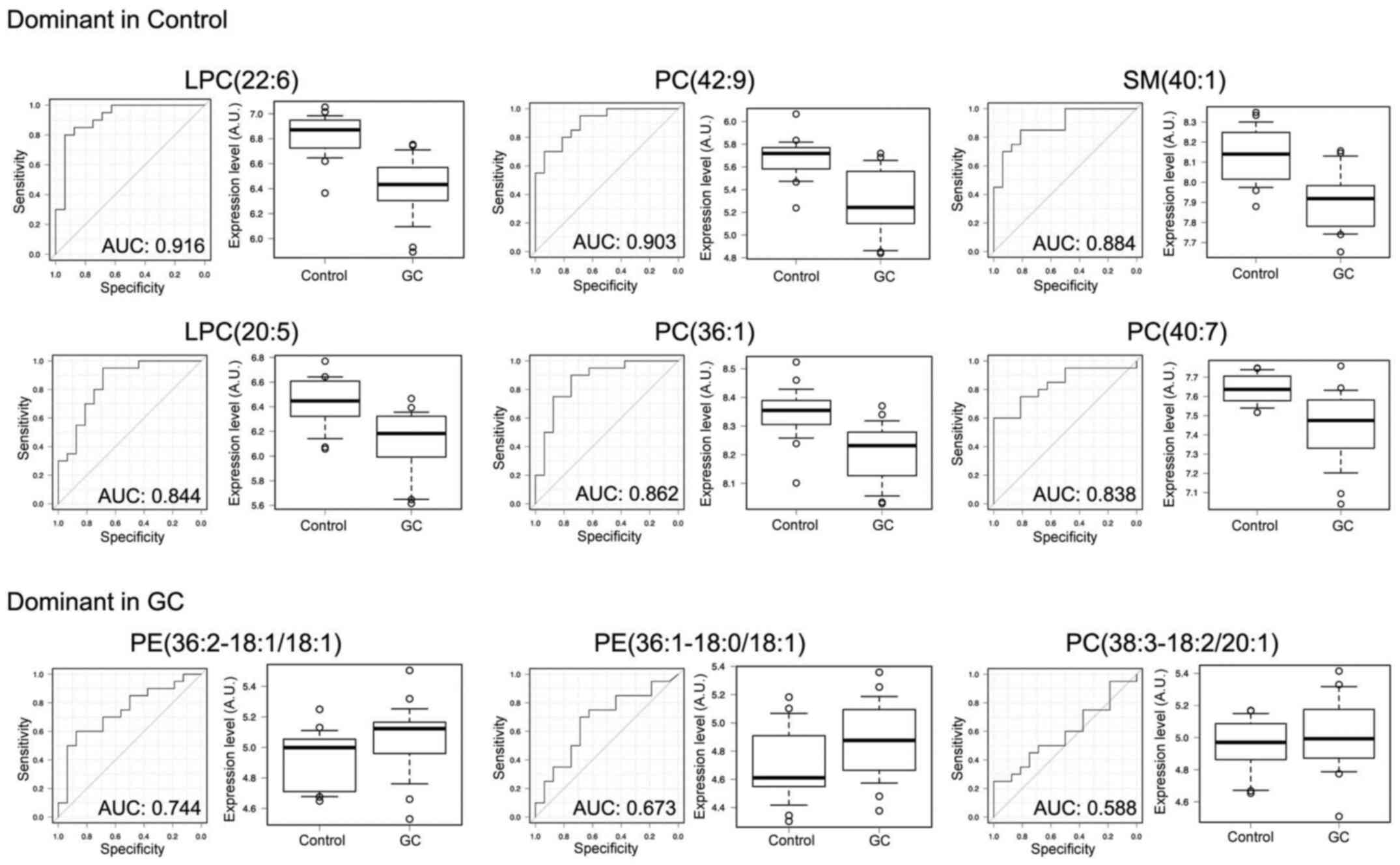
Lipid biomarkers of GC
The simultaneous analysis of the plasma lipidome
using LC/ESI-MS identified and quantified a total of 236
phospholipid molecules. The top ten molecules, which were up- or
downregulated in the GC group, are listed in Table II. Of these, PE(36:2-18:1/18:1)
showed the most remarkable upregulation in the GC group. By
contrast, LPC(38:2) exhibited the most marked difference in
relative expression in the control group, as a result of its
suppression in the GC group. Overall, there was a downregulation
trend for the ion intensities of the majority of molecules in GC
plasma. The results of detailed analyses comparing the ion
intensities between the GC and control groups, and the ROC curves
with each area under the curve are shown in Fig. 1. These results suggested that certain
lipid molecules, particularly the downregulated lipids, can be used
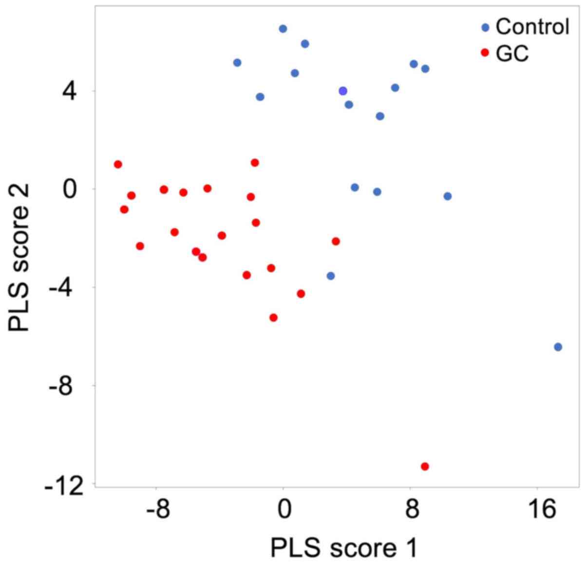
as biomarkers for GC. In addition, the results of the PLS score
plot indicated that the lipid composition of GC plasma is
specifically changed, and can be used in discriminant analysis
(Fig. 2).
| Table II.Candidate markers of phospholipids
for GC. |
Table II.
Candidate markers of phospholipids
for GC.
| Candidate
molecule | Relative expression
levels (GC/control) | P-value
(−log10) |
|---|
| Dominant in
control |
|
|
|
LPC(22:6) | 0.424 | 6.410 |
|
PC(42:9) | 0.399 | 4.720 |
|
SM(40:1) | 0.579 | 4.680 |
|
LPC(20:5) | 0.502 | 3.970 |
|
PC(36:1) | 0.732 | 3.850 |
|
PC(40:7) | 0.680 | 3.600 |
|
PC(40:1) | 0.329 | 3.590 |
|
LPC(20:0) | 0.646 | 3.460 |
|
LPC(18:2) | 0.644 | 3.290 |
|
LPC(22:0) | 0.572 | 3.290 |
| Dominant in GC |
|
|
|
PE(36:2-18:1/18:1) | 1.590 | 1.800 |
|
PE(36:1-18:0/18:1) | 1.508 | 0.970 |
|
PC(38:3-18:2/20:1) | 1.253 | 0.790 |
|
PC(34:3-16:1/18:2) | 1.235 | 0.720 |
|
PE(34:2-16:0/18:2) | 1.270 | 0.560 |
|
PE(34:1) | 1.180 | 0.550 |
|
PE(34:2) | 1.207 | 0.490 |
|
PE(34:3) | 1.397 | 0.480 |
|
PE(36:3) | 1.272 | 0.420 |
|
PC(34:2) | 1.053 | 0.380 |
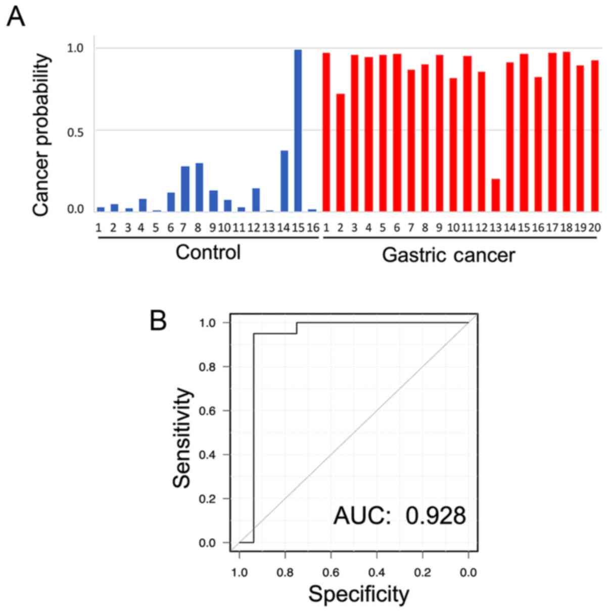
Discrimination of GC by machine
learning
Fig. 3A and Table SI show the individual value of
cancer probability in each plasma sample by using discriminant
analysis with LOOCV. When a threshold of 0.5 was set for the
probability of a sample being cancerous, each characteristic of
each patient was distinguished. Although the correct
characteristics of only one patient in each group could not be
detected, a correct classification of subjects in the GC and
control groups was achieved with an accuracy of 94.4% for almost
all patients in both groups. The specificity and sensitivity were
93.8 and 95.0%, respectively. Furthermore, the area under the ROC
curve was 0.928 (Fig. 3B).
Discussion
The present study analyzed the expression levels of
phospholipids, including phosphatidylcholine, sphingomyelins and
phosphatidylethanolamine, in peripheral blood samples. Although
these molecules are universally present in the whole body, certain
plasma lipid molecules showed pronounced differences between the
cancer-free controls and the patients with GC. However, the
discrimination probabilities for each individual molecule were
insufficient as an independent diagnostic tool. Therefore, an
integrative analysis of all these molecules using a machine
learning method was performed, aiming to establish more useful
diagnostic systems with a higher accuracy. Compared with the
results obtained using conventional GC markers, such as CEA and
CA19-9, the results of integrative analysis showed a much higher
sensitivity and specificity.
In discrimination analysis using LOOCV, correct
results were not achieved for all the patients in each group.
Regarding patient GC no. 13 (Fig. 3;
Table SI), who was not determined
as a correct cancerous characteristic, the patient had no specific
characteristics observed besides pathological findings of T3N2M0
and non-elevation of both CEA and CA19-9. In the control group,
patient control no. 15 (Fig. 3;
Table SI), who did not display
non-cancerous characteristics after discrimination, was after
endoscopic resection for early surficial esophageal cancer. This
could be one of the reasons for the inaccuracy of the results.
Although further comparative analyses with strict setting of
control specimens may be necessary to establish clinically useful
diagnostic algorithms with a higher accuracy, our results suggest
that, at least for advanced GC, using machine learning methods may
be useful for the detection of GC.
To achieve significant accuracy in studies comparing
patients with and without cancer, it is crucial to use the
appropriate methodology for selecting the control specimens.
Control specimens are frequently collected from ‘HVs’; however,
there is often an age difference between patients with and without
cancer, as the latter tend to be much younger than the former. This
can significantly affect the results of the analysis, particularly
in a lipidomic study, since the differences in the systemic
metabolism associated with the patients' background and age are
likely to be large (29). Previous
studies have reported lipidomic approaches for cancer diagnosis
using blood samples. For example, Guo et al (18) demonstrated that serum phospholipids
were useful biomarkers for the different pathophysiological states
of lung, gastric, intestinal and pancreatic cancer. Consistent with
our results, the authors found that PC(34:2) was one of the six
molecules that was increased in patients with GC. By contrast, Lee
et al (19), reported that
patients with GC had increased levels of LPC(18:2), and decreased
levels of PC(34:2) and PE(36:3). One of the reasons for this
discrepancy may be the control groups used in the different
studies. In fact, in one report, the individuals in the control
group were ~10 years younger than the patients in the GC group.
To prevent such a background bias, the present study
used control plasma samples from patients who had underwent
endoscopic resection for their early cancer months before the
collection of biological samples for the SHINGEN biobank. For these
patients, the clinical guidelines recommend additional surgical
resection for their risk of lymph node metastases, and indeed this
is commonly performed (30–32). However, the risk of metastases was
generally low, and in the present study, there was no remaining
cancer or metastasis in postoperative pathological findings.
Therefore, in the present study, these cancer-free patients could
be used as the control group in the different comparisons against
the group of patients with advanced GC. By selecting this control
group, the patients' background, including age and gender, were
similar between the two groups. In addition, patients' status such
as nutritional status, liver disorder or lipidemic disorder may
affect the levels of plasma phospholipids. To investigate the
effects of these factors, stratified analyses were performed for
presence or absence of liver or lipidemic disorders. As a result,
there were no obvious differences between patients with and without
liver or lipidemic disorders in terms of specific phospholipid
expression levels in the present study (data not shown). However,
this should be further investigated in future studies using a
large-scale cohort.
Concerning the control group settings, we previously
performed the similar examinations to those described in the
present study using plasma samples derived from HVs. Although each
indicated molecule showed various expression levels in the HV group
compared with those of the control and GC samples (Fig. S3), PLS analysis for the three groups
(control, GC and HV) showed different characteristics for each
group and good probability of discriminating each other (Fig. S4). This result means that
amplification of the database to include HV samples should be
considered for cancer screening during health checks, and that the
integration analyses using machine learning methods presented in
the current study contribute to discriminating each group. It
appears to be reasonable that the cancer high-risk group was set as
a control in the present study, and research including HV samples
should be performed in future studies. To detect microresidual
tumors or recurrent microtumors after surgery, comparison between
pre- and postoperative conditions in the same patient is important,
and this will be investigated in future studies.
To establish a novel diagnostic method for GC,
previous studies have focused on the analysis of low molecular
weight metabolites found in blood, serum or plasma, including
lipids, primary metabolites and cell-free nucleic acids (33–36).
During the preparation of serum, blood clots are formed by using
serum separating agents and/or coagulation accelerators. In this
process, numerous components in platelets, such as intracellular
messengers/mediators, cell membrane and organelles, are released
into the serum from disrupted platelets (37). The released components affect the MS
results of the molecular composition of serum, particularly the
phospholipid results. To avoid this problem, the plasma used in the
present study was obtained by employing the anticoagulant agent
EDTA and by centrifugation, without platelet disruption. This
methodology may have contributed to the present results. Thus, the
authors recommend the use of this methodology for similar studies
in this field.
In summary, the novel cancer diagnosis approach
employed in the present study may contribute to the development of
relevant desirable biomarkers in the near future. The present study
has certain limitations that need to be acknowledged. First, it is
difficult to derive a definitive conclusion due to the small number
of patients included in the study. Second, the patients in the
control group, although they were cancer-free at the time of plasma
collection and for months before sample collection, they had a
previous cancer history. Despite certain limitations, the results
of the present study strongly suggest that this new approach has a
promising future as a diagnostic tool for GC. In conclusion, the
present study suggests the diagnostic prospects of a plasma
lipidomic and machine learning approach for GC. By accumulating
more reliable data, this novel methodology may also be able to
predict the efficacy of each therapy.
Supplementary Material
Supporting Data
Acknowledgements
The authors would like to thank Ms. Arisa Ogihara
(First Department of Surgery, Faculty of Medicine, University of
Yamanashi, Yamanashi, Japan), Ms. Ayumi Manita (Department of
Anatomy and Cell Biology, Faculty of Medicine, University of
Yamanashi, Yamanashi, Japan) and Ms. Masumi Tanzawa (Department of
Anatomy and Cell Biology, Faculty of Medicine, University of
Yamanashi, Yamanashi, Japan) for their technical assistance. The
authors would like to thank Shimadzu Corporation (Kyoto) (to which
TM is affiliated) for lending the Mass Spectrometry instruments to
ST.
Funding
The present study was performed mainly with expenses
grants of the University of Yamanashi, and partially supported by
JSPS KAKENHI (grant nos. 17K10601 and 20K09031 to DI).
Availability of data and materials
The datasets used and/or analyzed during the current
study are available from the corresponding author on reasonable
request.
Authors' contributions
RS and KY performed the majority of the experiments
and wrote the initial draft of the manuscript. RS, KY, KS, SF, HA,
YK, TM, KO, TI, DI and ST designed the study and contributed to
analysis and interpretation of data, and assisted in the
preparation of the manuscript. RS, KY and TI were responsible for
confirming the authenticity of all the raw data. All other authors
have contributed to data collection and interpretation, and
critically reviewed the manuscript. All authors approved the final
version of the manuscript, and agree to be accountable for all
aspects of the work in ensuring that questions related to the
accuracy or integrity of any part of the work are appropriately
investigated and resolved. All authors read and approved the final
manuscript.
Ethics approval and consent to
participate
The biological sample collection in the SHINGEN
(#1665) biobank and the study design (#2192) were approved by the
Ethics Committee of the University of Yamanashi (Yamanashi, Japan).
All procedures followed were in accordance with the ethical
standards of the responsible committee on human experimentation
(institutional and national) and with the Helsinki Declaration of
1964 and later versions. Written informed consent to be included in
the study, or the equivalent, was obtained from all patients.
Patient consent for publication
Although detailed clinical data of individual
patients were not included in the present study, consent for
publication was obtained from all patients included in the present
study.
Competing interests
The authors declare that they have no competing
interests.
Glossary
Abbreviations
Abbreviations:
|
AUC
|
area under the curve
|
|
CA19-9
|
carbohydrate antigen 19-9
|
|
CEA
|
carcinoembryonic antigen
|
|
EDTA
|
ethylenediamine tetraacetic acid
|
|
GC
|
gastric cancer
|
|
HV
|
healthy volunteer
|
|
LC/ESI-MS
|
liquid chromatography/electrospray
ionization-mass spectrometry
|
|
LOOCV
|
leave-one-out cross validation
|
|
LR
|
logistic regression
|
|
MS
|
mass spectrometry
|
|
PLS
|
partial least squares
|
|
ROC
|
receiver operating characteristics
|
|
SHINGEN
|
Yamanashi Biobank of
Gastroenterological Cancers
|
|
UICC
|
Union for International Cancer
Control
|
References
|
1
|
Ferlay J, Colombet M, Soerjomataram I,
Mathers C, Parkin DM, Piñeros M, Znaor A and Bray F: Estimating the
global cancer incidence and mortality in 2018: GLOBOCAN sources and
methods. Int J Cancer. 144:1941–1953. 2019. View Article : Google Scholar : PubMed/NCBI
|
|
2
|
Ito S, Sano T, Mizusawa J, Takahari D,
Katayama H, Katai H, Kawashima Y, Kinoshita T, Terashima M,
Nashimoto A, et al: A phase II study of preoperative chemotherapy
with docetaxel, cisplatin, and S-1 followed by gastrectomy with D2
plus para-aortic lymph node dissection for gastric cancer with
extensive lymph node metastasis: JCOG1002. Gastric Cancer.
20:322–331. 2017. View Article : Google Scholar : PubMed/NCBI
|
|
3
|
Sasako M, Sano T, Yamamoto S, Kurokawa Y,
Nashimoto A, Kurita A, Hiratsuka M, Tsujinaka T, Kinoshita T, Arai
K, et al: D2 lymphadenectomy alone or with para-aortic nodal
dissection for gastric cancer. N Engl J Med. 359:453–462. 2008.
View Article : Google Scholar : PubMed/NCBI
|
|
4
|
Kim DH, Oh SJ, Oh CA, Choi MG, Noh JH,
Sohn TS, Bae JM and Kim S: The relationships between perioperative
CEA, CA 19-9, and CA 72-4 and recurrence in gastric cancer patients
after curative radical gastrectomy. J Surg Oncol. 104:585–591.
2011. View Article : Google Scholar : PubMed/NCBI
|
|
5
|
Shimada H, Noie T, Ohashi M, Oba K and
Takahashi Y: Clinical significance of serum tumor markers for
gastric cancer: A systematic review of literature by the Task Force
of the Japanese Gastric Cancer Association. Gastric Cancer.
17:26–33. 2014. View Article : Google Scholar : PubMed/NCBI
|
|
6
|
Shoda K, Ichikawa D, Fujita Y, Masuda K,
Hiramoto H, Hamada J, Arita T, Konishi H, Komatsu S, Shiozaki A, et
al: Monitoring the HER2 copy number status in circulating tumor DNA
by droplet digital PCR in patients with gastric cancer. Gastric
Cancer. 20:126–135. 2017. View Article : Google Scholar : PubMed/NCBI
|
|
7
|
Vaysse PM, Heeren RMA, Porta T and Balluff
B: Mass spectrometry imaging for clinical research-latest
developments, applications, and current limitations. Analyst.
142:2690–2712. 2017. View Article : Google Scholar : PubMed/NCBI
|
|
8
|
Zhou Z, Tu J and Zhu ZJ: Advancing the
large-scale CCS database for metabolomics and lipidomics at the
machine-learning era. Curr Opin Chem Biol. 42:34–41. 2018.
View Article : Google Scholar : PubMed/NCBI
|
|
9
|
Liebal UW, Phan ANT, Sudhakar M, Raman K
and Blank LM: Machine learning applications for mass
spectrometry-based metabolomics. Metabolites. 10:2432020.
View Article : Google Scholar
|
|
10
|
Enjoji M, Kohjima M, Ohtsu K, Matsunaga K,
Murata Y, Nakamuta M, Imamura K, Tanabe H, Iwashita A, Nagahama T
and Yao K: Intracellular mechanisms underlying lipid accumulation
(white opaque substance) in gastric epithelial neoplasms: A pilot
study of expression profiles of lipid-metabolism-associated genes.
J Gastroenterol Hepatol. 31:776–781. 2016. View Article : Google Scholar : PubMed/NCBI
|
|
11
|
Yao K, Iwashita A, Tanabe H, Nishimata N,
Nagahama T, Maki S, Takaki Y, Hirai F, Hisabe T, Nishimura T and
Matsui T: White opaque substance within superficial elevated
gastric neoplasia as visualized by magnification endoscopy with
narrow-band imaging: A new optical sign for differentiating between
adenoma and carcinoma. Gastrointest Endosc. 68:574–580. 2008.
View Article : Google Scholar : PubMed/NCBI
|
|
12
|
de Carvalho AC, Kowalski LP, Campos AH,
Soares FA, Carvalho AL and Vettore AL: Clinical significance of
molecular alterations in histologically negative surgical margins
of head and neck cancer patients. Oral Oncol. 48:240–248. 2012.
View Article : Google Scholar : PubMed/NCBI
|
|
13
|
Xu J, Wang X, Xu P, Liu S, Teng F, Liu X,
Zhu Q, Hua X, Gong Z and Jia X: Mass spectrometry-based peptidome
profiling of human serous ovarian cancer tissues. Int J Biochem
Cell Biol. 107:53–61. 2019. View Article : Google Scholar : PubMed/NCBI
|
|
14
|
Kerian KS, Jarmusch AK, Pirro V, Koch MO,
Masterson TA, Cheng L and Cooks RG: Differentiation of prostate
cancer from normal tissue in radical prostatectomy specimens by
desorption electrospray ionization and touch spray ionization mass
spectrometry. Analyst. 140:1090–1098. 2015. View Article : Google Scholar : PubMed/NCBI
|
|
15
|
Iwano T, Yoshimura K, Inoue S, Odate T,
Ogata K, Funatsu S, Tanihata H, Kondo T, Ichikawa D and Takeda S:
Breast cancer diagnosis based on lipid profiling by probe
electrospray ionization mass spectrometry. Br J Surg. 107:632–635.
2020. View Article : Google Scholar : PubMed/NCBI
|
|
16
|
Ishii H, Saitoh M, Sakamoto K, Sakamoto K,
Saigusa D, Kasai H, Ashizawa K, Miyazawa K, Takeda S, Masuyama K
and Yoshimura K: Lipidome-based rapid diagnosis with machine
learning for detection of TGF-β signalling activated area in head
and neck cancer. Br J Cancer. 122:995–1004. 2020. View Article : Google Scholar : PubMed/NCBI
|
|
17
|
Taguchi R, Nishijima M and Shimizu T:
Basic analytical systems for lipidomics by mass spectrometry in
Japan. Methods Enzymol. 432:185–211. 2007. View Article : Google Scholar : PubMed/NCBI
|
|
18
|
Guo Y, Ren J, Li X, Liu X, Liu N, Wang Y
and Li Z: Simultaneous quantification of serum multi-phospholipids
as potential biomarkers for differentiating different
pathophysiological states of lung, stomach, intestine, and
pancreas. J Cancer. 8:2191–2204. 2017. View Article : Google Scholar : PubMed/NCBI
|
|
19
|
Lee GB, Lee JC and Moon MH: Plasma lipid
profile comparison of five different cancers by nanoflow ultrahigh
performance liquid chromatography-tandem mass spectrometry. Anal
Chim Acta. 1063:117–126. 2019. View Article : Google Scholar : PubMed/NCBI
|
|
20
|
Yang M, Forbes ME, Bitting RL, O'Neill SS,
Chou PC, Topaloglu U, Miller LD, Hawkins GA, Grant SC, DeYoung BR,
et al: Incorporating blood-based liquid biopsy information into
cancer staging: Time for a TNMB system? Ann Oncol. 29:311–323.
2018. View Article : Google Scholar : PubMed/NCBI
|
|
21
|
Vaidyanathan R, Soon RH, Zhang P, Jiang K
and Lim CT: Cancer diagnosis: From tumor to liquid biopsy and
beyond. Lab Chip. 19:11–34. 2018.PubMed/NCBI
|
|
22
|
Ponti G, Manfredini M and Tomasi A:
Non-blood sources of cell-free DNA for cancer molecular profiling
in clinical pathology and oncology. Crit Rev Oncol Hematol.
141:36–42. 2019. View Article : Google Scholar : PubMed/NCBI
|
|
23
|
Kaczor-Urbanowicz KE, Wei F, Rao SL, Kim
J, Shin H, Cheng J, Tu M, Wong DTW and Kim Y: Clinical validity of
saliva and novel technology for cancer detection. Biochim Biophys
Acta Rev Cancer. 1872:49–59. 2019. View Article : Google Scholar : PubMed/NCBI
|
|
24
|
Union for International Cancer Control, .
TNM classification of malignant tumours. 8th edition. Brierley JD,
Gospodarowicz MK and Wittekind C: John Wiley & Sons, Inc.; New
York: 2017
|
|
25
|
World Medical Association, . World medical
association declaration of Helsinki: Ethical principles for medical
research involving human subjects. JAMA. 310:2191–2194. 2013.
View Article : Google Scholar : PubMed/NCBI
|
|
26
|
Kanda Y: Investigation of the freely
available easy-to-use software ‘EZR’ for medical statistics. Bone
Marrow Transplant. 48:452–458. 2013. View Article : Google Scholar : PubMed/NCBI
|
|
27
|
Johno H, Yoshimura K, Mori Y, Kimura T,
Niimi M, Yamada M, Tanigawa T, Fan J and Takeda S: Detection of
potential new biomarkers of atherosclerosis by probe electrospray
ionization mass spectrometry. Metabolomics. 14:382018. View Article : Google Scholar : PubMed/NCBI
|
|
28
|
Bousquet O and Elisseeff A: Stability and
generalization. J Mach Learn Res. 2:499–526. 2002.
|
|
29
|
Singh R, Sharma S, Singh RK, Mahdi AA,
Singh RK, Lee Gierke C and Cornelissen G: Effect of gender, age,
diet and smoking status on chronomics of circulating plasma lipid
components in healthy Indians. Clin Chim Acta. 459:10–18. 2016.
View Article : Google Scholar : PubMed/NCBI
|
|
30
|
Japanese Gastric Cancer Association, .
Japanese gastric cancer treatment guidelines 2018 (5th edition).
Gastric Cancer. 24:1–21. 2021. View Article : Google Scholar : PubMed/NCBI
|
|
31
|
Kitagawa Y, Uno T, Oyama T, Kato K, Kato
H, Kawakubo H, Kawamura O, Kusano M, Kuwano H, Takeuchi H, et al:
Esophageal cancer practice guidelines 2017 edited by the Japan
esophageal society: Part 2. Esophagus. 16:25–43. 2019. View Article : Google Scholar : PubMed/NCBI
|
|
32
|
Hashiguchi Y, Muro K, Saito Y, Ito Y,
Ajioka Y, Hamaguchi T, Hasegawa K, Hotta K, Ishida H, Ishiguro M,
et al: Japanese Society for Cancer of the Colon and Rectum (JSCCR)
guidelines 2019 for the treatment of colorectal cancer. Int J Clin
Oncol. 25:1–42. 2020. View Article : Google Scholar : PubMed/NCBI
|
|
33
|
Ros-Mazurczyk M, Jelonek K, Marczyk M,
Binczyk F, Pietrowska M, Polanska J, Dziadziuszko R, Jassem J,
Rzyman W and Widlak P: Serum lipid profile discriminates patients
with early lung cancer from healthy controls. Lung Cancer.
112:69–74. 2017. View Article : Google Scholar : PubMed/NCBI
|
|
34
|
Banales JM, Iñarrairaegui M, Arbelaiz A,
Milkiewicz P, Muntané J, Muñoz-Bellvis L, La Casta A, Gonzalez LM,
Arretxe E, Alonso C, et al: Serum metabolites as diagnostic
biomarkers for cholangiocarcinoma, hepatocellular carcinoma, and
primary sclerosing cholangitis. Hepatology. 70:547–562. 2019.
View Article : Google Scholar : PubMed/NCBI
|
|
35
|
Otandault A, Anker P, Al Amir Dache Z,
Guillaumon V, Meddeb R, Pastor B, Pisareva E, Sanchez C, Tanos R,
Tousch G, et al: Recent advances in circulating nucleic acids in
oncology. Ann Oncol. 30:374–384. 2019. View Article : Google Scholar : PubMed/NCBI
|
|
36
|
Kahlert C: Liquid biopsy: Is there an
advantage to analyzing circulating exosomal DNA compared to cfDNA
or are they the same? Cancer Res. 79:2462–2465. 2019. View Article : Google Scholar : PubMed/NCBI
|
|
37
|
Provost P: The clinical significance of
platelet microparticle-associated microRNAs. Clin Chem Lab Med.
55:657–666. 2017. View Article : Google Scholar : PubMed/NCBI
|