Introduction
Hepatocellular carcinoma (HCC) is a common tumor of
the digestive system and the second leading cause of tumor deaths
worldwide (1). The pathogenesis of
HCC is complex, involving environmental factors, dietary habits and
familial inheritance, and infection with hepatitis B or C virus
remains a major risk factor (2).
Chronic hepatitis develops into cirrhosis and eventually leads to
HCC. In China, which is one of the highly endemic countries,
hepatitis B virus carriers account for 20% of the total population,
with a 7–8% prevalence rate of hepatitis B (3,4). HCC has
no specificity in the early stage and is a rapidly progressive
disease (5). Numerous patients are
diagnosed in the middle and late stages, with poor therapeutic
effects and prognosis (3).
Current clinical applications include hepatectomy,
liver transplantation, radiofrequency ablation (RFA), transarterial
chemoembolization (TACE), and sorafenib (6,7). TACE
includes different combinations of chemotherapeutic and
embolization drugs. Injection of an embolization agent can obstruct
the artery supplying the tumor, resulting in ischemic necrosis of
the tumor tissue (8).
Chemoembolization agents include anticancer drugs, such as
mitomycin and carboplatin or drug microspheres, iodinated oil
emulsions and gelatin sponges (6).
TACE treatment has been demonstrated to have a positive effect on
survival outcomes (9). TACE is a
first-line treatment for mid-term HCC, such as asymptomatic,
unresectable nodular lesions, no vascular invasion or extrahepatic
diffusion and well-preserved liver function (10).
Lei et al (3)
compared the effects of oxaliplatin alone and oxaliplatin combined
with endostar on the immune function of patients with primary liver
cancer. Their study revealed that oxaliplatin combined with
endostar was effective in prolonging patient survival time,
improving T lymphocyte subgroup levels and enhancing patient
immunity (3). Ito et al
(11) reported that RFA improved the
efficacy of adaptive T cell therapy in murine models, and this can
be supplemented by immunotherapy to achieve more effective results.
However, Löffler et al (12)
demonstrated that RFA-induced immune effects are not sufficient
against tumors. Therefore, the combination of traditional therapy
and immunotherapy is worthy of further study. The aim of the
present study was to investigate whether TACE therapy was
associated with immune function in patients with HCC.
Materials and methods
Patients
A total of 114 patients (age range, 35–91 years old)
with HCC were enrolled in the present study at Qingdao Sixth
People's Hospital (Qingdao, China). Date of the first diagnosis of
HCC was from April 2013 to February 2020. Date of the last
follow-up was April 2020. A total of 50 patients were treated by
surgery, 10 by biopsies and the rest had neither surgery nor
biopsy. The following inclusion criteria were used for the
patients: i) First TACE treatment; ii) conformed to the indications
of TACE treatment, with no history of radiotherapy or systemic
chemotherapy before TACE treatment; iii) no autoimmune diseases
present or no drugs affecting immune function were taken 3 months
before treatment. The exclusion criteria used were as follows: i)
Patients with metastatic liver cancer; ii) patients with serious
heart, brain, lung, kidney and blood system diseases; iii) pregnant
and lactating women; iv) patients had communication disorders, such
as cases with hepatic encephalopathy or depression; and v)
unwilling to participate.
The criteria for diagnosis included clinical
manifestations, imageology, α-fetoprotein (AFP) levels and
pathological examination, which met the diagnostic criteria of the
Chinese Society of Clinical Oncology guidelines for the diagnosis
and treatment of primary liver cancer (v1; 2018) (13). All patients provided signed informed
consent for surgery and were treated with TACE. Clinical data were
collected by the hospital system. Fresh peripheral blood was
collected and stored in tubes with EDTA anticoagulation for
assessment of immune function before intervention and 1 month after
treatment. Written informed consent was signed by all patients and
healthy donors before participation in the study. The present study
was approved by the Ethics Committee of Qingdao Sixth People's
Hospital (Qingdao, China).
Liver function assessment
criteria
Child-Pugh grading system (14) was used to evaluate patient liver
function. A total of five indices (including the hepatic
encephalopathy, ascites, total bilirubin, albumin and prothrombin
time) of patients were divided into 3 levels, corresponding to
1,2,3, respectively. Finally, scores were added up and a high score
indicated the severity of liver damage. See Table I for details.
 | Table I.Child-Pugh grading system. |
Table I.
Child-Pugh grading system.
|
| Score |
|---|
|
|
|
|---|
| Indices | 1 | 2 | 3 |
|---|
| Total bilirubin
(µmol/l) | <34.2 | 34.2–51.3 | >51.3 |
| Albumin (g/l) | ≥35 | 28–35 | <28 |
| Prothrombin time
(sec) | 1–4 | 4–6 | >6 |
| Hepatic
encephalopathy (grade) | None | 1 or 2 | 3 or 4 |
| Ascites | None | Mild | Moderate |
TACE procedure and treatment response
evaluation
All patients were treated with TACE using the
Seldinger technique (15).
Ultrasound-guided percutaneous insertion of a retrograde catheter
was performed from the femoral artery through the celiac trunk and
hepatic artery, and the final location of the catheter was
determined by the tumor location. Chemotherapeutic drugs, including
fluorouracil, mitomycin and carboplatin were mixed with iodized oil
to form a pingyangmycin lipiodol emulsion. The mixture was injected
into gelfoam to block blood supply to the tumor as embolic agents.
The dose and type of chemotherapeutic drug depended on the
patient's age, weight, and liver and kidney function. In addition,
patients also underwent concomitant therapy, such as disodium
cantharidinate (0.25 g; once daily; intravenous injection) for
antitumor therapy, tiopronin (0.2 g; once daily; intravenous
injection) for liver protection and supportive treatment, such as
hemostasis and rehydration to prevent electrolyte disturbances and
prophylactic antibiotics for preventing infection in patients after
surgery. According to the Response Evaluation Criteria In Solid
Tumors (16), the therapeutic effect
was evaluated as follows: Complete remission (CR), where all target
lesions had disappeared; partial response (PR), where the sum of
the longest diameter of target lesions decreased by ≥30%; stable
disease (SD), indicating neither PR nor progressive disease (PD);
and PD, where the sum of the longest diameters of the target
lesions increased by ≥20% or new lesions or metastases appeared. CR
and PR were defined as a satisfactory response, while SD and PD
were regarded as a poor response. Overall survival was defined as
the time interval between patients undergoing TACE and death or the
last follow-up.
Diagnosis of liver cirrhosis
The diagnostic criteria for cirrhosis included: i)
Pseudolobules were diagnosed through percutaneous liver biopsy; and
ii) liver stiffness measurement ≥17.5 kPa using FibroScan
(transient elastography). Patients with either of the
aforementioned criteria were diagnosed with liver cirrhosis.
Isolation of human peripheral blood
mononuclear cells (PBMCs)
Peripheral blood was collected from 114 patients
with HCC who received TACE intervention and 20 healthy donors. The
recruitment of healthy blood donors at the Sixth People's Hospital
of Qingdao (Qingdao, China) began in February 2014 and ended in
October 2017. The age range of the healthy controls was 21–67
years. There were 14 men and 4 women. The inclusion criteria used
for the healthy contacts were as follows: i) Body mass index (BMI)
between 19–26; >45 kg for women, >50 kg for men; iii)
understood fully the purpose of the study and voluntarily signed
the informed consent; and iv) had no history of chronic or serious
diseases, such as cardiovascular disease, and were in good general
health. The exclusion criteria were as follows: i) Had taken any
medication within 3 months; ii) Had any medical condition; PBMCs
were separated using the Ficoll-Hypaque density gradient deposition
method (17). At room temperature,
the blood diluted by PBS was centrifuged at 300 × g for 20 min.
Cells of the mononuclear cell layer were collected and cleaned with
PBS at room temperature at 300 × g for 10 min. The precipitated
cells were suspended in the medium for later use. PBMCs were washed
for 3 min using PBS at room temperature and further analyzed by
flow cytometry or reverse transcription-quantitative PCR
(RT-qPCR).
Flow cytometric analysis
Flow cytometry was used to detect CD3+,
CD4+ and CD8+ cells. PBMCs were collected,
washed for 3 min using PBS at room temperature and centrifuged at
200 × g for 5 min at room temperature and the supernatant was
discarded. FITC-anti-CD3 (1:200; cat. no. 561806; BD Biosciences),
phycoerythrin-anti-CD4 (1:200; cat. no. 561843; BD Biosciences) and
allophycocyanin-Cy7-anti-CD8 (1:200; cat. no. 557760; BD
Biosciences) antibodies were added, followed by incubation at 4°C
for 20 min. PBMCs were washed for 5 min using PBS at room
temperature. Following centrifugation at 200 × g for 5 min at room
temperature, the PBMCs were resuspended with PBS containing 1%
paraformaldehyde for fixation at room temperature, detected
(FACSCanto II; BD Biosciences) and analyzed by CellQuest software
v.5.1 (BD Biosciences).
RNA extraction and RT-qPCR
Total RNA was extracted from isolated cells using
TRIzol® reagent (Thermo Fisher Scientific Inc.)
according to the manufacturer's protocol. Extracted total RNA was
then reverse transcribed into cDNA. The reaction tube containing
total RNA was first held at 37°C for 15 min for reverse
transcription, then heated at 85°C for 5 min for inactivation of
reverse transcriptase, and cooled to 4°C for storage. cDNA was
analyzed by qPCR according to the Takara two-step method (Takara
Bio, Inc.). The thermocycling conditions were as follows: Firstly,
1 cycle at 95°C for 30 sec for initial denaturation; followed by 40
cycles at 95°C for 5 sec for denaturation and at last 60°C for
30–34 sec for annealing. The primer sequences of programmed cell
death 1 ligand (PD-L1) and programmed cell death protein 1 (PD1)
are shown in Table II.
| Table II.Primer sequences. |
Table II.
Primer sequences.
| Gene | Forward primer
(5′-3′) | Reverse primer
(5′-3′) |
|---|
| PD-L1 |
GAGGGAATGCGTATTTTGGGT |
AGGTTGTTCTTGTGTCACCTG |
| PD1 |
TCGTCCACTGCGGCTTTTTA |
GCAAGGTCGGGTATTTAAGCA |
| GAPDH |
TGTGGGCATCAATGGATTTGG |
ACACCATGTATTCCGGGTCAAT |
The 2−ΔΔCq method (18) was used to quantify gene expression.
GAPDH was used as the internal reference gene.
Statistical analysis
All experiments were performed three times.
Quantitative data are presented as the mean ± standard deviation,
and categorical data is shown as the number of cases. Comparisons
between before and after treatment were performed using a paired
t-test. The association between the levels of PD1/PD-L1 and the
efficacy of TACE was assessed using one-way ANOVA followed by
Tukey's multiple comparisons test. The association between
PD1/PD-L1 expression and survival time was analyzed by Kaplan-Meier
analysis with a log-rank test. All data were analyzed using SPSS
v22.0 (IBM Corp.) and GraphPad Prism 8.0 software (GraphPad
Software, Inc.). P<0.05 was considered to indicate a
statistically significant difference.
Results
Clinicopathological characteristics of
patients with HCC
A total of 114 patients with HCC, including 84 male
and 30 female patients, were included in the present study
(Table III). The mean age was
66.5±12.6 years. There were 54 patients who were >65 years old
and the remaining 60 patients were ≤65 years old. A total of 97
patients had hepatitis B, while 17 patients had hepatitis C.
According to the Child-Pugh grading system (14), 89 cases were classified as grade A,
20 cases were classified as grade B and 5 cases were classified as
grade C.
| Table III.Clinicopathologic characteristics of
patients with hepatocellular carcinoma (n=114). |
Table III.
Clinicopathologic characteristics of
patients with hepatocellular carcinoma (n=114).
|
Characteristics | Value |
|---|
| Sex, n |
|
|
Male | 84 |
|
Female | 30 |
| Mean age ± std,
years | 66.5±12.6 |
| Age, n |
|
| >65
years | 54 |
| ≤65
years | 60 |
| Etiology, n |
|
|
Hepatitis B | 97 |
|
Hepatitis C | 17 |
| Child-Pugh
classification, n |
|
| A | 89 |
| B | 20 |
| C | 5 |
| Liver cirrhosis,
n |
|
|
Present | 100 |
|
Absent | 14 |
| Tumor size, n |
|
| ≤5
cm | 59 |
| >5
cm | 55 |
| Tumor number,
n |
|
|
Single | 80 |
|
Multiple | 34 |
| Median maximum
diameter of multiple tumors, cm (range) | 3.4 (0.8–7.5) |
| Maximum diameter of
multiple tumors, n |
|
| <2
cm | 12 |
| 2–5
cm | 12 |
| >5
cm | 10 |
| Number of multiple
tumors, n |
|
| 2 | 20 |
| 3 | 8 |
| 4 | 3 |
| 5 | 2 |
|
>5 | 1 |
| AFP before TACE
treatment, n (ng/ml) |
|
|
<200 | 54 |
|
≥200 | 60 |
| AFP after TACE
treatment, n (ng/ml) |
|
|
<200 | 99 |
|
≥200 | 15 |
| TACE response,
n |
|
|
Poor | 55 |
|
Well | 59 |
| Median follow-up,
months (range) | 23.5 (3–82) |
| Overall
mortality | 37 (32.5%) |
A total of 100 cases were diagnosed with liver
cirrhosis. The maximum tumor diameter was >5 cm in 55 cases and
≤5 cm in 59 cases. There were 80 cases presenting with a single
tumor and 34 cases presenting with multifocal tumors (Table III). Among patients with multifocal
tumors, the maximum diameter of multiple tumors ranged between 0.8
and 7.5 cm, with a median of 3.4 cm. A total of 20 patients had two
tumors, 8 patients had three tumors, 3 patients had four tumors, 2
patients had five tumors and 1 patient had more than five tumors
(seven tumors). The levels of AFP before TACE treatment were
<200 ng/ml in 54 cases, while 60 patients exceeded these levels.
After TACE treatment, AFP levels in 99 cases were <200 ng/ml and
15 cases remained at levels of ≥200 ng/ml. A total of 59 patients
responded well to TACE, while the remaining 55 patients responded
poorly. The end date of the follow-up was April 2020, with a median
of 23.5 months (range, 3–82 months). A total of 37 patients died
and the overall mortality rate was 32.5% (Table III).
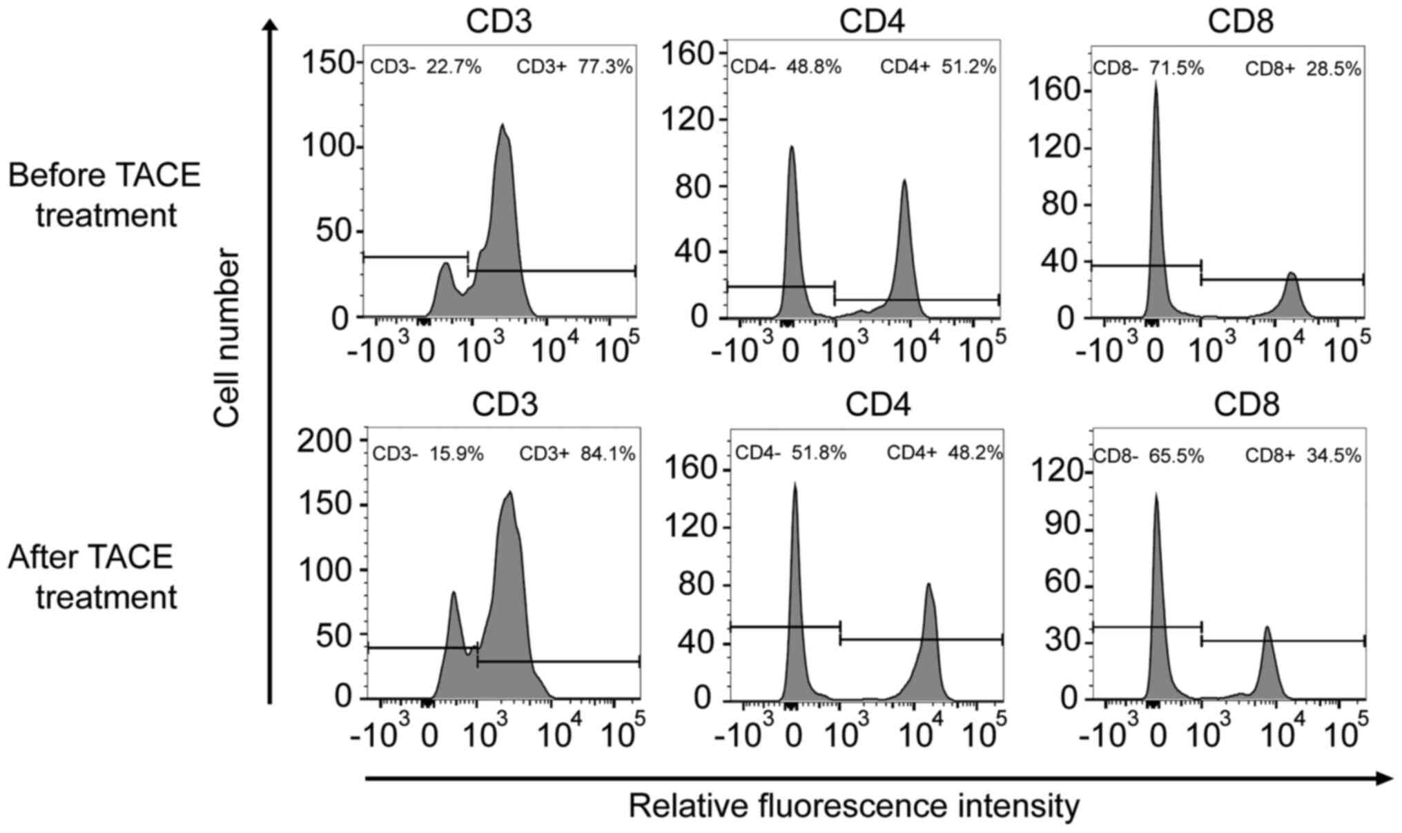
Effects of TACE treatment on the
immune function of patients
To further verify the effects of TACE treatment on
the immune function of patients, the immune status of patients was
examined before and after TACE treatment. The levels of
CD3+, CD4+/CD8+, PD1 and PD-L1
were selected as indices to evaluate immune function (Table IV, Fig.
1). Before patients underwent TACE treatment, the mean values
of PD1 and PD-L1 mRNA expression levels were 2.496 and 2.853,
respectively. After TACE treatment was performed, the mean values
of PD1 and PD-L1 mRNA expression levels were 4.312 and 3.11,
respectively. CD4+/CD8+ significantly
decreased after TACE treatment (P<0.001). Notably, the relative
mRNA expression levels of PD1 were significantly upregulated
(P<0.001). The mRNA expression levels of PD-L1 were also
increased after TACE treatment; however, this difference was not
significant (P=0.083). In addition, there was no significant
difference observed in CD3+ levels (P=0.167).
| Table IV.Immune parameters in patients with
hepatocellular carcinoma before and after TACE treatment. |
Table IV.
Immune parameters in patients with
hepatocellular carcinoma before and after TACE treatment.
| Parameter | Before TACE
treatment | After TACE
treatment | t | P-value |
|---|
|
CD4+/CD8+ (mean ±
std) | 1.818±0.720 | 1.462±0.717 | 4.141 | <0.001 |
| CD3+
(mean ± std) (µl) | 81.632±6.141 | 82.658±5.867 | −1.392 | 0.167 |
| PD1 relative mRNA
level | 2.496±0.939 | 4.312±0.558 | −17.801 | <0.001 |
| PD-L1 relative mRNA
level | 2.853±1.048 | 3.113±1.209 | −1.679 | 0.083 |
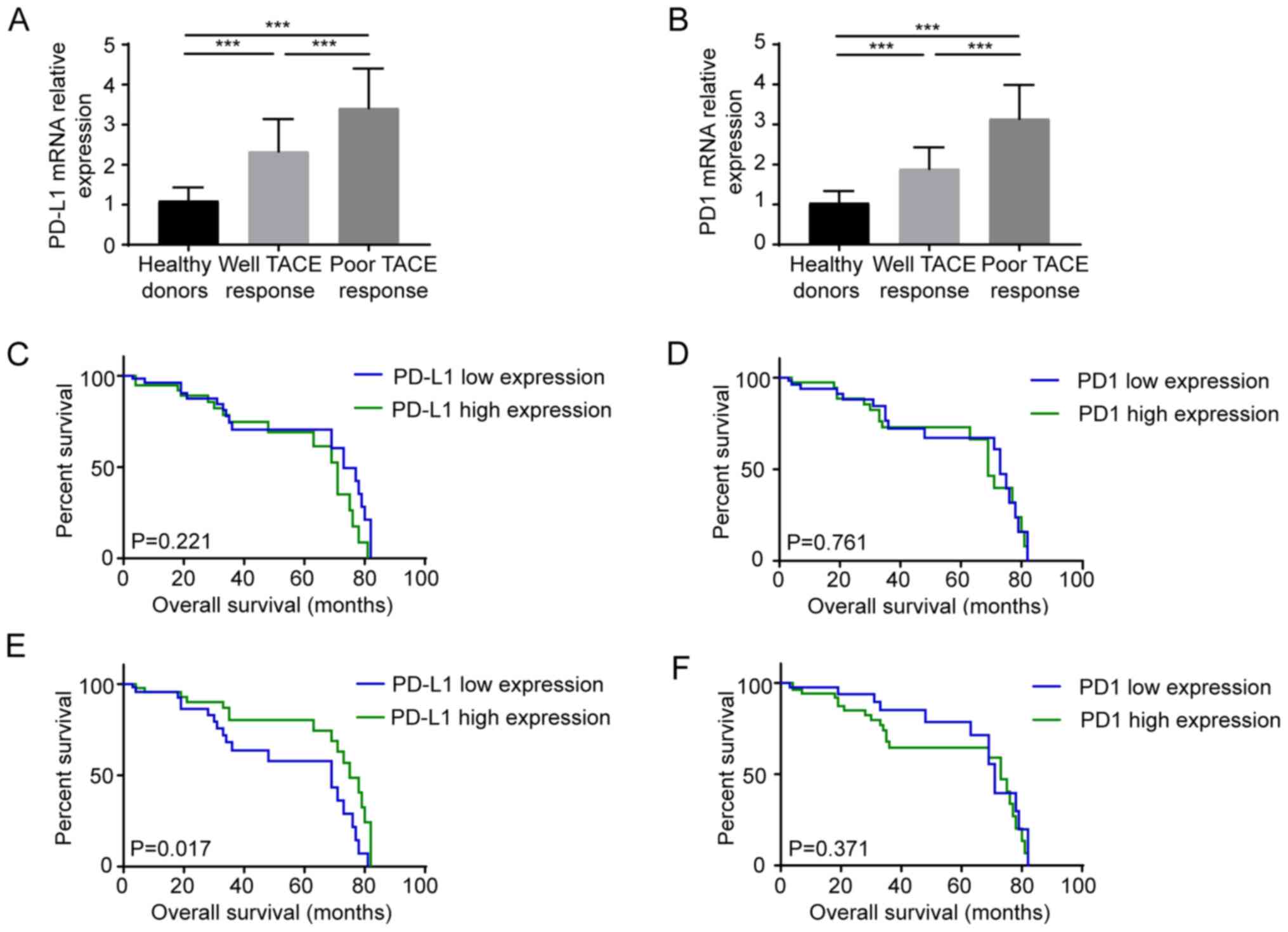
To further clarify the association between the
therapeutic effects of TACE and PD1/PD-L1 expression, the mRNA
expression levels of PD-L1/PD1 were detected in healthy donors and
patients, which were divided into well or poor response to TACE
groups (Fig. 2A and B). Compared
with healthy subjects, the expression levels of PD-L1/PD1 in
patients with HCC were significantly increased (P<0.001). The
mRNA expression levels of both PD-L1 (Fig. 2A) and PD1 (Fig. 2B) in patients with HCC with poor TACE
response were higher compared with those in patients with
satisfactory TACE response (all P<0.001). Kaplan-Meier curves
were plotted to analyze the survival of patients with high and low
expression levels of PD1/PD-L1. According to the average mentioned
in Table IV, patients were divided
into two groups for further analysis. Before patients underwent
TACE treatment, PD1 mRNA levels >2.5 and PD-L1 mRNA levels
>3.0 were regarded as high PD1/PD-L1 expression levels,
respectively. After TACE treatment was performed, PD1 mRNA
expression >4.0 and PD-L1 mRNA expression >3.0 were
considered as high PD1/PD-L1 expression levels, respectively. There
was no statistical difference in survival prognosis between the
high expression PD1 (P=0.761)/PD-L1 (P=0.221) group and the low
expression group before TACE treatment (Fig. 2C and D). However, after TACE
treatment, PD-L1 expression could be used to predict the prognosis
of patients with HCC. After TACE treatment, compared with high
PD-L1 expression, high PD-L1 expression significantly indicated
improved prognosis (P=0.017), while the expression level of PD1
(P=0.371) could not predict the prognosis before and after TACE
treatment (Fig. 2E and F).
Discussion
The immune system serves an important role in tumor
progression (19). The interaction
between the tumor and immune system can be divided into three
stages: Immune elimination, immune balance and immune escape
(20). In the early stages of
tumorigenesis, natural killer cells recognize and kill cancer
cells, and the debris is consumed by macrophages. Dendritic cells
exposed to cancer cell antigens secrete inflammatory cytokines and
present neoantigens to T cells (20,21). T
cells and B cells are then activated, resulting in the activation
of cellular immunity, which results in protection against
inflammation in the body (22). A
study by Ge et al (23) on
the influence of immune cell infiltration on the prognosis of
patients with colon cancer indicated that CD4+ T cell
infiltration exerted marked antitumor effects. A high density of
CD8+ T cells in advanced gastric cancer has been
considered to be associated with favorable prognosis (24). In the present study, PBMCs from 114
patients with HCC were collected to determine the effects of TACE
therapy on the immune system. The CD4+/CD8+
ratio decreased after TACE treatment, indicating that the
CD8+ cells were increased and immune function of the
patients was restored (25).
Tumor cells can evade immune surveillance through a
variety of mechanisms, including activating immune checkpoints to
suppress the antitumor immune response (26). PD1 is expressed on activated T cells,
B lymphocytes and natural killer cells. PD1 binds to tumor cells
with its cognate ligands (PD-L1), thereby inhibiting T cell
proliferation and response, suppressing patient immunity and
helping tumor immune escape (27).
Studies have revealed that PD1/PD-L1 expression in non-small cell
lung cancer, glioblastoma and lymphoma is associated with patient
prognosis (28–31). High PD1 expression has been
demonstrated to predict improved prognosis in patients with breast
cancer (32). A recent study
indicated that PD1 has different effects on the prognosis of
different subtypes of lymphoma (33). In the present study, the association
between PD1/PD-L1 expression and TACE was also analyzed. After TACE
treatment, the mRNA expression levels of both PD1 and PD-L1 were
increased, and patients with low PD-L1 mRNA expression had a poor
prognosis. This result is consistent with that of Hanna et
al (34), which showed the high
expression of PD-L1 was associated with a good prognosis in young
women with oral cavity squamous cell carcinoma. It has been
reported that PD-L1 upregulation is associated with antitumor
inflammatory response induced by CD8 cell infiltration (34,35).
However, the effect of TACE treatment on PD-L1 expression and
function of immune cells remains unclear. The association between
PD-L1 and prognosis and the intrinsic mechanism require further
investigation.
In recent years, immunotherapy has developed rapidly
and obtained remarkable achievements. Lee et al (36) revealed that the combination of
cytokine-induced killer cell therapy with RFA or TACE could improve
patient outcomes. Lee et al (37) reported a positive association between
PD-L1 and sorafenib resistance. These results suggested that PD-L1
inhibitors combined with sorafenib may be a novel strategy to
prevent or overcome sorafenib-acquired resistance in patients with
HCC (38). In the present study, the
expression levels of PD-L1 and PD1 in patients with poor response
were higher than those in patients with good response before TACE
treatment. In addition, the mRNA expression levels of both PD1 and
PD-L1 were increased after TACE treatment. Therefore, further
studies are required to determine the therapeutic effects of TACE
and immunotherapy combination strategy.
Although the present results indicated that TACE
combined with immunotherapy has potential clinical value for
patients with HCC, there were also several limitations in the
present study. Firstly, the specific mechanism of the increase in
PD1 and PD-L1 in PBMCs was not investigated. As for the mechanism
of TACE-induced PD1/PD-L1 mRNA upregulation, related studies have
reported that, following chemotherapy and targeted drug treatment,
residual tumor cells can restore the stemness and enhance immune
escape, which is one of the reasons for the failure of existing
treatment models (39–41). Furthermore, residual tumor cells have
been demonstrated to reshape the surrounding immune
microenvironment by upregulating the levels of inhibitory
cytokines, such as IL-1 and MMP family proteins, to force
suppressive immune cells in the immune microenvironment to complete
the immune escape and promote the recurrence of tumor cells
(40,42). Due to abundant blood flow in the
liver, suppressive cytokines and immune cells may affect the
PD1/PD-L1 levels in peripheral blood lymphocytes (43,44).
Further studies will be required to evaluate the intrinsic
mechanism. Secondly, the present study used RT-qPCR to detect the
expression levels of PD1/PD-L1 instead of flow cytometry based on
previous studies (45–47), which performed qPCR to detect the
expression levels of PD1/PD-L1, it was hypothesized that qPCR is a
reliable way for detecting PD1/PD-L1 expression. However, this
meant that the present study only detected the mRNA levels of
PD1/PD-L1. In addition, more samples should be included in future
studies to determine the effect of TACE treatment on immune
function.
In summary, the present data demonstrated that TACE
could improve the proportion of CD8+ cells and the mRNA
expression level of PD1 in patients with HCC. Before TACE
treatment, high PD1 and PD-L1 mRNA levels were significantly
associated with poor therapeutic response to TACE. After TACE
treatment, high PD-L1 mRNA expression was significantly associated
with improved patient prognosis. TACE combined with immunotherapy
has potential clinical value for patients with HCC.
Acknowledgements
Not applicable.
Funding
No funding was received.
Availability of data and materials
All data generated or analyzed during this study are
included in this published article.
Authors' contributions
RW designed the study. YH and ZJ collected and
analyzed the data. JG and SW performed the experiments, prepared
figures and tables and drafted the manuscript. JG and RW confirmed
the authenticity of all the raw data. All authors have read and
approved the final manuscript.
Ethics approval and consent to
participate
The present study was approved by the Ethics
Committee of Qingdao Sixth People's Hospital (Qingdao, China;
approval no. 2013-R-0012). Written informed consent was provided by
patients or their guardians prior to the study start.
Patient consent for publication
Not applicable.
Competing interests
The authors declare that they have no competing
interests.
References
|
1
|
Park JW, Chen M, Colombo M, Roberts LR,
Schwartz M, Chen PJ, Kudo M, Johnson P, Wagner S, Orsini LS and
Sherman M: Global patterns of hepatocellular carcinoma management
from diagnosis to death: The bridge study. Liver Int. 35:2155–2166.
2015. View Article : Google Scholar : PubMed/NCBI
|
|
2
|
Wallace MC, Preen D, Jeffrey GP and Adams
LA: The evolving epidemiology of hepatocellular carcinoma: A global
perspective. Expert Rev Gastroenterol Hepatol. 9:765–779. 2015.
View Article : Google Scholar : PubMed/NCBI
|
|
3
|
Lei C, Ren D, Fu M, Sun C, Ren H, Pan Q
and Li Y: Curative effect of endostar combined with oxaliplatin in
the treatment of primary hepatic carcinoma and its influence on
immune cells. Oncol Lett. 17:3665–3670. 2019.PubMed/NCBI
|
|
4
|
Trépo C, Chan HL and Lok A: Hepatitis B
virus infection. Lancet. 384:2053–2063. 2014. View Article : Google Scholar
|
|
5
|
Ichikawa T, Sano K and Morisaka H:
Diagnosis of pathologically early HCC with EOB-MRI: Experiences and
current consensus. Liver Cancer. 3:97–107. 2014. View Article : Google Scholar : PubMed/NCBI
|
|
6
|
Hartke J, Johnson M and Ghabril M: The
diagnosis and treatment of hepatocellular carcinoma. Semin Diagn
Pathol. 34:153–159. 2017. View Article : Google Scholar : PubMed/NCBI
|
|
7
|
Zhu ZX, Huang JW, Liao MH and Zeng Y:
Treatment strategy for hepatocellular carcinoma in China:
Radiofrequency ablation versus liver resection. Jpn J Clin Oncol.
46:1075–1080. 2016.PubMed/NCBI
|
|
8
|
Tsurusaki M and Murakami T: Surgical and
locoregional therapy of HCC: TACE. Liver Cancer. 4:165–175. 2015.
View Article : Google Scholar : PubMed/NCBI
|
|
9
|
Llovet JM and Bruix J: Systematic review
of randomized trials for unresectable hepatocellular carcinoma:
Chemoembolization improves survival. Hepatology. 37:429–442. 2003.
View Article : Google Scholar : PubMed/NCBI
|
|
10
|
Raoul JL, Forner A, Bolondi L, Cheung TT,
Kloeckner R and de Baere T: Updated use of TACE for hepatocellular
carcinoma treatment: How and when to use it based on clinical
evidence. Cancer Treat Rev. 72:28–36. 2019. View Article : Google Scholar : PubMed/NCBI
|
|
11
|
Ito F, Vardam TD, Appenheimer MM, Eng KH,
Gollnick SO, Muhitch JB and Evans SS: In situ thermal ablation
augments antitumor efficacy of adoptive T cell therapy. Int J
Hyperthermia. 36 (Suppl 1):S22–S36. 2019. View Article : Google Scholar : PubMed/NCBI
|
|
12
|
Löffler MW, Nussbaum B, Jäger G,
Jurmeister PS, Budczies J, Pereira PL, Clasen S, Kowalewski DJ,
Mühlenbruch L, Königsrainer I, et al: A non-interventional clinical
trial assessing immune responses after radiofrequency ablation of
liver metastases from colorectal cancer. Front Immunol.
10:25262019. View Article : Google Scholar
|
|
13
|
Zhou J, Sun HC, Wang Z, Cong WM, Wang JH,
Zeng MS, Yang JM, Bie P, Liu LX, Wen TF, et al: Guidelines for
diagnosis and treatment of primary liver cancer in China (2017
edition). Liver Cancer. 7:235–260. 2018. View Article : Google Scholar : PubMed/NCBI
|
|
14
|
Zhao S, Wang M, Yang Z, Tan K, Zheng D, Du
X and Liu L: Comparison between child-pugh score and
albumin-bilirubin grade in the prognosis of patients with HCC after
liver resection using time-dependent ROC. Ann Transl Med.
8:5392020. View Article : Google Scholar : PubMed/NCBI
|
|
15
|
Shimura S, Odagiri S, Furuya H, Okada K,
Ozawa K, Nagase H, Yamaguchi M and Cho Y: Echocardiography-guided
aortic cannulation by the Seldinger technique for type A dissection
with cerebral malperfusion. J Thorac Cardiovasc Surg. 159:784–793.
2020. View Article : Google Scholar : PubMed/NCBI
|
|
16
|
Eisenhauer EA, Therasse P, Bogaerts J,
Schwartz LH, Sargent D, Ford R, Dancey J, Arbuck S, Gwyther S,
Mooney M, et al: New response evaluation criteria in solid tumours:
Revised RECIST guideline (version 1.1). Eur J Cancer. 45:228–247.
2009. View Article : Google Scholar : PubMed/NCBI
|
|
17
|
Shi B, Sun A and Zhang X: Influence of
different ex vivo cell culture methods on the proliferation and
anti-tumor activity of cytokine-induced killer cells from gastric
cancer patients. Onco Targets Ther. 11:2657–2672. 2018. View Article : Google Scholar : PubMed/NCBI
|
|
18
|
Livak KJ and Schmittgen TD: Analysis of
relative gene expression data using real-time quantitative PCR and
the 2(-Delta Delta C(T)) method. Methods. 25:402–408. 2001.
View Article : Google Scholar : PubMed/NCBI
|
|
19
|
Gonzalez H, Hagerling C and Werb Z: Roles
of the immune system in cancer: from tumor initiation to metastatic
progression. Genes Dev. 32:1267–1284. 2018. View Article : Google Scholar : PubMed/NCBI
|
|
20
|
Wu X, Peng M, Huang B, Zhang H, Wang H,
Huang B, Xue Z, Zhang L, Da Y, Yang D, et al: Immune
microenvironment profiles of tumor immune equilibrium and immune
escape states of mouse sarcoma. Cancer Lett. 340:124–133. 2013.
View Article : Google Scholar : PubMed/NCBI
|
|
21
|
Quezada SA, Peggs KS, Simpson TR and
Allison JP: Shifting the equilibrium in cancer immunoediting: From
tumor tolerance to eradication. Immunol Rev. 241:104–118. 2011.
View Article : Google Scholar : PubMed/NCBI
|
|
22
|
Ferrari SM, Fallahi P, Galdiero MR,
Ruffilli I, Elia G, Ragusa F, Paparo SR, Patrizio A, Mazzi V,
Varricchi G, et al: Immune and Inflammatory Cells in Thyroid Cancer
Microenvironment. Int J Mol Sci. 20:2019. View Article : Google Scholar
|
|
23
|
Ge P, Wang W, Li L, Zhang G, Gao Z, Tang
Z, Dang X and Wu Y: Profiles of immune cell infiltration and
immune-related genes in the tumor microenvironment of colorectal
cancer. Biomed Pharmacother. 118:1092282019. View Article : Google Scholar : PubMed/NCBI
|
|
24
|
Wang Y, Zhu C, Song W, Li J, Zhao G and
Cao H: PD-L1 expression and CD8(+) T cell infiltration predict a
favorable prognosis in advanced gastric cancer. J Immunol Res.
2018:41805172018. View Article : Google Scholar : PubMed/NCBI
|
|
25
|
Zerbini A, Pilli M, Penna A, Pelosi G,
Schianchi C, Molinari A, Schivazappa S, Zibera C, Fagnoni FF,
Ferrari C and Missale G: Radiofrequency thermal ablation of
hepatocellular carcinoma liver nodules can activate and enhance
tumor-specific T-cell responses. Cancer Res. 66:1139–1146. 2006.
View Article : Google Scholar : PubMed/NCBI
|
|
26
|
Darvin P, Toor SM, Nair VS and Elkord E:
Immune checkpoint inhibitors: recent progress and potential
biomarkers. Exp Mol Med. 50:1–11. 2018. View Article : Google Scholar : PubMed/NCBI
|
|
27
|
Abril-Rodriguez G and Ribas A: SnapShot:
immune checkpoint inhibitors. Cancer Cell. 31:848–848 e841. 2017.
View Article : Google Scholar : PubMed/NCBI
|
|
28
|
Mazieres J, Drilon A, Lusque A, Mhanna L,
Cortot AB, Mezquita L, Thai AA, Mascaux C, Couraud S, Veillon R, et
al: Immune checkpoint inhibitors for patients with advanced lung
cancer and oncogenic driver alterations: Results from the
IMMUNOTARGET registry. Ann Oncol. 30:1321–1328. 2019. View Article : Google Scholar : PubMed/NCBI
|
|
29
|
Wang Z, Zhang C, Liu X, Wang Z, Sun L, Li
G, Liang J, Hu H, Liu Y, Zhang W and Jiang T: Molecular and
clinical characterization of PD-L1 expression at transcriptional
level via 976 samples of brain glioma. Oncoimmunology.
5:e11963102016. View Article : Google Scholar : PubMed/NCBI
|
|
30
|
Veloza L, Teixido C, Castrejon N, Climent
F, Carrió A, Marginet M, Soldini D, González-Farré B,
Ribera-Cortada I, Lopez-Guillermo A, et al: Clinicopathological
evaluation of the programmed cell death 1 (PD1)/programmed cell
death-ligand 1 (PD-L1) axis in post-transplant lymphoproliferative
disorders: Association with Epstein-Barr virus, PD-L1 copy number
alterations, and outcome. Histopathology. 75:799–812. 2019.
View Article : Google Scholar : PubMed/NCBI
|
|
31
|
Ferrara R, Mezquita L, Texier M, Lahmar J,
Audigier-Valette C, Tessonnier L, Mazieres J, Zalcman G, Brosseau
S, Le Moulec S, et al: Hyperprogressive disease in patients with
advanced non-small cell lung cancer treated with PD-1/PD-L1
inhibitors or with single-agent chemotherapy. JAMA Oncol.
4:1543–1552. 2018. View Article : Google Scholar : PubMed/NCBI
|
|
32
|
Jiang C, Cao S, Li N, Jiang L and Sun T:
PD-1 and PD-L1 correlated gene expression profiles and their
association with clinical outcomes of breast cancer. Cancer Cell
Int. 19:2332019. View Article : Google Scholar : PubMed/NCBI
|
|
33
|
Xie M, Huang X, Ye X and Qian W:
Prognostic and clinicopathological significance of PD-1/PD-L1
expression in the tumor microenvironment and neoplastic cells for
lymphoma. Int Immunopharmacol. 77:1059992019. View Article : Google Scholar : PubMed/NCBI
|
|
34
|
Hanna GJ, Woo SB, Li YY, Barletta JA,
Hammerman PS and Lorch JH: Tumor PD-L1 expression is associated
with improved survival and lower recurrence risk in young women
with oral cavity squamous cell carcinoma. Int J Oral Maxillofac
Surg. 47:568–577. 2018. View Article : Google Scholar : PubMed/NCBI
|
|
35
|
Dong H, Strome SE, Salomao DR, Tamura H,
Hirano F, Flies DB, Roche PC, Lu J, Zhu G, Tamada K, et al:
Tumor-associated B7-H1 promotes T-cell apoptosis: A potential
mechanism of immune evasion. Nat Med. 8:793–800. 2002. View Article : Google Scholar : PubMed/NCBI
|
|
36
|
Lee JH, Lee JH, Lim YS, Yeon JE, Song TJ,
Yu SJ, Gwak GY, Kim KM, Kim YJ, Lee JW and Yoon JH: Adjuvant
immunotherapy with autologous cytokine-induced killer cells for
hepatocellular carcinoma. Gastroenterology. 148:1383–1391.e6. 2015.
View Article : Google Scholar : PubMed/NCBI
|
|
37
|
Lee JH, Lee JH, Lim YS, Yeon JE, Song TJ,
Yu SJ, Gwak GY, Kim KM, Kim YJ, Lee JW and Yoon JH: Sustained
efficacy of adjuvant immunotherapy with cytokine-induced killer
cells for hepatocellular carcinoma: An extended 5-year follow-up.
Cancer Immunol Immunother. 68:23–32. 2018. View Article : Google Scholar : PubMed/NCBI
|
|
38
|
Liu J, Liu Y, Meng L, Liu K and Ji B:
Targeting the PD-L1/DNMT1 axis in acquired resistance to sorafenib
in human hepatocellular carcinoma. Oncol Rep. 38:899–907. 2017.
View Article : Google Scholar : PubMed/NCBI
|
|
39
|
Naruse T, Yanamoto S, Okuyama K, Ohmori K,
Tsuchihashi H, Furukawa K, Yamada SI and Umeda M:
Immunohistochemical study of PD-1/PD-L1 axis expression in oral
tongue squamous cell carcinomas: Effect of neoadjuvant chemotherapy
on local recurrence. Pathol Oncol Res. 26:735–742. 2020. View Article : Google Scholar : PubMed/NCBI
|
|
40
|
Walcher L, Kistenmacher AK, Suo H, Dluczek
S, Strauß A, Blaudszun AR, Yevsa T, Fricke S and Kossatz-Boehlert
U: Cancer stem cells-origins and biomarkers: Perspectives for
targeted personalized therapies. Front Immunol. 11:12802020.
View Article : Google Scholar : PubMed/NCBI
|
|
41
|
Oh SJ, Ahn EJ, Kim O, Kim D, Jung TY, Jung
S, Lee JH, Kim KK, Kim H, Kim EH, et al: The role played by SLUG,
an epithelial-mesenchymal transition factor, in invasion and
therapeutic resistance of malignant glioma. Cell Mol Neurobiol.
39:769–782. 2019. View Article : Google Scholar : PubMed/NCBI
|
|
42
|
Peng J, Hamanishi J, Matsumura N, Abiko K,
Murat K, Baba T, Yamaguchi K, Horikawa N, Hosoe Y, Murphy SK, et
al: Chemotherapy induces programmed cell death-ligand 1
overexpression via the nuclear factor-κB to foster an
immunosuppressive tumor microenvironment in ovarian cancer. Cancer
Res. 75:5034–5045. 2015. View Article : Google Scholar : PubMed/NCBI
|
|
43
|
Baraibar I, Melero I, Ponz-Sarvise M and
Castanon E: Safety and tolerability of immune checkpoint inhibitors
(PD-1 and PD-L1) in cancer. Drug Saf. 42:281–294. 2019. View Article : Google Scholar : PubMed/NCBI
|
|
44
|
Chen S, Crabill GA, Pritchard TS, McMiller
TL, Wei P, Pardoll DM, Pan F and Topalian SL: Mechanisms regulating
PD-L1 expression on tumor and immune cells. J Immunother Cancer.
7:3052019. View Article : Google Scholar : PubMed/NCBI
|
|
45
|
Ma LJ, Feng FL, Dong LQ, Zhang Z, Duan M,
Liu LZ, Shi JY, Yang LX, Wang ZC, Zhang S, et al: Clinical
significance of PD-1/PD-Ls gene amplification and overexpression in
patients with hepatocellular carcinoma. Theranostics. 8:5690–5702.
2018. View Article : Google Scholar : PubMed/NCBI
|
|
46
|
Kawahara T, Ishiguro Y, Ohtake S, Kato I,
Ito Y, Ito H, Makiyama K, Kondo K, Miyoshi Y, Yumura Y, et al: PD-1
and PD-L1 are more highly expressed in high-grade bladder cancer
than in low-grade cases: PD-L1 might function as a mediator of
stage progression in bladder cancer. BMC Urol. 18:972018.
View Article : Google Scholar : PubMed/NCBI
|
|
47
|
D'Alterio C, Nasti G, Polimeno M, Ottaiano
A, Conson M, Circelli L, Botti G, Scognamiglio G, Santagata S, De
Divitiis C, et al: CXCR4-CXCL12-CXCR7, TLR2-TLR4, and PD-1/PD-L1 in
colorectal cancer liver metastases from neoadjuvant-treated
patients. Oncoimmunology. 5:e12543132016. View Article : Google Scholar : PubMed/NCBI
|